Scaricare la presentazione
La presentazione è in caricamento. Aspetta per favore

2
Factors Affecting the Sedimentation Rate
Elevating the ESR: Inflammatory Diseases Cytokine driven processes that elevate fibrinogen ie TB, Pneumonia, rheumatoid arthritis Relative/Absolute Increase in Globulin Proteins Loss of albumin ie nephrotic syndrome or increase in globulins ie multiple myeloma Extensive Tissue Necrosis Myocardial infarction, trauma, tumors Other Causes Pregnancy (increase in fibrinogen, anemia), anemia, age, heparinized blood Lowering the ESR: Increased Plasma Viscosity Waldenstrom's macroglobulinemia Red Cell Number or Shape Polycythemia vera, sickle cell disease Decreased Plasma Proteins Hepatic necrosis, hypofibrinogenemia Others Causes Trichinosis
, anemia, age, heparinized blood. Lowering the ESR: Increased Plasma Viscosity Waldenstrom s macroglobulinemia. Red Cell Number or Shape Polycythemia vera, sickle cell disease. Decreased Plasma Proteins Hepatic necrosis, hypofibrinogenemia. Others Causes Trichinosis.")
3
Range - The range of normal SRE is 0-15 mm/hr for men and 0-20 mm/hr for women. Many articles have detailed the elevation of the ESR with age and some have suggested the formula of age divided by 2 for men and age plus 10 divided by 2 for women although this has not been universally adopted. Utility - Although still widely used, the sedimentation rate has limited use as a diagnostic test. It is useful for predicting prognosis in diseases such as rheumatoid arthritis and Hodgkin's disease and it has utility as a marker of treatment efficacy in many diseases such as rheumatoid arthritis, the vasculitides, collagen vascular diseases, and septic arthritis.

4
CRP (Proteina C reattiva)
The C-reactive protein owes its name to the ability of this protein to precipitate pneumococcal C-polysaccharide in the presence of calcium. It was first discovered in 1930 by Tillet and Frances. a positive CRP may indicate any of a number of things: Rheumatoid arthritis Rheumatic fever Cancer Tubercolosis Pneumococcal pneumonia Myocardial infartion Systemic Lupus Erithematosus Positive CRP results also occur during the last half of pregnancy or with the use of oral contraceptives.

5
CRP Utility - Because the CRP is a direct measure of inflammation and it is becoming easier and cheaper to do, there may be a time the CRP supersedes the ESR (although the same was said for the measure of plasma viscosity 10 years ago). It is as useful as the ESR in most cases and more accurately reflects the current level of inflammation.
. It is as useful as the ESR in most cases and more accurately reflects the current level of inflammation.")
6
PRINCIPALI MALATTIE AUTOIMMUNI
SISTEMICHE INTERMEDIE ORGANO-SPECIFICHE T. HASHIMOTO LES S. GOODPASTURE ADDISON IDIOPATICO AR UVEITE FACOGENICA LUPUS DISCOIDE GASTRITE ATROFICA CIRROSI B. PRIM. DIABETE GIOVANILE OFTALMIA SIMPATICA SCLERODERMIA SCLEROSI MULTIPLA IFERTILITA' MASCH. D. MIOSITE M. DI SJOGREN S. PLURIENDOCRINE AUTOIMMUNI CONNETTIVITE MISTA MIASTENIA GRAVE COLITE ULCEROSA ANEMIA PERNICIOSA

7
Lupus eritematoso sistematico
Appare nelle donne tra 13 e 40 anni. Rapporto maschio-femmina 1:10 Caratterizzato da febbre, debolezza, artriti, disfunsioni renali I pazienti producono autoanticorpi verso il DNA, istoni, eritrociti, piastrine, leucociti, e fattori di coagulazione del sangue Gli immunocomplessi depositati lungo le pareti dei vasi sanguigni causano una ipersensibilità di tipo III, originano danno endoteliale che da luogo alle reazioni infiammatorie che generano vasculuiti e glomerulonefriti

8
Esami di Routine e Lupus
Incremento della VES e dei livelli di PCR Emocromo: Anemia (Sintomo Costante) Leucopenia (Linfocitopenia) Trombocitopenia Alterazioni della coagulazione (Lupus Anticoagulante) Esame Urine: Albuminuria (Microalbuminuria) Ematuria
 Leucopenia (Linfocitopenia) Trombocitopenia. Alterazioni della coagulazione (Lupus Anticoagulante) Esame Urine: Albuminuria (Microalbuminuria) Ematuria.")
9
Allo stato attuale la dimostrazione della positività della ricerca di autoanticorpi anti-nucleo (ANA) o la presenza di anticorpi anti-dsDNA o anti-Sm (uno degli antigeni nucleari estraibili, ENA) costituiscono 2 degli 11 criteri utilizzati da anni per la diagnosi di lupus eritematoso sistemico
 o la presenza di anticorpi anti-dsDNA o anti-Sm (uno degli antigeni nucleari estraibili, ENA) costituiscono 2 degli 11 criteri utilizzati da anni per la diagnosi di lupus eritematoso sistemico")
10
In generale il protocollo diagnostico iniziale, in pazienti sintomatici prevede la rilevazione degli anticorpi anti-nucleo in IFI; il pattern di fluorescenza nucleare o citoplasmatico determina la scelta successiva, rappresentata dalla ricerca di autoanticorpi diretti verso uno o più specifici autoantigeni intracellulari

11
UV light Autoimmunity

12
Standardizzata con linee cellulari epiteliali (HEp-2)
Determinazione degli ANA: Tecnica IFI Standardizzata con linee cellulari epiteliali (HEp-2) (esprimono antigeni umani presenti in tutte le fasi del ciclo cellulare) Ai fini diagnostici I titoli di 1:40 e di 1:160 sono considerati come livelli decisionali: Titolo soglia 1:40 (alta sensibilità/bassa specificità) <1:40 negativo. (Anticorpi antinucleo a basso titolo 1:40 - 1:80 possono essere presenti in soggetti sani, nelle gravide, in donne sopra i 40 anni, negli anziani) >1:40 e <1:160 basso positivo (in assenza di sintomi specifici, il protocollo diagnostico deve prevedere un monitoraggio in tempi successivi) >/=1:160 sono da considerare comunque suggestivi di patologia autoimmune.
 (esprimono antigeni umani presenti in tutte le fasi del ciclo cellulare) Ai fini diagnostici I titoli di 1:40 e di 1:160 sono considerati come livelli decisionali: Titolo soglia 1:40 (alta sensibilità/bassa specificità) <1:40 negativo. (Anticorpi antinucleo a basso titolo 1:40 - 1:80 possono essere presenti in soggetti sani, nelle gravide, in donne sopra i 40 anni, negli anziani) >1:40 e <1:160 basso positivo (in assenza di sintomi specifici, il protocollo diagnostico deve prevedere un monitoraggio in tempi successivi) >/=1:160 sono da considerare comunque suggestivi di patologia autoimmune.")
13
Pattern omogeneo Pattern periferico
Fluorescenza omogeneamente diffusa a tutto il nucleo con colorazione dei cromosomi delle cellule in mitosi. Gli Ab sono diretti contro desossiribonucleoproteine, istoni, dsDNA. Pattern periferico Forte fluorescenza alla periferia del nucleo debole al centro. Dovuta alla presenza di autoanticorpi diretti contro ds DNA o contro desossiribonucleoproteine

14
IL test IFI viene utilizzato anche per la valutazione degli anticorpi anti ds DNA questo test si fonda su una reazione a carico del DNA mitocondriale a doppia elica contenuto nel cinetoplasto di un emoflagellato non patogeno per l’uomo (Crithidia luciliae) Negativo Positivo

15
Limitazioni di quest’approccio:
Alcuni ENA (anti-Ro/SSA ed anti-Jo1) possono dare risultati falsamente negativi (ridotta espressione degli antigeni-bersaglio nelle cellule HEp-2; perdita e/o denaturazione degli antigeni in fase di allestimento dei vetrini Non univocità della modalità di refertazione Dipendenza dell’affidabilità del dato dall’esperienza del microscopista Difficoltà di reperimento di sieri standard di riferimento
 possono dare risultati falsamente negativi (ridotta espressione degli antigeni-bersaglio nelle cellule HEp-2; perdita e/o denaturazione degli antigeni in fase di allestimento dei vetrini. Non univocità della modalità di refertazione. Dipendenza dell’affidabilità del dato dall’esperienza del microscopista. Difficoltà di reperimento di sieri standard di riferimento.")
16
Gli anticorpi anti-DNA
Test EIA DNA a singola elica (denaturato, ssDNA; determinanti antigenici: zone ricche di G-C e A-T) DNA nativo a doppia elica (dsDNA, epitopi localizzati lungo lo scheletro glico-fosfato) Metodica IFI (Crithidia luciliae), DNA nativo a doppia elica Gli anticorpi anti-ssDNA non hanno una buona associazione con precisi quadri patologici. Gli anticorpi anti-dsDNA sono altamente specifici per il LES (10° criterio diagnostico del LES)
 DNA nativo a doppia elica (dsDNA, epitopi localizzati lungo lo scheletro glico-fosfato) Metodica IFI (Crithidia luciliae), DNA nativo a doppia elica. Gli anticorpi anti-ssDNA non hanno una buona associazione con precisi quadri patologici. Gli anticorpi anti-dsDNA sono altamente specifici per il LES (10° criterio diagnostico del LES)")
17
rappresentare il principale dato di laboratorio.
AutoantigeniENA: Ro/SS-A La/SS-B Sm RNP Topoisomerasi I (Scl-70) Istidil-tRNA sintetasi (Jo-1) Proteina B centromerica (CENP-B) rRNP Nucleosomi (cromatina) In presenza di segni clinici di S. di Sjogren o di Dermatomiosite/polimiosite possono rappresentare il principale dato di laboratorio.
 Istidil-tRNA sintetasi (Jo-1) Proteina B centromerica (CENP-B) rRNP. Nucleosomi (cromatina) In presenza di segni clinici di S. di Sjogren o di Dermatomiosite/polimiosite possono. rappresentare il principale dato di laboratorio.")
18
LUPUS Protocolli diagnostici in alcune malattie Autoimmuni
ANA Pattern omogeneo o periferico ad alto titolo Anti dsDNA Positivo ENA anti-Sm (altri inutili) AnticorpiAntifosfolipidi Incostantemente presenti e lupus Anticoagulante
 AnticorpiAntifosfolipidi Incostantemente presenti. e lupus Anticoagulante.")
19
Protocolli diagnostici di base in alcune malattie Autoimmuni
Sindrome di Sjogren IgG Incremento policlonale ANA 20% dei casi negativi 80% dei casi positivi 80% granulare 20% omogeneo Raramente Nucleolare Anti dsDNA (Inutili) ENA 80% Ssa/Ro 70% SSb/La
 ENA 80% Ssa/Ro. 70% SSb/La.")
20
Polimiosite/Dermatomiosite
Protocolli diagnostici di base in alcune malattie Autoimmuni Polimiosite/Dermatomiosite VES Molto Elevata Emocromo Lieve anemia, Eosinofilia ANA Pattern granulare Anti dsDNA (Inutili) ENA 15% dei casi positivo Jo1 CPK, LDH Generalmente aumentate Fattore Reumatoide 30% dei casi positivo
 ENA 15% dei casi positivo Jo1. CPK, LDH Generalmente aumentate. Fattore Reumatoide 30% dei casi positivo.")
21
Antiphospholipid antibodies: Can be present in 30% 0f SLE patients.
Antibodies directed against phosphorylated polysaccharide esters of fatty acids Include: lupus anticoagulant, b2-glycoprotein-I, anti-prothrombin Abs, and anticardiolipin Ab. False positive VDRL can be seen in 50% of patients aPL production can also be associated with: Medications Infections Neoplasms (lymphoma) VDRL measures flocculation of lipid particles (that contain cholesterol, and negatively charged phospholipid, cardiolipin) Antiphospholipid antibodies bind to cardiolipin and cause flocculation. Depending on the method used, PT can also be high in patients with lupus anticoagulant (Moll and Ortel) in these patients you should check: Chromogenic factor X and Prothrombin-proconvertin time Infection is associated with increased tissue factor release
 VDRL measures flocculation of lipid particles (that contain cholesterol, and negatively charged phospholipid, cardiolipin) Antiphospholipid antibodies bind to cardiolipin and cause flocculation. Depending on the method used, PT can also be high in patients with lupus anticoagulant (Moll and Ortel) in these patients you should check: Chromogenic factor X and Prothrombin-proconvertin time. Infection is associated with increased tissue factor release.")
22
ANCA Anti-Neutrophil Cytoplasmic Antibodies

23
ANCA: Abs directed against several proteins in cytoplasm of neutrophils in sera of patients with different Vascular Autoimmune disease and Systemic Autoimmune Disease . Measured by indirect immunofluorescence. ELISA is used to detect specific Abs to proteinase-3 , and myeloperoxidase (MPO)
")
24
ANCA Patterns: Cytoplasmic ANCA (C-ANCA): anti-proteinase -3
Perinuclear ANCA (p-ANCA): anti- myeloperoxidase (MPO), but also elastase and other proteins in the neutrophil granules Atypical Patterns: Ab to elastase, cathepsin G, lactoferrin, etc.
: anti- myeloperoxidase (MPO), but also elastase and other proteins in the neutrophil granules. Atypical Patterns: Ab to elastase, cathepsin G, lactoferrin, etc.")
25
Gli ANCA: patologie associate
Sono considerati i principali markers sierologici specifici delle vasculiti e delle malattie infiammatorie croniche,con un’incidenza dell’85-90% nelle prime e 20-70% nelle seconde ; Vasculite : infiammazione e Malattie infiammatorie croniche necrosi dei vasi con conseguente intestinali (MICI): modificazione del lume vasale ed con questo termine si indicano : alterazioni ischemiche dei tessuti - Rettocolice ulcerosa: irrorati; infiammazione del colon; quella maggiormente - Morbo di Crohn : distruzione rappresentata è il Morbo di Wegener. continua della parete dell’intestino,specialmente a carico del tenue.
: modificazione del lume vasale ed con questo termine si indicano : alterazioni ischemiche dei tessuti - Rettocolice ulcerosa: irrorati; infiammazione del colon; quella maggiormente - Morbo di Crohn : distruzione. rappresentata è il Morbo di Wegener. continua della parete dell’intestino,specialmente a carico del tenue.")
26
Classificazione p-ANCA o ANCA perinucleari : reagiscono principalmente con la mieloperossidasi (MPO) presente nei granuli α-azzurrofili, dando una fluorescenza di tipo perinucleare ; c-ANCA o ANCA citoplasmatici : diretti contro la proteinasi3 (PR3), dando una fluorescenza finemente granulare e diffusa per il citoplasma ; x-ANCA o ANCA atipici : - danno una fluorescenza sia citoplasmatica che nucleare - si osservano nei soggetti affetti da MICI,ma hanno una sensibilità sconosciuta e una specificità bassa.
 presente nei granuli α-azzurrofili, dando una fluorescenza di tipo perinucleare ; c-ANCA o ANCA citoplasmatici : diretti contro la proteinasi3 (PR3), dando una fluorescenza finemente granulare e diffusa per il citoplasma ; x-ANCA o ANCA atipici : - danno una fluorescenza sia citoplasmatica che nucleare. - si osservano nei soggetti affetti da MICI,ma hanno una sensibilità sconosciuta e una specificità bassa.")
27
C-ANCA (cytoplamsic-ANCA):
Diffuse staining of neutrophil cytoplasm in immunofluorescene. Recognizes PR3(Proteinase –3), a serine protease in primary granules of neutrophils. Is seen in 85% (range 30-90%) of patients with Wegeners Granulomatosis. Highly specific for WG (98%) Titer correlates with disease activity (in 60% of cases) and severity.
, a serine protease in primary granules of neutrophils. Is seen in 85% (range 30-90%) of patients with Wegeners Granulomatosis. Highly specific for WG (98%) Titer correlates with disease activity (in 60% of cases) and severity.")
28
P-ANCA: P-ANCA in the setting of vasculitis is Ab against MPO.
Vasculitidies associated with MPO include: Microscopic Polyangiitis (45-80%) Idiopathic Crescentic GN: (65%) Churg Strauss: (60%) PAN: (15%) Anti- MPO can also be due to medications: PTU, hydralazine, minocycline, D-penicillamine. Atypical P-ANCA can be seen with: RA, SLE, IBD, Chronic liver diseases.
 Idiopathic Crescentic GN: (65%) Churg Strauss: (60%) PAN: (15%) Anti- MPO can also be due to medications: PTU, hydralazine, minocycline, D-penicillamine. Atypical P-ANCA can be seen with: RA, SLE, IBD, Chronic liver diseases.")
29
Laboratory Testing, Principals and Guidelines
Rheumatoid Arthritis Laboratory Testing, Principals and Guidelines

30
Rheumatoid Factor (RF) Introduction:
RF is an antibody directed against the FC portion of IgG. RF was originally described by Waaler and Rose in 1940 The RF measured in laboratories is IgM RF, but IgG & IgA RF have been described. IgG and IgA RF are occasionally positive in RA patients who are IgM-RF negative. However, measurement of IgG, IgA, and 7S-IgM RFs may be prognostically importanat. Because they may be associated with more severe disease.

31
Conditions Associated with Positive RF:
Healthy Individuals: RF can be positive in up to 4% of young and healthy individuals. RF positivity is higher in elderly people without rheumatic diseases (ranging from 3-25% in different studies). RF titer in this setting is usually < 1:160 k
. RF titer in this setting is usually < 1:160. k.")
32
Conditions Associated with Positive RF: …cont.
II. Rheumatic Disorders: Rheumatoid Arthritis: %* Sjogren’s syndrome: 75-95% SLE: % Polymyositis/Dermatomyositis: 5-10% * Variable in different studies, depending on severity of disease in the study population cc

33
Non-Rheumatic Diseases associated with Positive RF
Astrix signifies the diseases with symptoms similar to RA Viral infections: Rubella, mumps, influenza, HIV Parasitic diseases: Chagas, Leishmaniasis, onchocerciasis, Schistosomiasis Malignancy: Leukemia and colon CA

34
Anti-Cyclic Citrullinated Peptide:
Target amino acid is citrulline, in filaggrin molecule derived from human skin. Citrulline is a post-translationally modified arginine residue. ELISA assay for anti-CCP may be useful in early stages of polyarthritis. [1] Sensitivity Specificity Anti-CCP % % IgM RF % % RF & anti-CCP % % [1] Bas et al. Rheumatoloy (Oxford) 2003; 62: 870 Anti CCP is of predictive value in RF NEGATIVE PATIENTS. Even severity of erosive disease was higher in those with positive CCP than those with positive RF and negative CCP
 2003; 62: 870. Anti CCP is of predictive value in RF NEGATIVE PATIENTS. Even severity of erosive disease was higher in those with positive CCP than those with positive RF and negative CCP.")
35
Serum Electrophoresis
Gel electrophoresis shows an increase in the globulins especially gamma (antibodies) and alpha-2-globulin There is often a decrease in albumin (7)
 and alpha-2-globulin. There is often a decrease in albumin (7)")
36
Complete Blood Count (CBC)
WBC in the peripheral blood often remained undisturbed RBC’s show a moderate normocytic hypochromic anemia of chronic disease A decrease in serum iron is common, Total Iron Binding Capacity (TIBC) and normal iron stores (ferritin) are essentially normal
 and normal iron stores (ferritin) are essentially normal.")
37
Creatinine Kinase (CK)
Serum CK is decreased below normal in >60% of RA patients. This is not to be confused with the CKMB (Myocardial) portion or CKMM (skeletal muscle) enzymes that are often measured during a MI or chronic muscular diseases (7)
 portion or CKMM (skeletal muscle) enzymes that are often measured during a MI or chronic muscular diseases (7)")
38
Synovial Biopsy Synovial fluid has high WBC and low viscosity, glucose is greatly diminished in the synovium RF usually present Characteristic rheumatoid nodules to aide in diagnosis Positive biopsy of subcutaneous rheumatoid nodule and synovia

39
American Rheumatism Association Diagnosis Criteria
Positive biopsy within diagnostic guidelines: criteria consists of: Poor mucin clotting of synovial fluid (4) Characteristic histological changes in synovium: mesothelial, macrophage, LE cells
 Characteristic histological changes in synovium: mesothelial, macrophage, LE cells.")
40
Testing non-indicative of RA
Other routine serological quantitative testing is normal. This group includes Calcium, Uric Acid, Alkaline Phosphorus, Phosphorus, and Antistreptolysin O titers

41
Diagnostica di laboratorio nello studio delle patologie immunoallergiche

42
La diagnostica dell’ asma bronchiale è costituita da:
Anamnesi ed esame obiettivo Test specifici in vivo e in vitro

43
VALUTAZIONE ANAMNESTICA
Predisposizione familiare (atopia) o altre patologie immunologiche; Attività lavorativa Abitudini di vita (sport, fumo, ambiente) Terapie in atto Stagionalità degli eventi allergici e modalità di insorgenza
 o altre patologie immunologiche; Attività lavorativa. Abitudini di vita (sport, fumo, ambiente) Terapie in atto. Stagionalità degli eventi allergici e modalità di insorgenza.")
44
Le reazioni di ipersensibilità IgE mediate determinano diversi tipi sintomatologie
L’allergia costituisce l’aspetto patologico, che si traduce in danno,con quadri clinici diversi secondo gli organi interessati: Rino-congiuntivite Asma Orticaria Allergie intestinali Shock anafilattico

45
I TEST UTILIZZATI SONO:
IN VIVO Test cutanei PRICK E PATCH TEST IN VITRO PRIST o IgE totali RAST o IgE specifiche

46
DOSAGGIO IgE TOTALI Soggetti normali,
IL RISCONTRO DI ELEVATI LIVELLI DI IgE TOTALI NON AUTORIZZA UNA DIAGNOSI DI ALLERGIA, POICHE’ POSSONO ESSERE ANCHE PRESENTI IN: Soggetti normali, Patologie Parassitarie Connettiviti, Infezioni batteriche croniche Malattie Linfoproliferative

47
Sistema di classificazione e cut-off relativi alla classe di IgE Specifiche
Elaborati in base alla curva standard e alla calibrazione, forniti in kU/L Classe kU/L <0, Assente o non rilevabile I ,35-0, Basso II ,70-3, Moderato III ,50- 17, Elevato IV ,5-52, Molto elevato V ,5-99, Molto elevato VI > Molto elevato

48
DOSAGGIO MEDIATORI E CITOCHINE
T-LINFOCITI IL-x - Mast cellule ISTAMINA PGs - LTs TRIPTASI EOS EOSINOFILI ECP PBM Basophil BASOFILO CHEMOCHINE

50
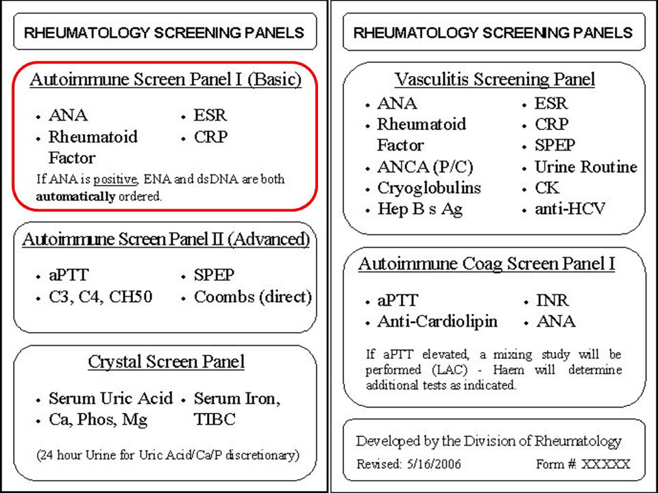
ANA: If the ANA is positive, the laboratory automatically performs Extractable Nuclear Antigens (ENA) and Double Stranded DNA Antibody (dsDNA) tests. If the ANA is negative, one may still request the Autoimmune Screen Panel II (Advanced) depending on the working diagnosis. Once the ANA has tested positive there is no diagnostic benefit in repeating this test. Please remember that an ANA may also be positive in up to 5% of normal young females and decisions to proceed with further testing should always take into account overall clinical and laboratory features.
 depending on the working diagnosis. Once the ANA has tested positive there is no diagnostic benefit in repeating this test. Please remember that an ANA may also be positive in up to 5% of normal young females and decisions to proceed with further testing should always take into account overall clinical and laboratory features.")
51
Rheumatoid Factor (RF): This test can be used alone or along with other diagnostic tests to identify specific autoimmune/rheumatologic or even infectious and malignant diseases. The test identifies the presence of a RF (an antibody {G,A,or M}) directed specifically against the CH2/3 domain of IgG. The titre may be relevant in some but not all conditions for disease monitoring (eg infections such as Tb), but a positive test is significant.
 directed specifically against the CH2/3 domain of IgG. The titre may be relevant in some but not all conditions for disease monitoring (eg infections such as Tb), but a positive test is significant.")
52
Serum electrophor

53
Autoimmune Screen Panel II
C3, C4, CH50: These tests measure the levels of complement proteins in a patient’s blood. The C4(classical pathway) and C3 (alternate) are static measures of the serum levels of these proteins, whereas the CH50 is a functional assay that correlates with the complement cascade activity. If the measured values are not within the reference range, this may be indicative of an autoimmune/rheumatologic disease with immune complex deposition. Coombs (direct): If an autoimmune/rheumatologic disease is suspected to be causing hemolytic anemia this test can check whether antibodies are indeed bound to red blood cells. This would indicate an autoimmune condition as the cause of the RBC destruction. SPEP (Serum protein electrophoresis): This test is used to monitor albumin and globulin protein levels in a patient’s blood. If an autoimmune/rheumatologic disease is affecting these levels, the test results may be out of the reference range. Lower albumin levels may reflect chronic illness, elevated acute phase reactants infection or inflammation, elevated gammaglobulins inflammation and if a spike present, possible gammopathy. aPTT: This test is used to measure how well the patient’s blood is clotting. It may be prolonged in association with autoimmune/rheumatologic disease and coagulopathies.
 and C3 (alternate) are static measures of the serum levels of these proteins, whereas the CH50 is a functional assay that correlates with the complement cascade activity. If the measured values are not within the reference range, this may be indicative of an autoimmune/rheumatologic disease with immune complex deposition. Coombs (direct): If an autoimmune/rheumatologic disease is suspected to be causing hemolytic anemia this test can check whether antibodies are indeed bound to red blood cells. This would indicate an autoimmune condition as the cause of the RBC destruction. SPEP (Serum protein electrophoresis): This test is used to monitor albumin and globulin protein levels in a patient’s blood. If an autoimmune/rheumatologic disease is affecting these levels, the test results may be out of the reference range. Lower albumin levels may reflect chronic illness, elevated acute phase reactants infection or inflammation, elevated gammaglobulins inflammation and if a spike present, possible gammopathy. aPTT: This test is used to measure how well the patient’s blood is clotting. It may be prolonged in association with autoimmune/rheumatologic disease and coagulopathies.")
55
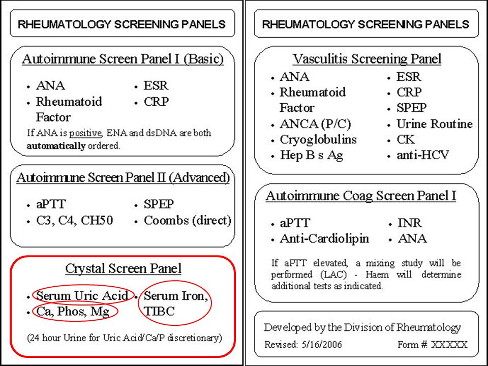
Crystal Screen Panel Serum Iron, TIBC: These tests are ordered together to test for iron levels and saturation in the patient’s blood. Serum iron tests the amount of iron in the patient’s serum while TIBC tests how much iron the patient’s transferrin plasma proteins can bind. An elevated serum iron and increased saturation will be seen in Hemochromatosis, which can be a cause of CPPD arthopathy (pseudogout). Serum Uric Acid: This test is used to detect the level of uric acid in the patient’s blood. High levels of uric acid can lead to monosodium urate crystal build up in joints which causes gout. Ca, Phos, Mg: These tests measure the amount of these minerals in the patient’s blood. Several crystal arthropathies may be associated with abnormal levels of these minerals.
. Serum Uric Acid: This test is used to detect the level of uric acid in the patient’s blood. High levels of uric acid can lead to monosodium urate crystal build up in joints which causes gout. Ca, Phos, Mg: These tests measure the amount of these minerals in the patient’s blood. Several crystal arthropathies may be associated with abnormal levels of these minerals.")
56
Immunodeficit PATOLOGIA MECCANISMI IMMUNODEFICIENZE IMMUNITA' UMORALE
Congenite Acquisite IMMUNITA' UMORALE IMMUNITA' CELLULARE

57
L'immunodeficienza può essere secondaria
perdita di proteine come si verifica nel caso di gravi ustioni, nelle enteropatie proteino-disperdenti, nella sindrome nefrosica. Le malattie da virus e batteri sono spesso accompagnate da immunodeficienza riguardante soprattutto i linfociti T. L’esempio più tipico è l’infezione da HIV Anche virus diversi dall’HIV alterano la responsività immunologica come il virus del morbillo e l’HTLV-1. lebbra lepromatosa Infezione da Mycobacterium tubercolosis Funghi o di parassiti come il plasmodio della malaria. L'immunodeficienza secondaria al morbillo può durare oltre due mesi e causare riattivazione tubercolare.

58
ALTRE IMMUNODEFICIENZE ACQUISITE
l’immunodeficit può essere la complicanza di un processo morboso l’immunodeficit può essere la conseguenza di una terapia, in tal caso si parla di immunodeficienza iatrogena. la senescenza La malnutrizione proteica e calorica La carenza di oligo elementi Le neoplasie,

59
L’immunodepressione iatrogena è dovuta nella maggior parte dei casi a terapie farmacologiche che uccidono o inattivano funzionalmente i linfociti Corticosteroidi, Ciclosporina A Radiazioni e farmaci inibitori della proliferazione cellulare

60
METODI DIAGNOSTICI IN CASO DI SOSPETTO IMMUNODEFICIT – PRIMO LIVELLO
ESAME EMOCROMOCITOMETRICO CON FORMULA (VALORI RELATIVI ED ASSOLUTI DELLE SINGOLE CELLULE) ELETTROFORESI SIERICA DOSAGGIO DELLE IMMUNOGLOBULINE (IgG, IgA E IgM) DOSAGGIO DEL COMPLEMENTO RICERCA DI AUTOANTICORPI NON-ORGANO SPECIFICI
 ELETTROFORESI SIERICA. DOSAGGIO DELLE IMMUNOGLOBULINE (IgG, IgA E IgM) DOSAGGIO DEL COMPLEMENTO. RICERCA DI AUTOANTICORPI NON-ORGANO SPECIFICI.")
61
Deficit Ig: What do we measure?
Protein levels Immunoglobulin levels Immunoglobulin subclasses Specific functional antibody levels Complement levels

62
Evaluating Lymphocyte Functionality by Immunoglobulin and Antibody Levels
Ig levels (preferably relative to serum albumen as a marker for loss) by RID, ELISA; low or elevated Existing titers Isohemagglutinins, anti A and B isoagglutinins, T independent B cell response heteroagglutinins/heterolysins (srbc) bactericidal (E. coli). IgG responses to vaccines (Never live ones!) T-dependent: DTP (tetanus), poliomyelitis, Hib-conjugate T-independent (over 5 years of age): Pneumococcal or Hib polysaccharide, typhoid Vi
 by RID, ELISA; low or elevated. Existing titers. Isohemagglutinins, anti A and B isoagglutinins, T independent B cell response. heteroagglutinins/heterolysins (srbc) bactericidal (E. coli). IgG responses to vaccines (Never live ones!) T-dependent: DTP (tetanus), poliomyelitis, Hib-conjugate. T-independent (over 5 years of age): Pneumococcal or Hib polysaccharide, typhoid Vi.")
63
What tests can we perform in cell mediated immunodeficiences?
Lymphocyte subset measurement Tests of cellular function Cytokine assays

64
By Presence/Absence/Counting
Complete blood count and differential totals and distribution of types of cells Fluorescent activated cell sorter (FACS) for subpopulation counts B lymphocytes: CD19 or CD20 T cells: CD3 (T and NK); CD4 and CD8 Phagocytic functional deficit
 for subpopulation counts. B lymphocytes: CD19 or CD20. T cells: CD3 (T and NK); CD4 and CD8. Phagocytic functional deficit.")
65
Flow cytometry results

66
In vitro Mitogenesis Leukocytes are collected from blood.
White cells are cultured in wells of microplates with appropriate stimulants.

67
In Vitro Proliferation Tests Compared

68
Nitroblue Tetrazolium Test
neutrophils tests phagocytosis, and dye reduction during superoxide production

Presentazioni simili


>")
