Scaricare la presentazione
La presentazione è in caricamento. Aspetta per favore

1
QUALI POSSIBILI APPROCCI ALLA PREVENZIONE DEI TUMORI?

2
DIETA & (CHEMO)PREVENZIONE
Effects of obesity on hormone production. Adipose tissue produces the enzymes aromotase and 17 -hydroxysteroid dehydrogenase (17 -HSD). So in obese individuals, there is typically an increased conversion of the androgens 4-androstenedione ( 4A) and testosterone (T) into the oestrogens oestrone (E1) and oestradiol (E2), respectively, by aromatase. 17 -HSD converts the less biologically active hormones 4A and E1 into the more active hormones T and E2, respectively. In parallel, obesity leads to hyperinsulinaemia, which in turn causes a reduction in the hepatic synthesis and circulating levels of sex-hormone-binding globulin (SHBG). The combined effect of increased formation of oestrone and testosterone, along with reduced levels of SHBG, leads to an increase in the bioavailable fractions of E2 and T that can diffuse to target cells, where they bind to oestrogen and androgen receptors. The effects of sex steroids binding their receptors can vary, depending on the tissue types, but in some tissues (for example, breast epithelium and endometrium) they promote cellular proliferation and inhibit apoptosis.
. So in obese individuals, there is typically an increased conversion of the androgens 4-androstenedione ( 4A) and testosterone (T) into the oestrogens oestrone (E1) and oestradiol (E2), respectively, by aromatase. 17 -HSD converts the less biologically active hormones 4A and E1 into the more active hormones T and E2, respectively. In parallel, obesity leads to hyperinsulinaemia, which in turn causes a reduction in the hepatic synthesis and circulating levels of sex-hormone-binding globulin (SHBG). The combined effect of increased formation of oestrone and testosterone, along with reduced levels of SHBG, leads to an increase in the bioavailable fractions of E2 and T that can diffuse to target cells, where they bind to oestrogen and androgen receptors. The effects of sex steroids binding their receptors can vary, depending on the tissue types, but in some tissues (for example, breast epithelium and endometrium) they promote cellular proliferation and inhibit apoptosis.")
3
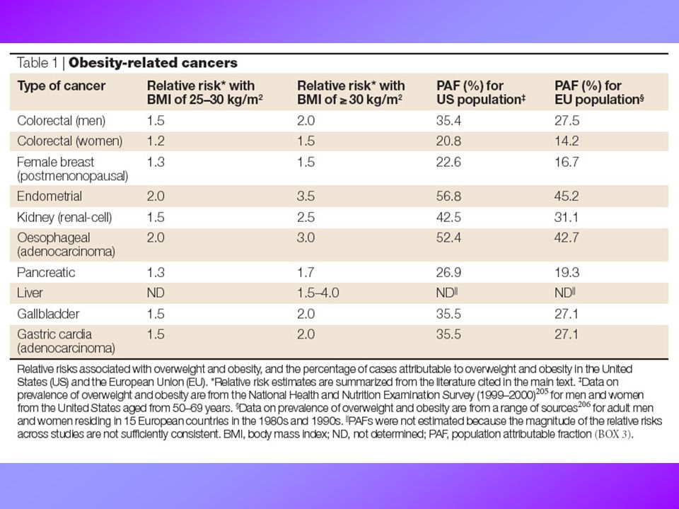
Contribution of overweight and obesity to mortality from cancer in the United States, Adapted from Adami H-O et al (N Engl J Med 2003;348:1623-4) McMillan, D. C et al. BMJ 2006;333: Copyright ©2006 BMJ Publishing Group Ltd.

5
Links between obesity, lifestyle factors, and risk for cancer
McMillan, D. C et al. BMJ 2006;333: Copyright ©2006 BMJ Publishing Group Ltd.

6
Effects of obesity on growth-factor production
Effects of obesity on growth-factor production. In obesity, increased release from adipose tissue of free fatty acids (FFA), tumour-necrosis factor- (TNF ) and resistin, and reduced release of adiponectin lead to the development of insulin resistance and compensatory, chronic hyperinsulinaemia (Box 4). Increased insulin levels, in turn, lead to reduced liver synthesis and blood levels of insulin-like growth factor binding protein 1 (IGFBP1), and probably also reduce IGFBP1 synthesis locally in other tissues. Increased fasting levels of insulin in the plasma are generally also associated with reduced levels of IGFBP2 in the blood. This results in increased levels of bioavailable IGF1. Insulin and IGF1 signal through the insulin receptors (IRs) and IGF1 receptor (IGF1R), respectively, to promote cellular proliferation and inhibit apoptosis in many tissue types. These effects might contribute to tumorigenesis.
, tumour-necrosis factor- (TNF ) and resistin, and reduced release of adiponectin lead to the development of insulin resistance and compensatory, chronic hyperinsulinaemia (Box 4). Increased insulin levels, in turn, lead to reduced liver synthesis and blood levels of insulin-like growth factor binding protein 1 (IGFBP1), and probably also reduce IGFBP1 synthesis locally in other tissues. Increased fasting levels of insulin in the plasma are generally also associated with reduced levels of IGFBP2 in the blood. This results in increased levels of bioavailable IGF1. Insulin and IGF1 signal through the insulin receptors (IRs) and IGF1 receptor (IGF1R), respectively, to promote cellular proliferation and inhibit apoptosis in many tissue types. These effects might contribute to tumorigenesis.")
7
Effects of obesity on hormone production
Effects of obesity on hormone production. Adipose tissue produces the enzymes aromotase and 17 -hydroxysteroid dehydrogenase (17 -HSD). So in obese individuals, there is typically an increased conversion of the androgens 4-androstenedione ( 4A) and testosterone (T) into the oestrogens oestrone (E1) and oestradiol (E2), respectively, by aromatase. 17 -HSD converts the less biologically active hormones 4A and E1 into the more active hormones T and E2, respectively. In parallel, obesity leads to hyperinsulinaemia, which in turn causes a reduction in the hepatic synthesis and circulating levels of sex-hormone-binding globulin (SHBG). The combined effect of increased formation of oestrone and testosterone, along with reduced levels of SHBG, leads to an increase in the bioavailable fractions of E2 and T that can diffuse to target cells, where they bind to oestrogen and androgen receptors. The effects of sex steroids binding their receptors can vary, depending on the tissue types, but in some tissues (for example, breast epithelium and endometrium) they promote cellular proliferation and inhibit apoptosis.
. So in obese individuals, there is typically an increased conversion of the androgens 4-androstenedione ( 4A) and testosterone (T) into the oestrogens oestrone (E1) and oestradiol (E2), respectively, by aromatase. 17 -HSD converts the less biologically active hormones 4A and E1 into the more active hormones T and E2, respectively. In parallel, obesity leads to hyperinsulinaemia, which in turn causes a reduction in the hepatic synthesis and circulating levels of sex-hormone-binding globulin (SHBG). The combined effect of increased formation of oestrone and testosterone, along with reduced levels of SHBG, leads to an increase in the bioavailable fractions of E2 and T that can diffuse to target cells, where they bind to oestrogen and androgen receptors. The effects of sex steroids binding their receptors can vary, depending on the tissue types, but in some tissues (for example, breast epithelium and endometrium) they promote cellular proliferation and inhibit apoptosis.")
8
Linee Guida della International Union Against Cancer
Adottare una dieta varia e ricca di alimenti di origine vegetale Ridurre l’assunzione di alimenti grassi Limitare o evitare il consumo di alcool Preparare e conservare gli alimenti in modo da ridurre potenziali contaminazioni Mantenere un equilibrio tra apporto e consumo energetico, evitando eccessivi aumenti o cali ponderali L’assunzione di integratori vitaminici e minerali non deve sostituire una dieta bilanciata e adeguata

9
Representative chemopreventive phytochemicals and their dietary sources

10
Dietary phytochemicals that block or suppress multistage carcinogenesis. Carcinogenesis is initiated with the transformation of the normal cell into a cancer cell (initiated cell). These cells undergo tumour promotion into preneoplastic cells, which progress to neoplastic cells. Phytochemicals can interfere with different steps of this process. Some chemopreventive phytochemicals inhibit metabolic activation of the procarcinogens to their ultimate electrophilic species, or their subsequent interaction with DNA. These agents therefore block tumour initiation (blocking agents). Alternatively, dietary blocking agents can stimulate the detoxification of carcinogens, leading to their secretion from the body. Other phytochemicals suppress the later steps (promotion and progression) of multistage carcinogenesis (suppressing agents). Some phytochemicals can act as both blocking and suppressing agents.
. These cells undergo tumour promotion into preneoplastic cells, which progress to neoplastic cells. Phytochemicals can interfere with different steps of this process. Some chemopreventive phytochemicals inhibit metabolic activation of the procarcinogens to their ultimate electrophilic species, or their subsequent interaction with DNA. These agents therefore block tumour initiation (blocking agents). Alternatively, dietary blocking agents can stimulate the detoxification of carcinogens, leading to their secretion from the body. Other phytochemicals suppress the later steps (promotion and progression) of multistage carcinogenesis (suppressing agents). Some phytochemicals can act as both blocking and suppressing agents..")
11
Fig. 1. Potential for intervention in carcinogenesis
Fig. 1. Potential for intervention in carcinogenesis. Chemopreventive agents can exert blocking or suppressing effects on different stages of the carcinogenic process. Blocking mechanisms prevent damage to DNA, whereas suppression slows down or inhibits the growth of transformed cells or new blood vessels. Abbreviation: ROS, reactive oxygen species.

12
Inflammation and carcinogenesis
Inflammation and carcinogenesis. Inflammatory processes include the production of reactive oxygen species (ROS) and nitric oxide (NO ) that can enhance carcinogenesis and tumour progression in multiple ways. ROS, in turn, can damage DNA and other cellular macromolecules. This damage results in apoptosis, increased proliferation, mutations and DNA-repair activities. Inflammatory processes can also increase angiogenesis by the induction of vascular endothelial growth factor and other growth factors. COX, cyclooxygenase; iNOS, inducible nitric-oxide synthase. Ulrich et al. Nature Reviews Cancer 6, 130–140 (February 2006) | doi: /nrc1801
 and nitric oxide (NO ) that can enhance carcinogenesis and tumour progression in multiple ways. ROS, in turn, can damage DNA and other cellular macromolecules. This damage results in apoptosis, increased proliferation, mutations and DNA-repair activities. Inflammatory processes can also increase angiogenesis by the induction of vascular endothelial growth factor and other growth factors. COX, cyclooxygenase; iNOS, inducible nitric-oxide synthase. Ulrich et al. Nature Reviews Cancer 6, 130–140 (February 2006) | doi: /nrc1801.")
13
Transcriptional activation by NRF2
Transcriptional activation by NRF2. NRF2 is a transcription factor that regulates expression of many detoxification or antioxidant enzymes. The Kelch-like-ECH-associated protein 1 (KEAP1) is a cytoplasmic repressor of NRF2 that inhibits its ability to translocate to the nucleus. These two proteins interact with each other through the double glycine-rich domains of KEAP1 and a hydrophilic region in the NEH2 domain of NRF2. KEAP1 contains many cysteine residues. Phase II enzyme inducers and/or prooxidants can cause oxidation or covalent modification (R) of these cysteine residues91. As a result, NRF2 is released from KEAP1. In addition, phosphorylation of NRF2 at serine (S) and threonine (T) residues by kinases such as phosphatidylinositol 3-kinase (PI3K), protein kinase C (PKC)131, c-Jun NH2-terminal kinase (JNK) and extracellular-signal-regulated kinase (ERK) is assumed to facilitate the dissociation of NRF2 from KEAP1 and subsequent translocation to the nucleus. p38 can both stimulate and inhibit the NRF2 nuclear translocation. In the nucleus, NRF2 associates with small MAF (the term is derived from musculoaponeurotic-fibrosarcoma virus), forming a heterodimer that binds to the antioxidant-responsive element (ARE) to stimulate gene expression. NRF2/MAF target genes encode phase II detoxification or antioxidant enzymes such as glutathione S-transferase 2 (GSTA2), NAD(P)H:quinone oxidoreductase (NQO1), -glutamate cysteine ligase ( -GCLC and -GCLM) and heme oxygenase-1 (HO-1). PI3K also phosphorylates the CCAAT/enhancer binding protein- (C/EBP ), inducing its translocation to the nucleus and binding to the CCAAT sequence of C/EBP- response element within the xenobiotic response element (XRE), in conjunction with NRF2 binding to ARE132. Transfection of human neuroblastoma cells with PI3K activates ARE, which is attenuated by a pharmacological inhibitor of PI3K or dominant-negative NRF2 (Ref. 133). Curcumin and caffeic acid phenethyl ester (CAPE) disrupt the NRF2–KEAP1 complex, leading to increased NRF2 binding to ARE99, 100. Sulphoraphane directly interacts with KEAP1 by covalent binding to its thiol groups91. 6-(Methylsulfinyl)hexyl isothiocyanate (6-HITC) — a sulphoraphane analogue from Japanese horseradish wasabi — stimulates nuclear translocation of NRF2, which subsequently activates ARE98.
 is a cytoplasmic repressor of NRF2 that inhibits its ability to translocate to the nucleus. These two proteins interact with each other through the double glycine-rich domains of KEAP1 and a hydrophilic region in the NEH2 domain of NRF2. KEAP1 contains many cysteine residues. Phase II enzyme inducers and/or prooxidants can cause oxidation or covalent modification (R) of these cysteine residues91. As a result, NRF2 is released from KEAP1. In addition, phosphorylation of NRF2 at serine (S) and threonine (T) residues by kinases such as phosphatidylinositol 3-kinase (PI3K), protein kinase C (PKC)131, c-Jun NH2-terminal kinase (JNK) and extracellular-signal-regulated kinase (ERK) is assumed to facilitate the dissociation of NRF2 from KEAP1 and subsequent translocation to the nucleus. p38 can both stimulate and inhibit the NRF2 nuclear translocation. In the nucleus, NRF2 associates with small MAF (the term is derived from musculoaponeurotic-fibrosarcoma virus), forming a heterodimer that binds to the antioxidant-responsive element (ARE) to stimulate gene expression. NRF2/MAF target genes encode phase II detoxification or antioxidant enzymes such as glutathione S-transferase 2 (GSTA2), NAD(P)H:quinone oxidoreductase (NQO1), -glutamate cysteine ligase ( -GCLC and -GCLM) and heme oxygenase-1 (HO-1). PI3K also phosphorylates the CCAAT/enhancer binding protein- (C/EBP ), inducing its translocation to the nucleus and binding to the CCAAT sequence of C/EBP- response element within the xenobiotic response element (XRE), in conjunction with NRF2 binding to ARE132. Transfection of human neuroblastoma cells with PI3K activates ARE, which is attenuated by a pharmacological inhibitor of PI3K or dominant-negative NRF2 (Ref. 133). Curcumin and caffeic acid phenethyl ester (CAPE) disrupt the NRF2–KEAP1 complex, leading to increased NRF2 binding to ARE99, 100. Sulphoraphane directly interacts with KEAP1 by covalent binding to its thiol groups91. 6-(Methylsulfinyl)hexyl isothiocyanate (6-HITC) — a sulphoraphane analogue from Japanese horseradish wasabi — stimulates nuclear translocation of NRF2, which subsequently activates ARE98.")
14
Effect of phytochemicals on activation of NF- B and AP1
Effect of phytochemicals on activation of NF- B and AP1. The NF- B signalling pathway converges on the multiprotein complex called the I B kinase (IKK) signalsome, leading to I B phosphorylation (P), ubiquitylation (Ub) and subsequent degradation by the 26S proteasome. NF- B is then released and translocated to the nucleus, where it binds to specific promoter regions of various genes. The IKK signalsome is activated by the NF- B-inducing kinase (NIK). Pathways that regulate NIK are likely to involve signalling through a family of mitogen-activated protein kinases (MAPKs), such as MAPK kinase kinase-1 (MEKK1) — a kinase that lies upstream of extracellular signal-regulated kinase (ERK) — MAPK/ERK kinase (MEK1/2) and p38 MAPK. Recent reports showed that NF- B activation is also regulated by the AKT signalling pathway58, 59, 129. Phosphatidylinositol 3-kinase (PI3K) activates AKT/protein kinase B via phosphorylation by 3-phosphoinositide-dependent protein kinase-1 (PDK1). Genistein specifically inhibits AKT activity and AKT-mediated NF- B activation58, 59. Epigallocatechin gallate (EGCG) can block the activities of PI3K and AKT49. There is crosstalk between the AKT and NF- B signalling pathways — AKT phosphorylation leads to activation of NF- B by stimulating I B kinase (IKK) activity129. IKK is also a target for chemopreventive phytochemicals, including curcumin24, 28, resveratrol71 and EGCG45, 130. The MAPK family proteins also regulate expression of AP1 — a heterogenous set of dimeric proteins made up of members of the c-JUN, c-FOS and ATF families. In this pathway, activation of ERK1/2 phosphorylates ELK1, c-JUN NH2-terminal kinase (JNK) phosphorylates c-JUN, and p38 phosphorylates both ELK1 and ATF2. This leads to transcriptional activation of target genes. External stimuli — including phorbol ester and ultraviolet radiation — activate specific isoforms of protein kinase C (PKC), which, in turn, leads to stimulation of the p21 RAS–ERK signalling pathway via RAF and MEK1/2. Activation of p38 and JNK is mediated by MAPK kinase-4 (MKK4), which is under control of the upstream kinase MEKK.
 signalsome, leading to I B phosphorylation (P), ubiquitylation (Ub) and subsequent degradation by the 26S proteasome. NF- B is then released and translocated to the nucleus, where it binds to specific promoter regions of various genes. The IKK signalsome is activated by the NF- B-inducing kinase (NIK). Pathways that regulate NIK are likely to involve signalling through a family of mitogen-activated protein kinases (MAPKs), such as MAPK kinase kinase-1 (MEKK1) — a kinase that lies upstream of extracellular signal-regulated kinase (ERK) — MAPK/ERK kinase (MEK1/2) and p38 MAPK. Recent reports showed that NF- B activation is also regulated by the AKT signalling pathway58, 59, 129. Phosphatidylinositol 3-kinase (PI3K) activates AKT/protein kinase B via phosphorylation by 3-phosphoinositide-dependent protein kinase-1 (PDK1). Genistein specifically inhibits AKT activity and AKT-mediated NF- B activation58, 59. Epigallocatechin gallate (EGCG) can block the activities of PI3K and AKT49. There is crosstalk between the AKT and NF- B signalling pathways — AKT phosphorylation leads to activation of NF- B by stimulating I B kinase (IKK) activity129. IKK is also a target for chemopreventive phytochemicals, including curcumin24, 28, resveratrol71 and EGCG45, 130. The MAPK family proteins also regulate expression of AP1 — a heterogenous set of dimeric proteins made up of members of the c-JUN, c-FOS and ATF families. In this pathway, activation of ERK1/2 phosphorylates ELK1, c-JUN NH2-terminal kinase (JNK) phosphorylates c-JUN, and p38 phosphorylates both ELK1 and ATF2. This leads to transcriptional activation of target genes. External stimuli — including phorbol ester and ultraviolet radiation — activate specific isoforms of protein kinase C (PKC), which, in turn, leads to stimulation of the p21 RAS–ERK signalling pathway via RAF and MEK1/2. Activation of p38 and JNK is mediated by MAPK kinase-4 (MKK4), which is under control of the upstream kinase MEKK.")
15
A standard study design for evaluating a new chemopreventive agent
A standard study design for evaluating a new chemopreventive agent. In the example shown, the rat model uses nitrosomethylurea as a carcinogen59. The importance of the latency period is discussed in the text and shown diagrammatically in Fig. 3.

16
Figure 3 | Extension of the latency period is important for chemoprevention in both animals and humans. a | If carcinogenic burden in an animal model is artefactually high, then it might seem that a chemopreventive agent might only delay the appearance of a cancer. b | However, if such carcinogenic burden is reduced to levels that are more relevant to human carcinogenesis, a more meaningful extension of latency can be obtained, which would allow a lifetime suppression of malignancy.

17
Punti fondamentali nella chemoprevenzione farmacologica
Gli agenti chemopreventivi devono avere una tossicità molto più bassa rispetto ai farmaci usati nella terapia dei tumori È necessario identificare pazienti con rischio elevato di sviluppare tumori che possano trarre il massimo beneficio da una strategia chemopreventiva È necessario identificare e validare biomarkers che consentano di valutare l’effettiva attività del trattamento

18
NSAIDs & COXibs

19
EVIDENZE CLINICHE DELL’EFFETTO DEI NSAIDs SUI TUMORI COLORETTALI
Regressione di polipi rettali in un paziente che assumeva indometacina e sulindac come analgesici Diminuzione del 40-50% del rischio di sviluppo di tumori del colon in popolazioni di pazienti trattati cronicamente per un periodo di anni con aspirina o altri NSAIDs Diminuzione del numero e delle dimensioni dei polipi in gruppi di pazienti con FAP (Familial Adenomatous Polyposis) trattati con sulindac
 trattati con sulindac.")
20
MODELLI ANIMALI PER LO STUDIO DEI TUMORI COLORETTALI
APCmin mouse: Topi ottenuti mediante trattamento con etilnitrosourea, seguito da selezione per trasmissione di mutazioni nella linea germinale. Missense mutation nel gene APC (Adenomatous Polyposis Coli) APC716 mouse modello simile al precedente, ma in questo caso il gene APC è troncato AOM-treated rat ratti trattati con azossimetano sviluppano lesioni preneoplastiche (aberrant crypt foci) modelli di xenograft nel topo nudo
 APC716 mouse. modello simile al precedente, ma in questo caso il gene APC è troncato. AOM-treated rat. ratti trattati con azossimetano sviluppano lesioni preneoplastiche (aberrant crypt foci) modelli di xenograft nel topo nudo.")
21
EFFETTO DEI NSAIDS SU MODELLI ANIMALI DI TUMORI COLORETTALI
Trattamento Esito APCmin mouse: numero di polipi Sulindac Piroxicam Celecoxib APC716 mouse rofecoxib AOM-treated rat Incidenza e numero di tumori Aspirina NS-398 Nimesulide Nude mouse xenograft Crescita di carcinomi del colon SC-58125 Meloxicam

22
COX enzymes in prostaglandin synthesis
COX enzymes in prostaglandin synthesis. Arachadonic acid is first liberated from membrane glycerophospholipids by the actions of the phospholipase A2 (PLA2) family of enzymes. The cyclooxygenase (COX) enzymes then catalyse the biosynthesis of the bicyclic endoperoxide intermediate PGG2, followed by reduction to PGH2. PGH2 is subsequently converted to one of several structurally related prostaglandins (PGs), including PGE2, PGD2, PGF2 , PGI2 and thromboxane A2 (TxA2), by the activity of specific PG synthases. Traditional non-steroidal anti-inflammatory drugs (NSAIDs) inhibit the activity of both COX-1 and COX-2, whereas the recently developed coxib family of NSAIDs selectively inhibit COX-2. COX-1 is responsible for 'housekeeping' PG biosynthesis and is constitutively produced in most tissues in the body. COX-2, on the other hand, is not normally produced in most tissues but is induced by a wide spectrum of growth factors and pro-inflammatory cytokines in specific pathophysiological conditions.
 family of enzymes. The cyclooxygenase (COX) enzymes then catalyse the biosynthesis of the bicyclic endoperoxide intermediate PGG2, followed by reduction to PGH2. PGH2 is subsequently converted to one of several structurally related prostaglandins (PGs), including PGE2, PGD2, PGF2 , PGI2 and thromboxane A2 (TxA2), by the activity of specific PG synthases. Traditional non-steroidal anti-inflammatory drugs (NSAIDs) inhibit the activity of both COX-1 and COX-2, whereas the recently developed coxib family of NSAIDs selectively inhibit COX-2. COX-1 is responsible for housekeeping PG biosynthesis and is constitutively produced in most tissues in the body. COX-2, on the other hand, is not normally produced in most tissues but is induced by a wide spectrum of growth factors and pro-inflammatory cytokines in specific pathophysiological conditions.")
23
NSAIDs, COX inhibition and prostaglandins
NSAIDs, COX inhibition and prostaglandins. The main targets of cyclooxygenase 2 (COX2)-specific inhibitors (COXibs) and non-steroidal anti-inflammatory drugs (NSAIDs) are COX1 and COX2 — enzymes that are central to the conversion of arachidonic acid to prostaglandins. An alternative pathway of arachidonic acid metabolism involves the lipoxygenases. Both the lipoxygenase and COX pathways are regulated by peroxide concentrations, with COX2 being induced at lower peroxide concentrations than COX1. Prostaglandins (PGs) influence angiogenesis, apoptosis, cell proliferation and migration. The balance between pro-thrombotic factors (for example, thromboxane A2) and anti-thrombotic factors (for example, prostacyclin) might be of particular relevance to cardiovascular toxicity. IL-6, interleukin 6; NF- B, nuclear factor- B; PDGF, platelet-derived growth factor; PLA2, phospholipase A2, which cleaves fatty acids, such as arichidonic acid, from phospholipids; VEGF, vascular endothelial growth factor.
-specific inhibitors (COXibs) and non-steroidal anti-inflammatory drugs (NSAIDs) are COX1 and COX2 — enzymes that are central to the conversion of arachidonic acid to prostaglandins. An alternative pathway of arachidonic acid metabolism involves the lipoxygenases. Both the lipoxygenase and COX pathways are regulated by peroxide concentrations, with COX2 being induced at lower peroxide concentrations than COX1. Prostaglandins (PGs) influence angiogenesis, apoptosis, cell proliferation and migration. The balance between pro-thrombotic factors (for example, thromboxane A2) and anti-thrombotic factors (for example, prostacyclin) might be of particular relevance to cardiovascular toxicity. IL-6, interleukin 6; NF- B, nuclear factor- B; PDGF, platelet-derived growth factor; PLA2, phospholipase A2, which cleaves fatty acids, such as arichidonic acid, from phospholipids; VEGF, vascular endothelial growth factor.")
24
Control of aromatase expression in breast carcinomas
Control of aromatase expression in breast carcinomas. The postulated interaction between breast cancer cells and associated fibroblasts and inflammatory cells in the adjacent stroma is shown. Aromatase seems to be expressed predominantly in the fibroblasts under the stimulatory influence of interleukin(IL)-6 and IL-11 and tumour-necrosis factor- (TNF ) released by inflammatory cells, and prostaglandin-E2 synthesized by cyclooxygenases (COX) in breast cancer cells and inflammatory cells. As such, COXs are targets for selective aromatase modulators (SAMs) that can indirectly modify the activity of aromatase.
-6 and IL-11 and tumour-necrosis factor- (TNF ) released by inflammatory cells, and prostaglandin-E2 synthesized by cyclooxygenases (COX) in breast cancer cells and inflammatory cells. As such, COXs are targets for selective aromatase modulators (SAMs) that can indirectly modify the activity of aromatase.")
Presentazioni simili


>")


