Scaricare la presentazione
La presentazione è in caricamento. Aspetta per favore

1
IMAGING DELLA PATOLOGIA
ESPANSIVA DEL SURRENE G. Angelelli 1

2
IDENTIFICAZIONE CARATTERIZZAZIONE 2

3
METODICHE DI STUDIO US TC RM MN

4
IDENTIFICAZIONE SENSIBILITÀ MOLTO ALTA POSSIBILI FALSI POSITIVI 4

5
“ Nodules are found commonly at autopsy… Some of these may represent localized compensatory overgrowth of adrenocortical cells… while others are true neoplasms” “ Size, weight and encapsulation are not reliable in differentiating adenomas from nodules” H. Sasano et al In tumours of endocrine organs Edited by RA DeLellis et al IARC PRESS. Lyon 2004; 5 5

7
CARATTERIZZAZIONE CISTI MIELOLIPOMA ADENOMA / NON ADENOMA 7

8
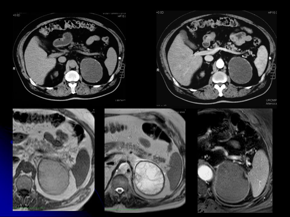
CISTI

11
MIELOLIPOMA

13
ADENOMA / NON ADENOMA 13

14
VALUTAZIONI CLINICHE CLINICA SIGNIFICATIVA NEOPLASIA PRIMITIVA NOTA
INCIDENTALOMA 14 14

15
CARATTERIZZAZIONE DELLA PATOLOGIA CRITERI MORFOLOGICI
CRITERI ISTOLOGICI CRITERI FISIOLOGICI 15

16
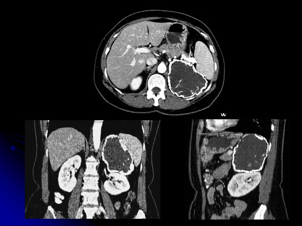
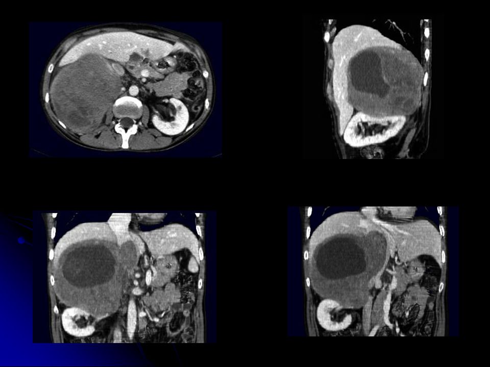
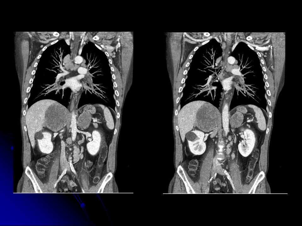
CRITERI MORFOLOGICI CONTORNI DIMENSIONI STRUTTURA ACCRESCIMENTO 16

17
17

18
DIMENSIONI DIAMETRI < 4cm adenomi
DIAMETRI > 4cm probabili lesioni maligne 18

19
“ For lesions larger than 4 – 5 cm, adrenal adenocarcinoma should be strongly considered, particularly if the patients has no other history of malignancy” DH Szolar et al Radiology 2005; 234: 19 19

20
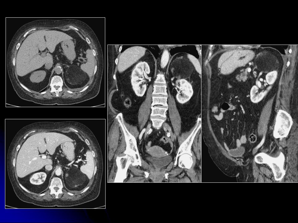
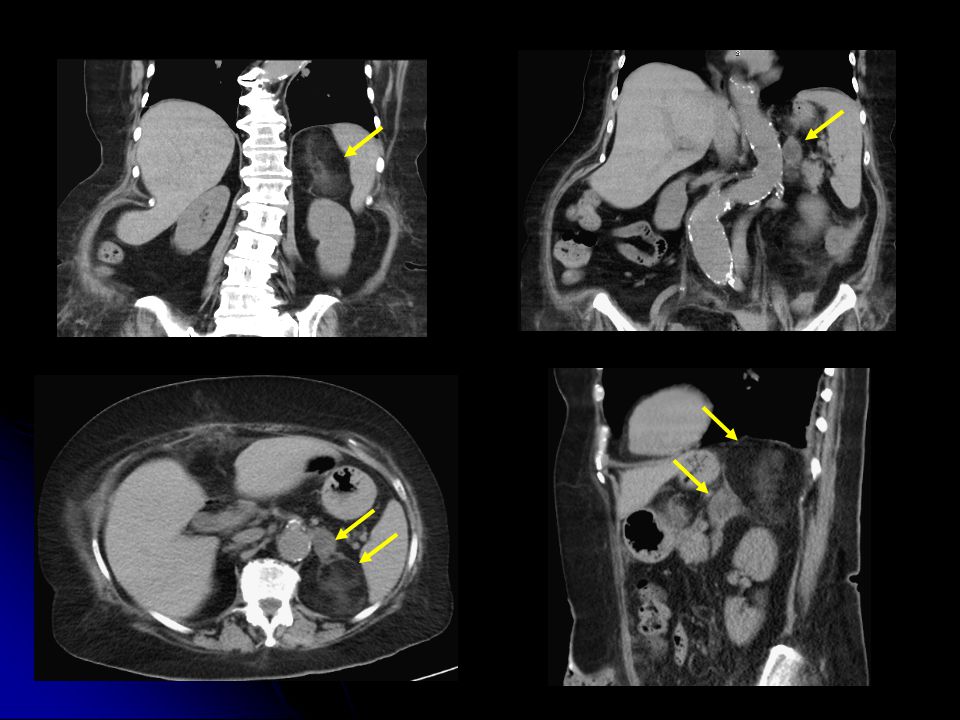
STRUTTURA

24
TEMPO DI ACCRESCIMENTO
LENTO NEGLI ADENOMI 24

25
Febbraio Maggio Luglio

26
LIPIDI INTRACITOPLASMATICI
CRITERI ISTOLOGICI LIPIDI INTRACITOPLASMATICI 26

27
ASPETTO IPODENSO <10 HU
ADENOMI (70%) TC SCANSIONI SENZA MdC ASPETTO IPODENSO <10 HU - 13 ± 19 27
 TC SCANSIONI SENZA MdC. ASPETTO IPODENSO <10 HU ±")
28
RM ADENOMA VS NON ADENOMA
“Voxels containing both fat and water will have signal cancellation and diminished signal intensity. This effect is helpful in identifying adenomas that contain relatively high cytoplasmatic lipid concentration…” Krebs TL et al Mag Reson Imaging Clin North Am 1997 PERDITA di segnale nelle sequenze T1 out-of-phase delle lesioni a contenuto lipidico (adenomi)
")
29
± 22.7

30
“ For lipid – rich adenomas, there is no difference between the tests, but chemical shift imaging might be superior when evaluating lipid – poor adenomas” GM Israel et al AJR 2004; 183: “ The results of both techniques were highly correlated and the adenomas that were indeterminate with one technique were also indeterminate with the other” EK Outwater et al Radiology 1996; 200: 30

31
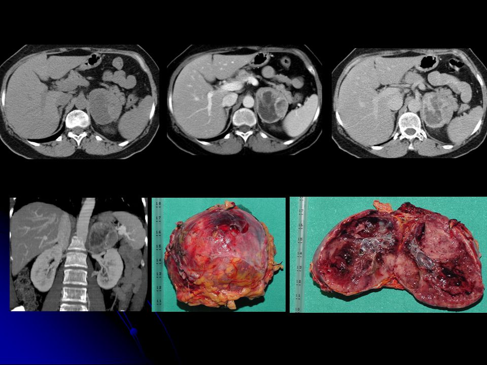
CRITERI ISTOLOGICI PUNTI CRITICI
“ Contrast – enhanced CT is usually performed in the portal venous phase…. ” WW Mayo - Smith et al Radiographics 2001; 21: “Approximately 30% of adenomas are lipid poor and overlap with other adrenal lesions…” S. Chong et Al. Radiographics 2006; 26: “Adrenocortical carcinoma can contain foci of intracytolasmatic lipid…” B Mackay et al Ultrastruct Pathol 1994; 18: 31

32
CRITERI FISIOLOGICI VASCOLARIZZAZIONE
32

33
VASCOLARIZZAZIONE DENSITOMETRIA (iniziale e tardiva)
WASH-OUT PERCENTUALE 33

34
DENSITOMETRIA INIZIALE ADENOMI – NON ADENOMI rapido incremento
DENSITOMETRIA TARDIVA Modalità Iniezione Contrasto Gettata Cardiaca 34

35
“ The more rapid washout of CT enhancement in adrenal adenomas … is a accurate method for use in the characterization of adrenal masses” M Korobkin et al Radiology 2000; 217: “ Wash-out of contrast media may be more specific than low attenuation at CT” CS Pena et al Radiology 2000; 217: 35

36
9.2 ± 22.3 39.2 ± 16.1 51.4 ± 14.0 41.3 ± 10.6

37
“ Five of six benign adrenal pheocromocytoma had enhancement washout curves similar to those of metastases rather than adenomas” DH Szolar et al Radiology 1998; 207: 37 37

38
17.9 ± 39.4 64.7 ± 31.0 40.0 ± 28.9

39
CRITERI FISIOLOGICI PUNTI CRITICI
“ Possibility of differentiating the small adrenal cortical carcinoma from the adrenal adenoma, using enhancement washout calculations not yet been systematically assessed” M Korobkin et al Radiology 2000; 217: “ Nonadenomas have a disturbed capillary permeability with prolonged retention of contrast material in the extracellular space” DM Szolar al Radiology 1997; 202: 39

40
CONCLUSIONI La Diagnostica per Immagini è molto sensibile nella dimostrazione dei processi espansivi surrenalici e la loro caratterizzazione è spesso possibile. Il ricorso alla biopsia può essere riservato a rarissimi casi.

41
GRAZIE PER L’ ATTENZIONE

42
ALGORITMO NELLE LESIONI SURRENALICHE
TC senza MdC < 10 HU > 10 HU adenoma controllo a 6 mesi rivalutazione endocrinologica a 12 mesi RM chemical shift wash-out perdita di segnale > 50% < 50% si no adenoma adenoma > 4cm < 4 cm > 4cm < 4 cm chirurgia PET/TC controllo TC (2 mesi) PET/TC controllo TC (2 mesi) chirurgia 42 42
 PET/TC. controllo TC. (2 mesi) chirurgia")
43
ALGORITMO NELLE LESIONI SURRENALICHE IN PAZIENTI NEOPLASTICI
TC senza MdC < 10 HU > 10 HU RM chemical shift benigna wash-out perdita di segnale > 50% < 50% si no benigna PET-TC/FNA benigna PET-TC/FNA 43

Presentazioni simili


>")


 A.Cristofoli (laureando.>")


>")


