Scaricare la presentazione
La presentazione è in caricamento. Aspetta per favore

1
Cos’è il Morbo di Parkinson
E’ una delle più comuni forme patologiche che interessano il movimento. Fu descritto per la prima volta, nel 1817, da James Parkinson: “moto tremolante involontario, con forza muscolare ridotta, di parti non in azione, anche quando vengono sorrette; con propensione a piegare il tronco in avanti ed a passare da un’andatura al passo alla corsa; assenza di alterazioni sensitive e dell’intelletto.”

2
Segni clinici del Morbo di Parkinson
Tremore a riposo: ritmico (tre-quattro cicli al minuto) che compare inizialmente in una mano ed è più evidente a riposo o durante situazioni di stress. Rigidità: resistenza al movimento passivo Bradicinesia: rallentamento nel compiere movimenti volontari Perdita dei riflessi posturali
 che compare inizialmente in una mano ed è più evidente a riposo o durante situazioni di stress. Rigidità: resistenza al movimento passivo. Bradicinesia: rallentamento nel compiere movimenti volontari. Perdita dei riflessi posturali.")
3
Cos’è il Morbo di Parkinson
E’ la più importante affezione del sistema extrapiramidale, caratterizzata dalla diminuzione del neurotrasmettitore DOPAMINA nel CORPO STRIATO del cervello per degenerazione della SUBSTANTIA NIGRA, pars compacta, che invia fibre efferenti allo STRIATO.

4
Sistema piramidale ed extrapiramidale
Le risposte motrici volontarie sono elaborate da particolari aree corticali, le aree motrici, ed inoltrate ai centri effettori del tronco e del midollo spinale, attraverso due sistemi discendenti: il sistema piramidale ed il sistema extrapiramidale. Il sistema piramidale collega le aree motrici della corteccia cerebrale con i neuroni motori del tronco encefalico e del midollo spinale. Le fibre del sistema piramidale originano da neuroni (motoneuroni superiori), detti cellule piramidali, contenuti nelle varie aree motrici della corteccia cerebrale. Nel bulbo, esse si collocano nella porzione ventrale, costituendo formazioni longitudinali, chiamate PIRAMIDI. Il sistema extrapiramidale è così chiamato, semplicemente perché è costituito da fibre filogeneticamente più antiche, che decorrono al di fuori del sistema piramidale e prevede anche alcuni centri intercalati tra aree corticali motrici e motoneuroni effettori.
, detti cellule piramidali, contenuti nelle varie aree motrici della corteccia cerebrale. Nel bulbo, esse si collocano nella porzione ventrale, costituendo formazioni longitudinali, chiamate PIRAMIDI. Il sistema extrapiramidale è così chiamato, semplicemente perché è costituito da fibre filogeneticamente più antiche, che decorrono al di fuori del sistema piramidale e prevede anche alcuni centri intercalati tra aree corticali motrici e motoneuroni effettori.")
5
The motor systems have three levels of control—the spinal cord, brain stem,
and forebrain—organized both serially and in parallel. The motor areas of the cerebral cortex can influence the spinal cord either directly or through the descending systems of the brain stem. All three levels of the motor systems receive sensory inputs and are also under the influence of two independent subcortical systems: the basal ganglia and the cerebellum. (The basal ganglia and cerebellum act on the cerebral cortex through relay nuclei in the thalamus, which are omitted from the diagram for clarity.)
")
6
A. The tennis player is watching the approaching ball
A. The tennis player is watching the approaching ball. He uses his visual cortex to identify the ball and judge its size, direction, and velocity. His premotor cortex develops a motor program that will allow him to approach the ball and hit it back. The amygdala adjusts the heart rate, respiration, and other homeostatic mechanisms to allow successful performance of the behavior. The amygdala also activates the hypothalamus to motivate the player to hit a good shot. B. To execute the shot the player must use all of the structures illustrated in A as well as others. The player's motor cortex must send signals to the spinal cord that will activate and inhibit many muscles in the arms and legs. The basal ganglia become involved in initiating motor patterns and perhaps in recalling learned movements to hit the ball properly. The cerebellum fine tunes the movements based on proprioceptive information from peripheral sensory receptors. The posterior parietal cortex provides the player with a sense of where his body is located in three-dimensional space and where his racket arm is located with respect to the rest of the body. During this entire process, brain stem nuclei are involved in regulating heart rate, respiration, and arousal. The hippocampus is not involved in hitting the ball, but it is involved in recording in memory all of the details of the point so that the player can brag about file:///E|it later. In fact, many other brain regions are also active during this simple behavior. The common sense notion that only a fraction of the brain is used at any one time is clearly wrong. It is more likely that virtually all of the brain is active in even simple behaviors such as hitting a tennis ball. In addition, the afferent information for the planned behavior recruits activity in the amygdala,

7
The four principal nuclei of the basal ganglia are (1) the striatum, (2) the globus pallidus
(or pallidum), (3) the substantia nigra (consisting of the pars reticulata and pars compacta), and (4) the subthalamic nucleus. The striatum consists of three important subdivisions: the caudate nucleus, the putamen, and the ventral striatum (which includes the nucleus accumbens). Except at its most anterior pole, the striatum is divided into the caudate nucleus and putamen by the internal capsule, a major collection of fibers that run between the neocortex and thalamus in both directions. All three subdivisions of the striatum have a common embryological origin.
, (3) the substantia nigra (consisting of the pars reticulata and pars compacta), and. (4) the subthalamic nucleus. The striatum consists of three important subdivisions: the caudate nucleus, the putamen, and the ventral striatum (which includes the. nucleus accumbens). Except at its most anterior pole, the striatum is divided into the. caudate nucleus and putamen by the internal capsule, a major collection of fibers that run. between the neocortex and thalamus in both directions. All three subdivisions of the striatum have. a common embryological origin.")
8
Sistema extrapiramidale: Nuclei della base e rapporti con le strutture circostanti

9
The anatomic connections of the basal ganglia-thalamocortical circuitry,
indicating the parallel direct and indirect pathways from the striatum to the basal ganglia output nuclei. Two types of dopamine receptors (D1 and D2) are located on different sets of output neurons in the striatum that give rise to the direct and indirect pathways. Inhibitory pathways are shown as gray arrows; excitatory pathways, as pink arrows. GPe = external segment of the globus pallidus; GPi = internal segment of the globus pallidus; SNc = substantia nigra pars compacta; STN = subthalamic nucleus. Connessioni anatomiche del circuito nuclei della base-talamo-corteccia: via diretta e via indiretta dallo striato ai nuclei della base.
 are located on different. sets of output neurons in the striatum that give rise to the direct and indirect pathways. Inhibitory. pathways are shown as gray arrows; excitatory pathways, as pink arrows. GPe = external. segment of the globus pallidus; GPi = internal segment of the globus pallidus; SNc = substantia. nigra pars compacta; STN = subthalamic nucleus. Connessioni anatomiche del circuito nuclei della base-talamo-corteccia: via diretta e via indiretta dallo striato ai nuclei della base.")
10
Le frecce grigie e nere indicano le connessioni inibitorie le frecce rosse e rosa quelle eccitatorie. Nel MP la degenerazione della via dopaminergica nigro-striatale produce modificazioni diverse dall’attività dei neuroni di proiezione strio-pallidali, indicate nel grado di inscurimento delle frecce che indicano le connessioni. Le frecce più scure indicano aumento dell’attività neuronale e le frecce più chiare una diminuzione. I segnali efferenti dei nuclei della base diretti al talamo aumentano nel morbo di Parkinso

11
Aree di neurodegenerazione e processi neurochimici coinvolti nel morbo di Parkinson.

12
samples Parkinsonian Midbrain Human Normal Midbrain
Our studies begin from here: from these “autoptic pieces”, as we say using the lab slang. On the right we see the human normal midbrain , where the subtantia nigra appears dark-pigmented, from neuromelanin-containing dopaminergic neurons and on the left we have a characteristic parkinsonian midbrain,whose the gross anatomical change is atrophy with nigrostriatal degeneration and an apparently suppressed neuromelanogenesis resulting as a bleching of the substantia nigra.due to the disappearence of dopaminergic neurons.The onset of motor dysfunction in PD is associated with a dramatic depletion in brain dopamine. Although histological abnormalities can be found in other dopaminergic and even non-dopaminergic cell groups, the most important site of changes is the origin of the dopaminergic nigrostriatal tract, the substantia nigra pars compacta, thus over the past years, the role of dopamine (DA) metabolism has been focus of neurodegeneration studies in PD. ). Post-mortem studies have consistently implicated oxidative damage in PD pathogenesis (2). The primary cause of oxidative stress, an imbalance between the production of reactive oxygen species (ROS) and the capacity of the cell to scavenge these toxic metabolites via its own antioxidant defence machine, has not been clear, but the leading candidate is the metabolism of dopamine itself, since it gives rise to various toxic species. Parkinsonian Midbrain Human Normal Midbrain
 metabolism has been focus of neurodegeneration studies in PD. ). Post-mortem studies have consistently implicated oxidative damage in PD pathogenesis (2). The primary cause of oxidative stress, an imbalance between the production of reactive oxygen species (ROS) and the capacity of the cell to scavenge these toxic metabolites via its own antioxidant defence machine, has not been clear, but the leading candidate is the metabolism of dopamine itself, since it gives rise to various toxic species. Parkinsonian Midbrain. Human Normal Midbrain.")
13
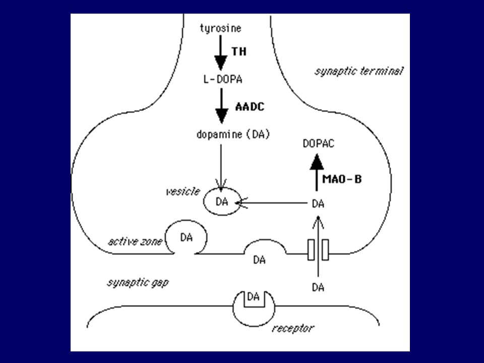
Tappe di sintesi della Dopamina

15
Catabolismo della Dopamina

16
Dopamina intracellulare

17
Neuromelanin formation
Neuromelanin formation. The pathway of the reactions converting dopamine to neuromelanin is a normal process in the substantia nigra, which it is tought to involve several steps:1) dopamine oxidation to dopamine o-quinone catalyzed by metals, oxygen, peroxynitrite or peroxidative activity of several enzymes (PX), such as prostaglandin H synthase, cytochrome P450, xantine oxidase 2) cyclization of dopamine o-quinone to dopaminochrome via an addition at physiological pH values, leading to the formation of unstable leukodopaminochrome and oxidation of leukodopaminochrome to dopaminochrome and polymerization of dopaminochrome to neuromelanin. (see the text)
 dopamine oxidation to dopamine o-quinone catalyzed by metals, oxygen, peroxynitrite or peroxidative activity of several enzymes (PX), such as prostaglandin H synthase, cytochrome P450, xantine oxidase 2) cyclization of dopamine o-quinone to dopaminochrome via an addition at physiological pH values, leading to the formation of unstable leukodopaminochrome and oxidation of leukodopaminochrome to dopaminochrome and polymerization of dopaminochrome to neuromelanin. (see the text)")
18
NEUROMELANINA: QUALI CERTEZZE
- nei neuroni dopaminergici dell’area tegmentale ventrale della substantia nigra nei neuroni noradrenergici del locus coeruleus - nella popolazione C2 del midollo allungato - CONTIENE: dopaminocromo (in substantia nigra) noradrenocromo (in locus coeruleus) E’ un complesso polimerico costituito da due classi di melanine: - eumelanine (monomeri indolici derivati dalla dopamina) insolubili di colore nero - feomelanine (monomeri benzotiazolici derivati dalla cisteinildopamina) solubili in alcali, di colore marrone. IMMAGAZZINA IL FERRO NELLA SUBSTANTIA NIGRA E’ presente in forma di granuli
 noradrenocromo (in locus coeruleus) E’ un complesso polimerico costituito da due classi di melanine: - eumelanine (monomeri indolici derivati dalla dopamina) insolubili di colore nero. - feomelanine (monomeri benzotiazolici derivati dalla cisteinildopamina) solubili in alcali, di colore marrone. IMMAGAZZINA IL FERRO NELLA SUBSTANTIA NIGRA. E’ presente in forma di granuli.")
19
NEUROMELANINA: QUALI IPOTESI.
PROBABILE RUOLO PROTETTIVO: contiene ferro, ma anche Zn, Mn, Cu Accumula composti organici tossici come l’1-metli-4-fenil-piridina (MPP+) e pesticidi, come il paraquat La sintesi di neuromelanina fornisce un meccanismo protettivo contro la tossicità della dopamina, prevenendo il suo accumulo nel citosol.
 e pesticidi, come il paraquat. La sintesi di neuromelanina fornisce un meccanismo protettivo contro la tossicità della dopamina, prevenendo il suo accumulo nel citosol.")
20
Formazione degli addotti : legame del Dopaminochinone con i gruppi sulfidrilici delle proteine.

21
Specie reattive dell’ossigeno e patogenesi del morbo di Parkinson

22
Alfa-sinucleina: lega vescicole citoplasmatiche
Parkinson’s disease: Divergent Causes, Convergent Mechanisms (Greenamyre and Hastings Science, 2004) Alfa-sinucleina: lega vescicole citoplasmatiche Parkina: E3Ligasi UHCL: ubiquitina idrolasi PinK1: chinasi mitocondriale DJ1: probabile ruolo nella trasmissione del segnale
 Alfa-sinucleina: lega vescicole citoplasmatiche. Parkina: E3Ligasi. UHCL: ubiquitina idrolasi. PinK1: chinasi mitocondriale. DJ1: probabile ruolo nella trasmissione del segnale.")
23
The potential pathways leading to dopaminergic cell death.
The potential pathways leading to dopaminergic cell death. In brief, reactive oxygen species, which are generated through the oxidative The potential pathways leading to dopaminergic cell death. In brief, reactive oxygen species, which are generated through the oxidative metabolism of DA or via the compromised oxidative phosphorylation in mitochondria by environmental toxins or genetic predispositions, cause modifications in protein homeostasis giving rise to abnormal intracellular protein aggregates, fibrillar deposits of a-synuclein in PD. The ubiquitin– proteosome system (UPS), a major cellular defense mechanism balancing against potential toxicity of protein aggregates, targets the aggregates for proteolytic degradation via the proteosome. Mutations in the parkin gene drift the balance toward the aggregate-prone state, otherwise parkin, an E3 ligase, ubiquitinates substrates for degradation via the UPS. Through the formation of its intermediate multimeric species, such as oligomers and protofibrils, conformational changes of a-synuclein caused by missense mutations or overexpression of wild-type not only show cellular toxicity by themselves but also bring about diverse consequences including defective proteolysis, oxidative stress and mitochondrial defects. In addition, several lines of recent evidence indicate that DJ-1 and PINK1 participate in antioxidative mechanisms and anti-apoptotic pathways, respectively, in a mitochondrial-dependent manner. Derangements occurring in any conceivable steps of these potential pathways seem to converge in detrimental outcomes leading to dopaminergic cell death. Abbreviations: DOPAC: 3,4-dihydroxyphenylacetic acid; DAQ: dopamine quinone; UCH-L1: ubiquitin C-terminal hydrolase; LRRK2: leucine-rich repeat kinase 2; SOD: superoxide dismutase metabolism of DA or via the compromised oxidative phosphorylation in mitochondria by environmental toxins or genetic predispositions, cause modifications in protein homeostasis giving rise to abnormal intracellular protein aggregates, fibrillar deposits of a-synuclein in PD. The ubiquitin– proteosome system (UPS), a major cellular defense mechanism balancing against potential toxicity of protein aggregates, targets the aggregates for proteolytic degradation via the proteosome. Mutations in the parkin gene drift the balance toward the aggregate-prone state, otherwise parkin, an E3 ligase, ubiquitinates substrates for degradation via the UPS. Through the formation of its intermediate multimeric species, such as oligomers and protofibrils, conformational changes of a-synuclein caused by missense mutations or overexpression of wild-type not only show cellular toxicity by themselves but also bring about diverse consequences including defective proteolysis, oxidative stress and mitochondrial defects. In addition, several lines of recent evidence indicate that DJ-1 and PINK1 participate in antioxidative mechanisms and anti-apoptotic pathways, respectively, in a mitochondrial-dependent manner. Derangements occurring in any conceivable steps of these potential pathways seem to converge in detrimental outcomes leading to dopaminergic cell death. Abbreviations: DOPAC: 3,4-dihydroxyphenylacetic acid; DAQ: dopamine quinone; UCH-L1: ubiquitin C-terminal hydrolase; LRRK2: leucine-rich repeat kinase 2; SOD: superoxide dismutase
, a major cellular defense mechanism balancing against potential toxicity of protein aggregates, targets the aggregates for proteolytic degradation via the proteosome. Mutations in the parkin gene drift the balance toward the aggregate-prone state, otherwise parkin, an E3 ligase, ubiquitinates substrates for degradation via the UPS. Through the formation of its intermediate multimeric species, such as oligomers and protofibrils, conformational changes of a-synuclein caused by missense mutations or overexpression of wild-type not only show cellular toxicity by themselves but also bring about diverse consequences including defective proteolysis, oxidative stress and mitochondrial defects. In addition, several lines of recent evidence indicate that DJ-1 and PINK1 participate in antioxidative mechanisms and anti-apoptotic pathways, respectively, in a mitochondrial-dependent manner. Derangements occurring in any conceivable steps of these potential pathways seem to converge in detrimental outcomes leading to dopaminergic cell death. Abbreviations: DOPAC: 3,4-dihydroxyphenylacetic acid; DAQ: dopamine quinone; UCH-L1: ubiquitin C-terminal hydrolase; LRRK2: leucine-rich repeat kinase 2; SOD: superoxide dismutase metabolism of DA or via the compromised oxidative phosphorylation in mitochondria by environmental toxins or genetic predispositions, cause modifications in protein homeostasis giving rise to abnormal intracellular protein aggregates, fibrillar deposits of a-synuclein in PD. The ubiquitin– proteosome system (UPS), a major cellular defense mechanism balancing against potential toxicity of protein aggregates, targets the aggregates for proteolytic degradation via the proteosome. Mutations in the parkin gene drift the balance toward the aggregate-prone state, otherwise parkin, an E3 ligase, ubiquitinates substrates for degradation via the UPS. Through the formation of its intermediate multimeric species, such as oligomers and protofibrils, conformational changes of a-synuclein caused by missense mutations or overexpression of wild-type not only show cellular toxicity by themselves but also bring about diverse consequences including defective proteolysis, oxidative stress and mitochondrial defects. In addition, several lines of recent evidence indicate that DJ-1 and PINK1 participate in antioxidative mechanisms and anti-apoptotic pathways, respectively, in a mitochondrial-dependent manner. Derangements occurring in any conceivable steps of these potential pathways seem to converge in detrimental outcomes leading to dopaminergic cell death. Abbreviations: DOPAC: 3,4-dihydroxyphenylacetic acid; DAQ: dopamine quinone; UCH-L1: ubiquitin C-terminal hydrolase; LRRK2: leucine-rich repeat kinase 2; SOD: superoxide dismutase.")
Presentazioni simili