Scaricare la presentazione
La presentazione è in caricamento. Aspetta per favore

1
bioimmagini di strutture cellulari
Segnali, chemiotassi e bioimmagini di strutture cellulari Sommario: Neurogenesi e guida assonale Reti neuronali del cervelletto Vasculogenesi 3D Trasduzione dei segnali biochimici nelle piastrine Diffusion Tensor Imaging

2
Neurogenesi e guida assonale nello sviluppo neurale
G. Aletti (UniMI), P. Causin (UniMI), M. Gozzo (UniMI), G. Merlo (Telethon), G. Naldi (UniMI), A. Puche (University Maryland), A. Zaghetto (Telethon, UniMI)
, P. Causin (UniMI), M. Gozzo (UniMI), G. Merlo (Telethon), G. Naldi (UniMI), A. Puche (University. Maryland), A. Zaghetto (Telethon, UniMI)")
3
Neurogenesi: una questione di … naso
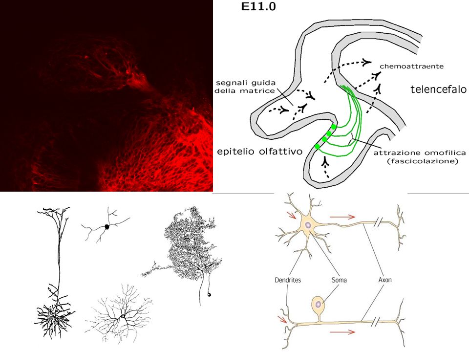
Neurogenesi e guida assonale nello sviluppo neurale Neurogenesi: una questione di … naso Recently, we are studying cellular and molecular mechanisms leading to the formation of functional neuronal networks in mammals. Formation of networks takes place during brain development where several cell and molecular mechanisms operate in a coordinated way to control successive steps identified as neurogenesis, cell migration, neural differentiation and neuronal connectivity. In the adult brain, these events are still occurring in precise areas or reactivated under pathological circumstances Un problema biologico: lo sviluppo del bulbo olfattivo Each primary olfactory neuron stochastically expresses one of ~1000 odorant receptors. The total population of these neurons therefore consists of ~1,000 distinct subpopulations, each of which are mosaically dispersed throughout one of four semi-annular zones in the nasal cavity. The axons of these different subpopulations are initially intermingled within the olfactory nerve. However, upon reaching the olfactory bulb, they sort out and converge so that axons expressing the same odorant receptor typically target one or two glomeruli.

4
Sviluppo del sistema olfattivo ovvero …. questione di naso.
Utilizzo di modelli computazionali per studiare la morfogenesi neurale del sistema olfattivo: come avviene il processo e l’organizzazione di questo sistema? Abbiamo varie scale: molecolare, cellulare, funzionale.

5
from J.A. St John et al., Int. J. Dev. Biol. 46: 639-647 (2002)
from A. Puche (Univ. Maryland)
")
7
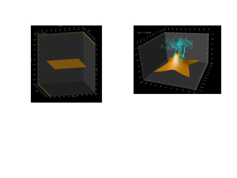
degradazione sorgente diffusione La diffusione di un fattore chemotattico si considera stazionaria

9
Con quali dati reali confrontarci?

10
Reti neuronali del cervelletto
Egidio D'Angelo (UniPV e INFM), Shyam Diwakar (UniMI) Giovanni Naldi (UniMI) Sergio Solinas (UniPV e INFM),
, Shyam Diwakar (UniMI) Giovanni Naldi (UniMI) Sergio Solinas (UniPV e INFM),")
11
Purkinje layer Granular layer
Ci siamo interessati del cervelletto, dati sperimentali relativi (in particolare) alla cellula granulare Purkinje layer Granular layer The Cerebellum Principles of Neural Science, Kandel et al., 4th Ed., 2000
 alla cellula granulare. Purkinje. layer. Granular. layer. The Cerebellum Principles of Neural Science, Kandel et al., 4th Ed.,")
12
Problema: sistema di equazioni di diffusione con dinamica non lineare
dei canali ionici e con opportune condizioni al bordo (di raccordo) tra i vari segmenti del neurone (nel caso di solo comportamento passivo si ha un’equazione di diffusione lineare). Si può dimostrare un Teorema di esistenza ed unicità. Nel caso di più neuroni ho un sistema di equazioni definite su un grafo. Nel caso di cellula granulare, molto compatta, si può pensare solo ad un sistema di equazioni differenziali ordinarie: concentriamo il modello nel soma. Linguaggio dei sistemi dinamici Studio numerico dei punti di equilibrio e della loro stabilità, analisi delle biforcazioni, studio nello spazio delle fasi dei fenomeni oscillatori.
 tra. i vari segmenti del neurone (nel caso di solo comportamento passivo. si ha un’equazione di diffusione lineare). Si può dimostrare un Teorema di esistenza ed unicità. Nel caso di più. neuroni ho un sistema di equazioni definite su un grafo. Nel caso di cellula granulare, molto compatta, si può pensare solo ad. un sistema di equazioni differenziali ordinarie: concentriamo il modello. nel soma. Linguaggio dei sistemi dinamici. Studio numerico dei punti di. equilibrio e della loro stabilità, analisi delle biforcazioni, studio. nello spazio delle fasi dei fenomeni. oscillatori.")
13
Training Signal Inputs Outputs
Granule cells Parallel fibers Parallel fiber Purkinje cell Inferior olive Training Signal Golgi cell Golgi axon inhibitory Granule cell Glomeruli Mossy fibers Inputs Cerebellar Nucleus Outputs Mossy fibers @ Copyright Coenen, 2002

14
Multi-Electrode Array
Nel prossimo futuro … Multi-Electrode Array Multi-Electrode Arrays (MEA) is being used to study the spatial distribution of signals into the cerebellum granular layer in acute slices. MEA consists of 60 conical platinum electrodes covered with glass and isolating material. MEA is used either to record or to stimulate slices, allowing the possibility of studying numerous aspects of the spatio-temporal dynamics of signals processing. The application of protocols to induce Long-term synaptic plasticity or the perfusion of specific receptors blockers allows to describe the spatial reconfiguration of signals, after which it is regulated by complex molecular mechanisms that cannot be analyzed by this technique.
 is being used to study the spatial distribution of signals into the cerebellum granular layer in acute slices. MEA consists of 60 conical platinum electrodes covered with glass and isolating material. MEA is used either to record or to stimulate slices, allowing the possibility of studying numerous aspects of the spatio-temporal dynamics of signals processing. The application of protocols to induce Long-term synaptic plasticity or the perfusion of specific receptors blockers allows to describe the spatial reconfiguration of signals, after which it is regulated by complex molecular mechanisms that cannot be analyzed by this technique.")
15
Vasculogenesi 3D Experimental data

16
Dentro la cellula: come si trasduce il segnale chemiotattico?

17
Vasculogenesi 3D Collaborazione:
UniMI (Giovanni Naldi, Matteo Semplice, Fausto Cavalli) PoliTO (Andrea Gamba) l'Istituto per la Ricerca e la Cura del Cancro (IRCC) di Candiolo (Guido Serini)
 PoliTO (Andrea Gamba) l Istituto per la Ricerca e la Cura del Cancro (IRCC) di Candiolo (Guido Serini)")
18
Trasduzione dei segnali biochimi nelle piastrine
La trombina attiva le piastrine tramite interazione con tre distinti recettori: PAR-1, PAR-4 ed il complesso glicoproteico (GP) Ib-IX-V.
 Ib-IX-V.")
19
Collaborazione con gruppo del
Prof. E. Di Benedetto (Vanderbilt University )
")
20
On platelet activation a number of responses can be shown to occur, including a change in shape from disk to sphere, platelet aggregation, granule release or secretion, and thromboxane (TxA2) production. Some of these products induce further platelet activation as a positive feedback. These include secreted ADP and TxA2.

21
AC, adenylyl cyclase; cAMP, cyclic adenosine monophosphate; CO, cyclooxygenase; DG, diacylglycerol; IP3, inositol 1,4,5 trisphosphate; MLC, myosin light-chain; PAF, platelet activating factor; PIP2, phosphatidylinositol bisphosphate; PKC, protein kinase C; PLA2, phospholipase A2; PLC, phospholipase C; R, receptor; vWD, von Willebrand Factor A number of physiologic agonists interact with specific receptors on the platelet surface to induce a series of responses. The agonists include ADP, epinephrine, TxA2, thrombin, PAF, collagen, vasopressin, and serotonin. Platelet activation initiates the production or release of several intracellular messenger molecules, including Ca2+ ions, products of phosphoinositide (PI) hydrolysis, DG, IP3, TxA2, and cyclic nucleotides [cAMP]. These modulate the various discernable platelet responses of Ca2+ mobilization, protein phosphorylation, aggregation, secretion, and liberation of arachidonic acid. The interaction between the agonist receptors on the platelet surface and the key intracellular effector enzymes (e.g. PLA2,PLC, and AC) are mediated by a group of GTP-binding proteins that are modulated by GTP. On platelet stimulation, phosphoinositides are hydrolyzed by PLC to DG and various inositol phosphates, including IP3. IP3 functions as a messenger to mobilize Ca2+ from an intracellular source. DG activates PKC and this results in the phosphorylation of a 47-kD protein, pleckstrin. On activation, platelets release arachidonic acid from phospholipids, which is mediated by PLA2. The free arachidonic acid is converted by COX to prostaglandins G2 and H2, and subsequently by thromboxane synthetase to TxA2. Together, these events lead to aggregation and secretion.
 hydrolysis, DG, IP3, TxA2, and cyclic nucleotides [cAMP]. These modulate the various discernable platelet responses of Ca2+ mobilization, protein phosphorylation, aggregation, secretion, and liberation of arachidonic acid. The interaction between the agonist receptors on the platelet surface and the key intracellular effector enzymes (e.g. PLA2,PLC, and AC) are mediated by a group of GTP-binding proteins that are modulated by GTP. On platelet stimulation, phosphoinositides are hydrolyzed by PLC to DG and various inositol phosphates, including IP3. IP3 functions as a messenger to mobilize Ca2+ from an intracellular source. DG activates PKC and this results in the phosphorylation of a 47-kD protein, pleckstrin. On activation, platelets release arachidonic acid from phospholipids, which is mediated by PLA2. The free arachidonic acid is converted by COX to prostaglandins G2 and H2, and subsequently by thromboxane synthetase to TxA2. Together, these events lead to aggregation and secretion.")
22
Diffusion Tensor Imaging (DTI)
Tecnica MR di imaging nella quale il contrasto è ottenuto dalla differente diffusione di molecole di acqua La DTI è utilizzata per identificare e generare mappe delle fibre di materia bianca nel cervello Giovanni Frisoni, Cristina Testa UniMI: Giovanni Naldi, Donatella Giuliani

23
DTI – Color-coding Based on Direction of Greatest Diffusion
Red = Left-Right Green = Anterior-Posterior Blue = Superior-Inferior

24
Trattografia di fibre: una nuova omica, la connectomica
Lazar et al., 2003 Problema: integrare immagini DTI con immagini in altri formati (PET, NMR, …).
.")
25
Problema: integrare immagini DTI con immagini in altri formati (PET, NMR, …).
.")
Presentazioni simili


>")


>")
 17 Ottobre 2009 DOPPIA ANTIAGGREGAZIONE PIASTRINICA.>")