Scaricare la presentazione
La presentazione è in caricamento. Aspetta per favore

1
L’ ‘epigenetica’ si riferisce a tutti i cambiamenti dell’espressione genica e dell’organizzazione della cromatina che sono indipendenti dalla sequenza del DNA.

2
Epigenetics -DNA methylation (5-mC, 5-hmC) -Histone PTMs (>100)
 -Histone PTMs (>100)")
3
Metilazione del DNA Nei vertebrati la metilazione interessa solamente la Citosina: l’enzima citosina metiltransferasi aggiunge un gruppo metile al C5 della citosina5-metilcitosina. mCpG (NEI VERTEBRATI NON IN DROSOPHILA) Non interferisce con il giusto appaiamento delle basi del dna Espressione genica,trasmissione di profili di espressione alle cellule figlie, sistema di difesa contro trasposoni Solo 3% delle C e’ metilata (CpG) 5meC e’ instabile e viene deaminata Timina Durante evoluzione il numero di dinucleotidi CpG nei vertebrati diminuisce CpG-TpG o CpA (escluse le CpGi) Cromatina in conformazione condensata (seq.ripetute) Altre basi deaminate producono basi anomale-attivazione del danno al dna Citosina nn metilata deaminata produce uracile che nel filamento del dna e’ una base anomala utilizzata per
 Non interferisce con il giusto appaiamento delle basi del dna. Espressione genica,trasmissione di profili di espressione alle cellule figlie, sistema di difesa contro trasposoni. Solo 3% delle C e’ metilata (CpG) 5meC e’ instabile e viene. deaminata Timina. Durante evoluzione il numero di dinucleotidi CpG nei vertebrati diminuisce CpG-TpG o CpA (escluse le CpGi) Cromatina in conformazione condensata (seq.ripetute) Altre basi deaminate producono basi anomale-attivazione del danno al dna. Citosina nn metilata deaminata produce uracile che nel filamento del dna e’ una base anomala utilizzata per.")
4
METODI PER LO STUDIO DELLE REGIONI metilate
1_Selezione DNA metilato MeDIP (ab anti 5meC) MCIp (MBD) MIRA (MBD+ his+colonna) MBD
 MCIp (MBD) MIRA (MBD+ his+colonna) MBD.")
5
METODI PER LO STUDIO DELLE REGIONI CpG
1_Analisi del DNA selezionato PCR Microarray (CpG island) Sequencing
 Sequencing.")
6
CpG ISLAND CpGisland sono state identificate tramite digestione del DNA di topo con HPAII(CCGG) Tratti di DNA ipometilato con frequenza di CG di oltre il 50% (frequenza attesa del dinucletide) Marcatori genici regioni trascrizionalmente attivestato permissivo della cromatina Cromatina in conformazione aperta Localizzate su Sito di Inizio della Trascrizione ma non solo CGIs were first identified by digestion of mouse genomic DNA using the methyl-CpG sensitive restriction enzyme HpaII (CCGG recognition site). A small portion of the genome, composed of very highly fragmented DNA, was found to be derived from sequences containing clusters of non-methylated CpG sites Promotori non ancora annotati
 Marcatori genici regioni trascrizionalmente attivestato permissivo della cromatina. Cromatina in conformazione aperta. Localizzate su Sito di Inizio della Trascrizione ma non solo. CGIs were first identified by digestion of mouse genomic DNA using the methyl-CpG sensitive restriction enzyme HpaII (CCGG recognition site). A small portion of the genome, composed of very highly fragmented DNA, was found to be derived from sequences containing clusters of non-methylated CpG sites. Promotori non ancora annotati.")
7
CpG islands e Regolazione della Trascrizione
Metilazione del Dna correla con silenziamento (GATA2 tessuto specifico) Stati trascizionali di molti geni non correlano con lo stato di metilazione dei singoli promotori TSS alternativi CpG island collocate dove ha origine un non-coding RNA CpGi intergeniche se metilate influiscono sull’espressione di interi locus (CTCF) 6. CGI methylation and transcription 6.1. CGI methylation and transcriptional regulation There is extensive evidence to support a functional role for pro- moter-CGI methylation in transcriptional repression (see, for example [10,72,80]). DNA methylation of the CpG-rich promoters of MASPIN and GATA2 correlates with tissue specific gene silencing [66,75]. In light of this evidence, it is tempting to hypothesise that the major function of CGI methylation is to repress transcription. However many genes display a relatively poor correlation between CGI hypermethylation and the transcriptional status of associated genes [29,63,81]. There are several potential explanations for this lack of corre- lation as illustrated in Fig. 3. In a simple example such as that de- picted in Fig. 3A, hypermethylation of the single promoter associated CGI would lead to stable transcriptional silencing. The majority of methylated CGIs are located within intragenic re- gions where the effect on transcription is less clear [1,29,63].1718 R.S. Illingworth, A.P. Bird / FEBS Letters 583 (2009) 1713–1720 Many genes can generate multiple transcripts by utilising alterna- tive transcription starts sites. Rauch and colleagues identified expression of PARP12 despite hypermethylation of its primary CGI promoter. Rapid amplification of cDNA ends (50 RACE), how- ever, identified transcription initiation from an intragenic pro- moter downstream of the methylated CGI [63]. Alternative promoters (e.g. P1–3 in Fig. 3B) could be inactivated by CGI meth- ylation (Fig. 3B – CGIs (i) and (ii)). Where intragenic islands do not associate with a known TSS, it is possible that their methylation could prevent spurious gene body transcription which could otherwise interfere with the cor- rect expression of the parent gene (Fig 3B – CGI (iii and iv)). As yet there is no evidence for this conjecture. There is evidence that Intragenic CGIs can localise to sites of antisense non-coding RNA (ncRNA) transcription initiation which negatively regulate the expression of the sense transcript (Fig. 3B – CGI (v)). Both the Air and Tsix ncRNA transcripts originate from CGIs and are involved in the regulation of the sense transcript [82–84]. The HOXD cluster is repressed in trans by the action of HOTAIR, a ncRNA transcribed from the HOXC locus [85]. In each case, CGI methylation results in the derepression of genes silenced by ncRNAs. Many hypermethylated CGIs are located in intergenic DNA out- side coding sequences and therefore have no obvious regulatory role in gene transcription (Fig. 3B – CGI (vi)). In the case of the H19/IGF2 imprinted locus however, parent specific methylation at an intergenic CGI upstream of the H19 ncRNA gene determines the expression of the imprinted locus. CGI methylation prevents the association of the insulator element CTCF and promotes expression of IGF2 from the paternal allele [58]. This illustrates a potential mechanism whereby hypermethylation of intergenic CGIs can illicit a transcriptional effect. These examples illustrate the complexity in determining the ef- fect of DNA methylation at CGIs. Characterisation of transcription initiation using RNA polymerase chromatin immunoprecipitation and RACE will provide a better understanding of the function of CGI methylation at these sites. Effetto complesso della Metilazione delle CpG islands
 Stati trascizionali di molti geni non correlano con lo stato di metilazione dei singoli promotori. TSS alternativi. CpG island collocate dove ha origine un non-coding RNA. CpGi intergeniche se metilate influiscono sull’espressione di interi locus (CTCF) 6. CGI methylation and transcription CGI methylation and transcriptional regulation. There is extensive evidence to support a functional role for pro- moter-CGI methylation in transcriptional repression (see, for example [10,72,80]). DNA methylation of the CpG-rich promoters of MASPIN and GATA2 correlates with tissue specific gene silencing [66,75]. In light of this evidence, it is tempting to hypothesise that the major function of CGI methylation is to repress transcription. However many genes display a relatively poor correlation between CGI hypermethylation and the transcriptional status of associated genes [29,63,81]. There are several potential explanations for this lack of corre- lation as illustrated in Fig. 3. In a simple example such as that de- picted in Fig. 3A, hypermethylation of the single promoter associated CGI would lead to stable transcriptional silencing. The majority of methylated CGIs are located within intragenic re- gions where the effect on transcription is less clear [1,29,63].1718 R.S. Illingworth, A.P. Bird / FEBS Letters 583 (2009) 1713–1720. Many genes can generate multiple transcripts by utilising alterna- tive transcription starts sites. Rauch and colleagues identified expression of PARP12 despite hypermethylation of its primary CGI promoter. Rapid amplification of cDNA ends (50 RACE), how- ever, identified transcription initiation from an intragenic pro- moter downstream of the methylated CGI [63]. Alternative promoters (e.g. P1–3 in Fig. 3B) could be inactivated by CGI meth- ylation (Fig. 3B – CGIs (i) and (ii)). Where intragenic islands do not associate with a known TSS, it is possible that their methylation could prevent spurious gene body transcription which could otherwise interfere with the cor- rect expression of the parent gene (Fig 3B – CGI (iii and iv)). As yet there is no evidence for this conjecture. There is evidence that Intragenic CGIs can localise to sites of antisense non-coding RNA (ncRNA) transcription initiation which negatively regulate the expression of the sense transcript (Fig. 3B – CGI (v)). Both the Air and Tsix ncRNA transcripts originate from CGIs and are involved in the regulation of the sense transcript [82–84]. The HOXD cluster is repressed in trans by the action of HOTAIR, a ncRNA transcribed from the HOXC locus [85]. In each case, CGI methylation results in the derepression of genes silenced by ncRNAs. Many hypermethylated CGIs are located in intergenic DNA out- side coding sequences and therefore have no obvious regulatory role in gene transcription (Fig. 3B – CGI (vi)). In the case of the H19/IGF2 imprinted locus however, parent specific methylation at an intergenic CGI upstream of the H19 ncRNA gene determines the expression of the imprinted locus. CGI methylation prevents the association of the insulator element CTCF and promotes expression of IGF2 from the paternal allele [58]. This illustrates a potential mechanism whereby hypermethylation of intergenic CGIs can illicit a transcriptional effect. These examples illustrate the complexity in determining the ef- fect of DNA methylation at CGIs. Characterisation of transcription initiation using RNA polymerase chromatin immunoprecipitation and RACE will provide a better understanding of the function of CGI methylation at these sites. Effetto complesso della Metilazione delle CpG islands.")
8
DNA Metiltransferasi (writers) e Fasi di metilazione
Metilazione differenziale de novo (Dnmt3a e Dnmt3b); Demetilazione passiva (natura sconosciuta); Mantenimento della metilazione e trasmissione alle cellule figlie (Dnmt1) dopo replicazione del DNA. Esiste un ulteriore DNA metilasi DNMT2 ma metila tRNA.
; Demetilazione passiva (natura sconosciuta); Mantenimento della metilazione e trasmissione alle cellule figlie (Dnmt1) dopo replicazione del DNA. Esiste un ulteriore DNA metilasi DNMT2 ma metila tRNA.")
9
Metilazione nello sviluppo dei mammiferi
Demetilazione enzimatica Demetilazione passiva DNA cellule uovo e spermatozoi altamente metilate e I profili di metilazione di queste cellule sono fortemente diversi dalle cellules omatiche I pronuclei hanno un genoma metilano in modo specifico gli spermatozoi prima della fusione subiscono un processo enzimatico di demetilazione Dopo la fusione dei pronuclei lo zigote subisce un rpocesso di demetilazione fino allo stato di blastula Allo stadio di blastocisti dove il processo di differenziamento comincia si ha un inzio di metilazione de novo del DNA Vriabile epr ogni tipo cellulare le cellule che differenziano e sono dette somatiche subiscono una forte metilazioneMentre le cellule derivative del trofoblasto Lr cellule germinali primordiali vengono demetilate e riprogrammate come sul lato sinistro del grafico Non si sa quanto questi drastici eveni influiscano sui vari meccaniscmi di differenziamento In C.elegance e Dhrosophila la metilazione e’ assente ma I processi di differenziamento sono simili Pero nei vertebrati sono importanti in fattianimali priv di ttivita metiltransferasica muoiono in vari stadi dello sviluppo Nelle staminali non c’’e metilazione e riescono a sopravvivere pero se si da un impulso di differenziamento vanno in apoptosi La demetilazione sere piu che altro ad azzerare l’informazione epigenetica della generazione precedente per poter acquisire uno stato di totipotenza versatilita’ all’inizio dello sviluppo Cancellazione dello stato epigenetico Rimetilazione e determinazione dell’imprinting Rimetilazione durante il differenziamento Demetilazione pssiva

10
Riprogrammazione epigenetica durante la gametogenesi
Esperimenti di knock-out Esperimenti di knock-out Istoni acetilati Epigenetic reprogramming in gametes Gametes are terminally differentiated and highly specialized cells that carry all the information that is nec- essary for the initiation of a new life cycle after normal fertilization. Nuclear-transplantation experiments have shown that both the maternal and paternal genomes are necessary for embryonic development as they are programmed differently and are functionally non-equivalent41,42. The functional differences between the paternal and maternal genomes are attributed to the differential expression of the paternal and maternal alleles of several dozen imprinted genes during development. These differences originate from the differential modification of the genome (or genomic imprinting) in the male and female gametes43. The paternal alleles of the H19 and Rasgrf1 genes are methylated in their 5′ upstream regions in the male germ cells during embryogenesis, whereas the other known imprinted genes, such as Igfr2 and Snprn, acquire their methylation imprints from the oocyte; the deletion of such DIFFERENTIALLY METHYLATED REGIONS results in the loss of imprinting44–49. The gamete-derived methylation patterns of imprinted genes are maintained in the somatic tissues throughout embryonic development, but are erased in the primordial germ cells50,51; therefore, genomic imprinting can be reversed in the germ line. Imprinting in oocytes. The genetic evidence that DNA methylation is an essential epigenetic mark for the establishment of genomic imprinting comes from recent studies of the Dnmt3-like (Dnmt3l) gene. Dnmt3l encodes a protein that shares homology with Dnmt3a and Dnmt3b in the PHD zinc-finger domain, but lacks the highly conserved methyltransferase motifs and has no enzymatic activity. Although Dnmt3l-deficient females produce mature and func- tional oocytes, embryos that are derived from these oocytes have neural-tube and placental abnormalities and die around midgestation52,53. The analysis of DNA- methylation patterns at several maternally methylated genes on different chromosomes, such as Igf2r, Peg1 and Peg3, and of several imprinted genes in the Snrpn locus has shown that Dnmt3l-deficient oocytes fail to establish maternal-specific methylation imprints. Significantly, a failure to establish maternal methyla- tion imprints in Dnmt3l-deficient oocytes results in the loss of the mono-allelic expression of all mater- nally imprinted genes that are examined in the off- spring. These results show that methylation imprints that are acquired during oocyte maturation serve as the maternal genomic imprints. Dnmt3l probably acts on imprinted genes through its interaction with the Dnmt3 family of DNA methyl- transferases. Dnmt3l binds to and co-localizes with Dnmt3a and Dnmt3b in the nuclei of cells in which both of these proteins are expressed53. These results indicate that Dnmt3l might cooperate with Dnmt3a or Dnmt3b to regulate the gamete-specific methylation of imprinted genes in the oocyte (FIG. 2). Consistent with this model, it has been shown that Dnmt3a/Dnmt3b- deficient females also fail to establish maternal methyla- tion imprints53. By contrast, inactivation of Dnmt1 in the oocyte does not perturb the establishment of methylation imprints, but affects the maintenance of imprinting in pre-implantation embryos54. The possible involvement of histone modification and chromatin remodelling in the establishment of genomic imprints during gametogenesis remains to be investigated. Methylation in spermatogenesis. In addition to its role in genomic imprinting in the female gamete, DNA methylation is also required for spermatogenesis. In Dnmt3a-knockout mice, the testes contain many abnormal spermatocytes at the meiosis prophase, but few mature sperms53. In the Dnmt3l-knockout mice, spermatogenesis is arrested at a time when the sper- matogonia enter meiosis, which results in complete lack of mature sperm52,53. Interestingly, histone modification is also crucial in spermatogenesis. Mice in which both histone methyltransferases Suv39h1 and Suv39h2 have been knocked out are sterile36. In Suv39h double-knockout mice, homologous chromosome pairing is impaired, which results in meiotic arrest at the pachytene stage. The timing for the requirement of DNA methylation and histone H3-K9 methylation during spermatogene- sis seems to correlate with histone deacetylation (FIG. 2). The core histones are hyperacetylated in spermatogonia and in pre-leptotene spermatocytes, but acetylated his- tones are not detected throughout meiosis in leptotene and pachytene spermatocytes, nor in most round sper- matids55. These findings indicate that DNA methylation and histone modification might suppress global gene expression when spermatocytes are undergoing meiosis. Alternatively, these epigenetic changes might have a role in regulating chromosome architecture, which changes dynamically during spermatogenesis. It is of great importance to understand the specific chromosomal changes that require DNA methylation and histone modification during spermatogenesis and meiosis. Genomic imprinting differenza nelle modificazioni del genoma dei gameti Alcuni geni ereditano l’imprinting (metilazione) dal genoma paterno e altri da quello materno Imprinting viene mantenuto fino alle cellule somatiche e perso nelle cellule germinali primordiali
 in the male and female gametes43. The paternal alleles of the H19 and Rasgrf1 genes are methylated in their 5′ upstream regions in the male germ cells during embryogenesis, whereas the other known imprinted genes, such as Igfr2 and Snprn, acquire their methylation imprints from the. oocyte; the deletion of such DIFFERENTIALLY METHYLATED REGIONS results in the loss of imprinting44–49. The gamete-derived methylation patterns of imprinted genes are maintained in the somatic tissues throughout embryonic development, but are erased in the primordial germ cells50,51; therefore, genomic imprinting can be reversed in the germ line. Imprinting in oocytes. The genetic evidence that DNA methylation is an essential epigenetic mark for the establishment of genomic imprinting comes from recent studies of the Dnmt3-like (Dnmt3l) gene. Dnmt3l encodes a protein that shares homology with Dnmt3a and Dnmt3b in the PHD zinc-finger domain, but lacks the highly conserved methyltransferase motifs and has no enzymatic activity. Although Dnmt3l-deficient females produce mature and func- tional oocytes, embryos that are derived from these oocytes have neural-tube and placental abnormalities and die around midgestation52,53. The analysis of DNA- methylation patterns at several maternally methylated genes on different chromosomes, such as Igf2r, Peg1 and Peg3, and of several imprinted genes in the Snrpn locus has shown that Dnmt3l-deficient oocytes fail to establish maternal-specific methylation imprints. Significantly, a failure to establish maternal methyla- tion imprints in Dnmt3l-deficient oocytes results in the loss of the mono-allelic expression of all mater- nally imprinted genes that are examined in the off- spring. These results show that methylation imprints that are acquired during oocyte maturation serve as the maternal genomic imprints. Dnmt3l probably acts on imprinted genes through its interaction with the Dnmt3 family of DNA methyl- transferases. Dnmt3l binds to and co-localizes with Dnmt3a and Dnmt3b in the nuclei of cells in which both of these proteins are expressed53. These results indicate that Dnmt3l might cooperate with Dnmt3a or Dnmt3b to regulate the gamete-specific methylation of imprinted genes in the oocyte (FIG. 2). Consistent with this model, it has been shown that Dnmt3a/Dnmt3b- deficient females also fail to establish maternal methyla- tion imprints53. By contrast, inactivation of Dnmt1 in the oocyte does not perturb the establishment of methylation imprints, but affects the maintenance of imprinting in pre-implantation embryos54. The possible involvement of histone modification and chromatin remodelling in the establishment of genomic imprints during gametogenesis remains to be investigated. Methylation in spermatogenesis. In addition to its role in genomic imprinting in the female gamete, DNA methylation is also required for spermatogenesis. In Dnmt3a-knockout mice, the testes contain many abnormal spermatocytes at the meiosis prophase, but few mature sperms53. In the Dnmt3l-knockout mice, spermatogenesis is arrested at a time when the sper- matogonia enter meiosis, which results in complete lack of mature sperm52,53. Interestingly, histone modification is also crucial in spermatogenesis. Mice in which both histone methyltransferases Suv39h1 and Suv39h2 have been knocked out are sterile36. In Suv39h double-knockout mice, homologous chromosome pairing is impaired, which results in meiotic arrest at the pachytene stage. The timing for the requirement of DNA methylation and histone H3-K9 methylation during spermatogene- sis seems to correlate with histone deacetylation (FIG. 2). The core histones are hyperacetylated in spermatogonia and in pre-leptotene spermatocytes, but acetylated his- tones are not detected throughout meiosis in leptotene and pachytene spermatocytes, nor in most round sper- matids55. These findings indicate that DNA methylation and histone modification might suppress global gene expression when spermatocytes are undergoing meiosis. Alternatively, these epigenetic changes might have a role in regulating chromosome architecture, which changes dynamically during spermatogenesis. It is of great importance to understand the specific chromosomal changes that require DNA methylation and histone modification during spermatogenesis and meiosis. Genomic imprinting differenza nelle modificazioni del genoma dei gameti. Alcuni geni ereditano l’imprinting (metilazione) dal genoma paterno e altri da quello materno. Imprinting viene mantenuto fino alle cellule somatiche e perso nelle cellule germinali primordiali.")
11
Metilazione del DNA nell’embrione durante stadi precoci
DEMETILAZIONE nell’EMBRIONE (methylated sites un-methylated sites) La trascrizione persiste e viene rimetilata la zona extra CpG island e poi il Gene viene silenziato con meccanismi non methyl –dipendentiPOTENZIALE TRASCRIZIONE ATTIVA Oppure la trascrizione viene bloccata con altri meccanismi e questo stato richiama metilazione de novo sia della CpG island che delle sequenze fiancheggiantiPOTENZIALE SILENZIAMENTO Many of the known biological effects of DNA methyl- ation are associated with CpG islands. It has been argued above that their methylation in the early embryo follows silencing events that are likely to be DNA methylation- independent. If transcriptional silence indeed triggers DNA methylation, then the corollary is that promoter activity early in development should create a methyl- ation-free CpG island (Fig. 2). In other words, unmethyl- ated CpG islands might be footprints of embryonic pro- moter activity. An obvious prediction of this model is that all unmethylated CpG islands, including those at promoters of highly tissue-specifically expressed genes, should contain promoters that function during early de- velopment when the methylation memory system is most active. Although very limited, the data so far favor this theory, because a CpG-island promoter whose prod- uct RNA is not expected to occur in the early embryo (-globin) is nevertheless expressed, whereas transcripts from a CpG-deficient promoter (-globin) are not de- tected (Daniels et al. 1997). Similarly, expression of the 68k neurofilament gene, which has a CpG-island pro- moter, was detected in ES cells, but opsin and casein genes, which are CpG-deficient genes, appeared to be silent (MacLeod et al. 1998). Why should active promoter regions escape de novo methylation? CpG islands often colocalize with origins of DNA replication (Delgado et al. 1998), and, according to one speculation, an early replication intermediate cre- ates the DNA methylation-free footprint (Antequera and Bird 1999). A more direct (but not mutually exclusive) mechanism would involve the sensing of chromatin states by the de novo methylation system as discussed above. Whereas histone H3 tails modified by methyl- ation on Lys 9 might recruit DNA methyltransferases (Tamaru and Selker 2001), modifications associated with active chromatin, such as acetylation of H3 or H4 or methylation of Lys 4 of histone H3, may actively exclude these enzymes. Biochemical evidence addressing this is- sue is eagerly awaited. Active demethylation of DNA Protection against de novo methylation by bound pro- teins or chromatin can ensure that DNA methylation never reaches a DNA sequence domain. Unmethylated domains could also arise by actively removing the modi- fication from DNA. This so-called active demethylation could be accomplished either by the thermodynamically unfavorable breakage of the carbon—carbon bond that links the pyrimidine to its methyl group, or by a repair- like process that excises the m5C base or nucleoside, leading to its replacement with C (Kress et al. 2001). Several laboratories have striven to isolate demethylase enzymes (for review, see Wolffe et al. 1999). The most impressive catalytic activity was shown by a fraction derived from human cells (Ramchandani et al. 1999) that was subsequently identified as MBD2 (Bhattacharya et al. 1999). The expressed protein reportedly showed ro- bust demethylation in vitro in the absence of added co- factors and released methanol as a by-product. Attempts to observe this property of MBD2 in other laboratories have not been successful. A cell extract showing demethylase activity was de- tected in rat myoblast cells (Weiss et al. 1996). Initial indications that the reaction was RNA-dependent were not sustained upon further enrichment of the activity (Swisher et al. 1998). An RNA-containing demethylating complex was, however, reported in chicken cells (Jost et al. 1997, 1999). These investigators searched for proteins with m5C-DNA glycosylase activity and identified the previously known thymine DNA glycosylase TDG, which can remove the pyrimidine base from T:G or U:G mismatches (Zhu et al. 2000b). MBD4, an unrelated DNA glycosylase with similar properties, was also found to be active against m5C:G pairs (Zhu et al. 2000a). As the efficiency of these reactions was much lower than that seen with the cognate mismatched substrates, it might be argued that the m5C glycosylase activity rep- resents a minor side reaction of little in vivo signifi- cance. Set against this is evidence that stable expression of a chicken TDG results in significant activation and concomitant demethylation of a reporter gene driven by a methylated ecdysone-retinoic acid-responsive pro- moter (Zhu et al. 2001). The normally silent reporter could also be activated by demethylation with 5-azacyti- dine, but generalized demethylation of the genome was not observed in TDG transfected cells. Previous studies showed an association between retinoid receptors and TDG, and implicated TDG in transcriptional activation (Um et al. 1998). Time will tell if the stimulation of retinoid-responsive promoters by TDG depends on its demethylating activity. The need to isolate demethylating enzymes has be- come more acute with the finding that the paternal ge- nome is subject to active demethylation soon after fer- tilization (Mayer et al. 2000; Oswald et al. 2000). Similar processes have been reported in pig and bovine embryos (Bourc’his et al. 2001; Kang et al. 2001a,b). This dramatic illustration of methylation loss in the absence of DNA replication raises questions about the prevalence of de- methylation by this mechanism. Interestingly, the ma- ternal genome, which also demethylates during early mouse development, does so by a different mechanism: passive failure to methylate progeny stands (Rougier et al. 1998). Why should maternal and paternal genomes choose such different routes to the same end? An intrigu- ing possibility is that the parental struggle over maternal resources for the embryo that is thought to underlie ge- nomic imprinting (Moore and Haig 1991) may be in- volved. The oocyte may be equipped to directly disarm the sperm genome of methylation imprints that might overexploit maternal resources (Reik and Walter 2001). It is even possible that the paternal genome, in delayed retaliation, may organize a campaign of interference with the maintenance methylation (e.g., by exporting maternal DNMTs to the cytoplasm). The extraordinary need for an oocyte variant of DNMT1 to translocate into the nucleus for only one cleavage cycle (the doubling from 8 to 16 cells; Howell et al. 2001) could represent maternal measures to compensate for interference of this kind.
 La trascrizione persiste e viene rimetilata la zona extra CpG island e poi il Gene viene silenziato con meccanismi non methyl –dipendentiPOTENZIALE TRASCRIZIONE ATTIVA. Oppure la trascrizione viene bloccata con altri meccanismi e questo stato richiama metilazione de novo sia della CpG island che delle sequenze fiancheggiantiPOTENZIALE SILENZIAMENTO. Many of the known biological effects of DNA methyl- ation are associated with CpG islands. It has been argued above that their methylation in the early embryo follows. silencing events that are likely to be DNA methylation- independent. If transcriptional silence indeed triggers DNA methylation, then the corollary is that promoter activity early in development should create a methyl- ation-free CpG island (Fig. 2). In other words, unmethyl- ated CpG islands might be footprints of embryonic pro- moter activity. An obvious prediction of this model is that all unmethylated CpG islands, including those at promoters of highly tissue-specifically expressed genes, should contain promoters that function during early de- velopment when the methylation memory system is most active. Although very limited, the data so far favor this theory, because a CpG-island promoter whose prod- uct RNA is not expected to occur in the early embryo (-globin) is nevertheless expressed, whereas transcripts from a CpG-deficient promoter (-globin) are not de- tected (Daniels et al. 1997). Similarly, expression of the 68k neurofilament gene, which has a CpG-island pro- moter, was detected in ES cells, but opsin and casein genes, which are CpG-deficient genes, appeared to be silent (MacLeod et al. 1998). Why should active promoter regions escape de novo methylation CpG islands often colocalize with origins of DNA replication (Delgado et al. 1998), and, according to one speculation, an early replication intermediate cre- ates the DNA methylation-free footprint (Antequera and Bird 1999). A more direct (but not mutually exclusive) mechanism would involve the sensing of chromatin states by the de novo methylation system as discussed above. Whereas histone H3 tails modified by methyl- ation on Lys 9 might recruit DNA methyltransferases (Tamaru and Selker 2001), modifications associated with active chromatin, such as acetylation of H3 or H4 or methylation of Lys 4 of histone H3, may actively exclude these enzymes. Biochemical evidence addressing this is- sue is eagerly awaited. Active demethylation of DNA. Protection against de novo methylation by bound pro- teins or chromatin can ensure that DNA methylation never reaches a DNA sequence domain. Unmethylated domains could also arise by actively removing the modi- fication from DNA. This so-called active demethylation could be accomplished either by the thermodynamically unfavorable breakage of the carbon—carbon bond that links the pyrimidine to its methyl group, or by a repair- like process that excises the m5C base or nucleoside, leading to its replacement with C (Kress et al. 2001). Several laboratories have striven to isolate demethylase enzymes (for review, see Wolffe et al. 1999). The most impressive catalytic activity was shown by a fraction derived from human cells (Ramchandani et al. 1999) that was subsequently identified as MBD2 (Bhattacharya et al. 1999). The expressed protein reportedly showed ro- bust demethylation in vitro in the absence of added co- factors and released methanol as a by-product. Attempts to observe this property of MBD2 in other laboratories have not been successful. A cell extract showing demethylase activity was de- tected in rat myoblast cells (Weiss et al. 1996). Initial indications that the reaction was RNA-dependent were not sustained upon further enrichment of the activity (Swisher et al. 1998). An RNA-containing demethylating complex was, however, reported in chicken cells (Jost et al. 1997, 1999). These investigators searched for proteins with m5C-DNA glycosylase activity and identified the previously known thymine DNA glycosylase TDG, which can remove the pyrimidine base from T:G or U:G mismatches (Zhu et al. 2000b). MBD4, an unrelated DNA glycosylase with similar properties, was also found to be active against m5C:G pairs (Zhu et al. 2000a). As the efficiency of these reactions was much lower than that seen with the cognate mismatched substrates, it might be argued that the m5C glycosylase activity rep- resents a minor side reaction of little in vivo signifi- cance. Set against this is evidence that stable expression of a chicken TDG results in significant activation and concomitant demethylation of a reporter gene driven by a methylated ecdysone-retinoic acid-responsive pro- moter (Zhu et al. 2001). The normally silent reporter could also be activated by demethylation with 5-azacyti- dine, but generalized demethylation of the genome was not observed in TDG transfected cells. Previous studies showed an association between retinoid receptors and TDG, and implicated TDG in transcriptional activation (Um et al. 1998). Time will tell if the stimulation of retinoid-responsive promoters by TDG depends on its demethylating activity. The need to isolate demethylating enzymes has be- come more acute with the finding that the paternal ge- nome is subject to active demethylation soon after fer- tilization (Mayer et al. 2000; Oswald et al. 2000). Similar processes have been reported in pig and bovine embryos (Bourc’his et al. 2001; Kang et al. 2001a,b). This dramatic illustration of methylation loss in the absence of DNA replication raises questions about the prevalence of de- methylation by this mechanism. Interestingly, the ma- ternal genome, which also demethylates during early mouse development, does so by a different mechanism: passive failure to methylate progeny stands (Rougier et al. 1998). Why should maternal and paternal genomes. choose such different routes to the same end An intrigu- ing possibility is that the parental struggle over maternal resources for the embryo that is thought to underlie ge- nomic imprinting (Moore and Haig 1991) may be in- volved. The oocyte may be equipped to directly disarm the sperm genome of methylation imprints that might overexploit maternal resources (Reik and Walter 2001). It is even possible that the paternal genome, in delayed retaliation, may organize a campaign of interference with the maintenance methylation (e.g., by exporting maternal DNMTs to the cytoplasm). The extraordinary need for an oocyte variant of DNMT1 to translocate into the nucleus for only one cleavage cycle (the doubling from 8 to 16 cells; Howell et al. 2001) could represent maternal measures to compensate for interference of this kind.")
12
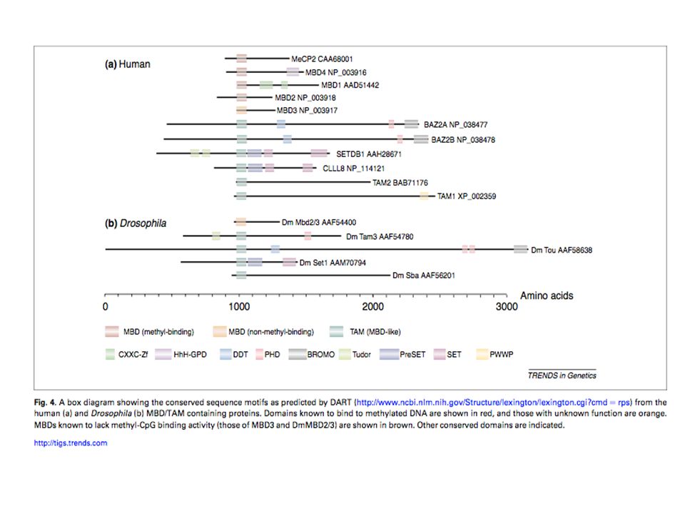
Methyl Binding Protein (MBD)
Riconoscono e legano le 5meCpG Reclutano altre proteine coinvolte nella repressione come HDAC(Histone Deacetylase) Per uomini e’ letale Perdita di funzione di MECP2 causa Sindrome di Rett (si trova sull’X) Nelle femmine si ha la Sindrome se negli X inattivati c’e’ al copia sana del gene (importante soprattutto nei neuroni), nel maschio e’ letale
 Per uomini e’ letale. Perdita di funzione di MECP2 causa Sindrome di Rett (si trova sull’X) Nelle femmine si ha la Sindrome se negli X inattivati c’e’ al copia sana del gene (importante soprattutto nei neuroni), nel maschio e’ letale.")
14
METILAZIONE E SILENZIAMENTO GENICO
TFs e Proteine Regolatrici proteggono il promotore dalla metilazione TFs e Proteine Regolatrici non sono piu’ legate per un meccanismo specifico (gene OFF but LEAKY) Metilazione delle CpG e reclutamento delle MBP che richiamano altri complessi di rimodellamento della cromatina e Istone Deacetilasi (gene COMPLETELY OFF)
 Metilazione delle CpG e reclutamento delle MBP che richiamano altri complessi di rimodellamento della cromatina e Istone Deacetilasi (gene COMPLETELY OFF)")
15
METILAZIONE E SILENZIAMENTO GENICO
reversibili Irreversibili (ETERO CROMATINA)
")
16
Metilazione del DNA e Repressione trascrizionale
Mechanisms of DNA methylation-mediated transcriptional repression Why does DNA methylation interfere with transcrip- tion? Two modes of repression can be envisaged, and it is likely that both are biologically relevant. The first mode involves direct interference of the methyl group in bind- ing of a protein to its cognate DNA sequence (Fig. 1). Many factors are known to bind CpG-containing se- quences, and some of these fail to bind when the CpG is methylated. Strong evidence for involvement of this mechanism in gene regulation comes from studies of the role of the CTCF protein in imprinting at the H19/Igf2 locus in mice (Bell and Felsenfeld 2000; Hark et al. 2000; Szabo et al. 2000; Holmgren et al. 2001). CTCF is asso- ciated with transcriptional domain boundaries (Bell et al. 1999) and can insulate a promoter from the influence of remote enhancers. The maternally derived copy of the Igf2 gene is silent owing to the binding of CTCF between its promoter and a downstream enhancer. At the pater- nal locus, however, these CpG-rich binding sites are methylated, preventing CTCF binding and thereby al- lowing the downstream enhancer to activate Igf2 expres- sion. Although there is evidence that H19/Igf2 imprint- ing involves additional processes (Ferguson-Smith and Surani 2001), the role of CTCF represents one of the clearest examples of transcriptional regulation by DNA methylation. The second mode of repression is opposite to the first, as it involves proteins that are attracted to, rather than repelled by, methyl-CpG (Fig. 1). A family of five methyl- CpG-binding proteins has been characterized that each contains a region closely related to the methyl-CpG- binding domain (MBD) of MeCP2 (Nan et al. 1993, 1997; Cross et al. 1997; Hendrich and Bird 1998). Four of these proteins—MBD1, MBD2, MBD3, and MeCP2—have been implicated in methylation-dependent repression of transcription (for review, see Bird and Wolffe 1999). An unrelated protein Kaiso has also recently been shown to bind methylated DNA and bring about methylation-de- pendent repression in model systems (Prokhortchouk et al. 2001). In vitro, Kaiso requires a 5 m5CGm5CG motif, and binding is highly dependent on the presence of meth- ylation. The presence of multiple methyl-CpG-binding proteins with repressive properties supports the argu- ment that these may be important mediators of the methylation signal, but their involvement in specific processes that require transduction of the DNA methyl- ation signal has yet to be shown. Targeted mutation of the gene for MeCP2 is, however, associated with neuro- logical dysfunction in humans and mice (Amir et al. 1999; Chen et al. 2001; Guy et al. 2001), and mutation of the mouse Mbd2 gene leads to a maternal behavior de- fect (Hendrich et al. 2001). A_TFs non riescono a legare le CpG se metilate (CTCF si lega al DNA e isola azione di attivazione di Enhancer metilazione DNA impedisce legame di CTCF ) B_MBD protein: MBD1, MBD2, MBD3, and MeCP2 and KAISO che possono riconoscere 5meCpG e richiamare altre proteine (silenziamento)
. Many factors are known to bind CpG-containing se- quences, and some of these fail to bind when the CpG is methylated. Strong evidence for involvement of this mechanism in gene regulation comes from studies of the role of the CTCF protein in imprinting at the H19/Igf2 locus in mice (Bell and Felsenfeld 2000; Hark et al. 2000; Szabo et al. 2000; Holmgren et al. 2001). CTCF is asso- ciated with transcriptional domain boundaries (Bell et al. 1999) and can insulate a promoter from the influence of remote enhancers. The maternally derived copy of the Igf2 gene is silent owing to the binding of CTCF between its promoter and a downstream enhancer. At the pater- nal locus, however, these CpG-rich binding sites are methylated, preventing CTCF binding and thereby al- lowing the downstream enhancer to activate Igf2 expres- sion. Although there is evidence that H19/Igf2 imprint- ing involves additional processes (Ferguson-Smith and Surani 2001), the role of CTCF represents one of the clearest examples of transcriptional regulation by DNA methylation. The second mode of repression is opposite to the first, as it involves proteins that are attracted to, rather than repelled by, methyl-CpG (Fig. 1). A family of five methyl- CpG-binding proteins has been characterized that each contains a region closely related to the methyl-CpG- binding domain (MBD) of MeCP2 (Nan et al. 1993, 1997; Cross et al. 1997; Hendrich and Bird 1998). Four of these proteins—MBD1, MBD2, MBD3, and MeCP2—have been implicated in methylation-dependent repression of transcription (for review, see Bird and Wolffe 1999). An unrelated protein Kaiso has also recently been shown to bind methylated DNA and bring about methylation-de- pendent repression in model systems (Prokhortchouk et al. 2001). In vitro, Kaiso requires a 5 m5CGm5CG motif, and binding is highly dependent on the presence of meth- ylation. The presence of multiple methyl-CpG-binding proteins with repressive properties supports the argu- ment that these may be important mediators of the methylation signal, but their involvement in specific processes that require transduction of the DNA methyl- ation signal has yet to be shown. Targeted mutation of the gene for MeCP2 is, however, associated with neuro- logical dysfunction in humans and mice (Amir et al. 1999; Chen et al. 2001; Guy et al. 2001), and mutation of the mouse Mbd2 gene leads to a maternal behavior de- fect (Hendrich et al. 2001). A_TFs non riescono a legare le CpG se metilate (CTCF si lega al DNA e isola azione di attivazione di Enhancer metilazione DNA impedisce legame di CTCF ) B_MBD protein: MBD1, MBD2, MBD3, and MeCP2 and KAISO che possono riconoscere 5meCpG e richiamare altre proteine (silenziamento)")
17
Ipometilazione delle CpGisland durante de novo metilazione 3 IPOTESI
CpGi refrattarie a de novo metilation (DNMT) a causa della loro sequenza CpGi sono target di un meccanismo di demetilazione del DNA che rimuove specificamente la 5meC I TFs legati alle CpGi precludono il legame alle DNMT (esperimento con mutazione del sito di SP1 in APRT promoterde novo METHYLATION) The mechanism by which CGIs remain hypomethylated during the period of global de novo methylation during early development remains unclear [35,36]. The characteristic clustering of CpG sites is a consequence of immunity against de novo methylation during the earliest stages of mammalian development. A simple sugges- tion would be that CGIs are intrinsically refractory to de novo methylation by DNA methyltransferases (DNMT) due to their DNA sequence (Fig. 2A). This seems unlikely however, as CGIs con- tain a substantially elevated density of CpG sites, the preferred substrate of the DNMT enzymes [37]. Moreover, CGIs located on the female inactive X chromosome and those of certain cultured mammalian cells readily acquire DNA methylation [2,38]. A second possibility is that CGIs are targeted by a DNA demeth- ylation mechanism, which specifically removes the methyl moiety from the cytosine base (Fig. 2B) [39]. Various protein factors, including CGBP (CpG-binding protein) possess a CXXC domain, which can specifically bind to non-methylated CpG sites [40,41]. This protein has been shown to associate with the MLL complex, which mediates the formation of transcriptionally permissive chromatin via histone modifying activities [42]. It is possible, that an equivalent recruitment mechanism could target a demethyla- tion activity to CGIs. However, no such demethylase activity has thus far been identified in somatic tissues. A plausible alternative is that bound transcription factors steri- cally preclude DNMT association at CGI sequences (Fig. 2C) [43]. Evidence for such a mechanism is supported by mouse transgenic experiments in which ablation of binding sites for the ubiquitous transcription factor Sp1 was shown to facilitate de novo methyla- tion of the APRT promoter CGI [44,45]. Consistent with this idea, a-globin is transcribed in the embryo and contains a promoter CGI whilst the related, but transcriptionally silent b-globin gene
 a causa della loro sequenza. CpGi sono target di un meccanismo di demetilazione del DNA che rimuove specificamente la 5meC. I TFs legati alle CpGi precludono il legame alle DNMT (esperimento con mutazione del sito di SP1 in APRT promoterde novo METHYLATION) The mechanism by which CGIs remain hypomethylated during the period of global de novo methylation during early development remains unclear [35,36]. The characteristic clustering of CpG sites is a consequence of immunity against de novo methylation during the earliest stages of mammalian development. A simple sugges- tion would be that CGIs are intrinsically refractory to de novo methylation by DNA methyltransferases (DNMT) due to their DNA sequence (Fig. 2A). This seems unlikely however, as CGIs con- tain a substantially elevated density of CpG sites, the preferred substrate of the DNMT enzymes [37]. Moreover, CGIs located on the female inactive X chromosome and those of certain cultured mammalian cells readily acquire DNA methylation [2,38]. A second possibility is that CGIs are targeted by a DNA demeth- ylation mechanism, which specifically removes the methyl moiety from the cytosine base (Fig. 2B) [39]. Various protein factors, including CGBP (CpG-binding protein) possess a CXXC domain, which can specifically bind to non-methylated CpG sites [40,41]. This protein has been shown to associate with the MLL complex, which mediates the formation of transcriptionally permissive chromatin via histone modifying activities [42]. It is possible, that an equivalent recruitment mechanism could target a demethyla- tion activity to CGIs. However, no such demethylase activity has thus far been identified in somatic tissues. A plausible alternative is that bound transcription factors steri- cally preclude DNMT association at CGI sequences (Fig. 2C) [43]. Evidence for such a mechanism is supported by mouse transgenic experiments in which ablation of binding sites for the ubiquitous transcription factor Sp1 was shown to facilitate de novo methyla- tion of the APRT promoter CGI [44,45]. Consistent with this idea, a-globin is transcribed in the embryo and contains a promoter CGI whilst the related, but transcriptionally silent b-globin gene.")
18
STATI EPIGENETICI NELLE CELLULE STAMINALI EMBRIONALI MURINE
Fig. 2. Epigenetic states and transcriptional regulation in mouse embryonic stem cells. (A) Methylation state (blue gradient) relative to CpG density (red bar). (B) DNA methylation and state of H3K4m3/H3K27m3 “bivalency” in undifferentiated mouse ES cells. The figure was composed from data reported in PcG refers to the polycomb group repressor complex (PRC) 2, which methylates H3K27 through activity of the histone methyltransferase E DNA methylation in undifferentiated mouse ESCs occurs primarily in ICPs and LCPs, or in non-CpG island regions of HCPs [26] (Fig. 2A).
 Methylation state (blue gradient) relative to CpG density (red bar). (B) DNA methylation and state of H3K4m3/H3K27m3 bivalency in undifferentiated mouse ES cells. The figure was composed from data reported in PcG refers to the polycomb group repressor complex (PRC) 2, which methylates H3K27 through activity of the histone methyltransferase E. DNA methylation in undifferentiated mouse ESCs occurs primarily in ICPs and LCPs, or in non-CpG island regions of HCPs [26] (Fig. 2A).")
19
ACUTE MYELOID LEUKEMIA
HELP for AML: Methylation Profiling Opens New Avenues Figure 1. A Decade of ‘‘-omics’’ Research in AML At the beginning of this decade, microarray-based gene expression profiling studies began providing new insight into leukemia pathogenesis. Since then, novel SNP microarray and sequencing-based genomics studies have unraveled novel findings, and now, genome-wide epigenomics promises to provide a deeper understanding of leukemia. Proteomics approaches are currently also under development. Future efforts will aim toward integrated data analyses, thereby leading to a more comprehensive view of the biology of this disease, a prerequisite for refined AML classification and improved patient management Profili di digestione da confrontare dove viene digerito con HpaII sensibile all ametilazione e MSP1 non sensibile poi amplificati solo I frammenti digeriti di bp There is growing evidence that aberrant gene expression in cancer is linked to epigenetic deregulation like promoter cytosine methylation in CpG-islands. In this issue of Cancer Cell, Figueroa et al. show that genome-wide promoter DNA methylation profiling reveals unique AML subgroups and methylation patterns that are associated with clinical outcome. The importance of aberrant promoter cytosine methylation in CpG islands and the resulting gene silencing has been shown to be involved in cancer development (oncosoppressor genes). Genome-wide promoter DNA methylation profiling to a large cohort of 344 newly diagnosed primary AML samples using the recently developed HELP (HpaII tiny fragment enrichment by ligation-mediated PCR) assay
. Genome-wide promoter DNA methylation profiling to a large cohort of 344 newly diagnosed primary AML samples using the recently developed HELP (HpaII tiny fragment enrichment by ligation-mediated PCR) assay.")
20
Hypermethylated genes in different cancers
Pathways Representative hypermethylated genes DNA repair hMLH1, MGMT, WRN, BRCA1 Hormone response Estrogen, progesterone, androgen, prolactin and thyroid-stimulating hormone receptors Vitamin response RARB2, CRBP1, Ras signaling RASSFIA, NOREIA Cell cycle p16INK4a, p15INK4b, Rb P53 network p14ARF, p73, HIC-1 Cell adherence and invasion E-cadherin, H-cadherin, FAT cadherin, EXT-1, SLIT2, EMP3 Apoptosis TMS1, DAPK1, WIF-1, SFRP1 Wnt signaling APC, DKK-1, IGFBP-3 Tyrosine kinase cascades SOCS-1, SOCS-3, SYK Transcription factors GATA-4, GATA-5, ID4 Homeobox genes PAX6, HOXA9 Other pathways GSTP1, LKB1/STK11, THBS-14, COX-2, SRBC, RIZ1, TPEF/HPP1, SLC5A8, Lamin A/C microRNAs miR-127 (targeting BCL6), miR-124a (targeting
, miR-124a (targeting.")
21
Metilazione della Citosina, solitamente CpG, deaminazione non riconosciuta dal apparato di riparazione del DNA, funzione. Esperimenti per lo studio della Metilazione MeDip, MCIp, MIRA Analisi del DNA PCR, Microarray (probe che rappresentano CpG island), sequencing CpG regioni con alta densita’ GC ipometilate solitamente nei promotori dei geni ma non solo, al’interno dei geni o in regioni intergeniche. Geni hanno diversi promotori alternativi e cpg island possono essere diverse ed eessere in stati di metilazione diversa, cpg island in regioni dove viene trascritto un ncrna oppure in regioni lontane dai geni ma che regolano l’espressione delll’intero locus. DNA metiltransferasi: Metilazione de novo, Mantenimento delle metilazione, Demetilazione passiva. Stadi di Metilazione nello sviluppo dei mammiferi Dna metil transferasi importanti nello sviluppo dell ovocita e dello spermatozoo(insieme a metilatori e deacetilatori degli istoni) Meccanismi di metilazione dopo la demetilazione embrionicadue stati di metilazione ereditabili dalle cellule figlie Methyl Binding Protein:riconoscono il DNA metilato (dominio conservato che riconosce dna metilato al di fuori di MBD3); Metilazione=condensamento della cromatinasilenziamento Diversi stati della cromatina: reversibili (solo alcune modificazioni istoniche), irreversibile(metilazione del DNA e modificazione istoniche)
, sequencing. CpG regioni con alta densita’ GC ipometilate solitamente nei promotori dei geni ma non solo, al’interno dei geni o in regioni intergeniche. Geni hanno diversi promotori alternativi e cpg island possono essere diverse ed eessere in stati di metilazione diversa, cpg island in regioni dove viene trascritto un ncrna oppure in regioni lontane dai geni ma che regolano l’espressione delll’intero locus. DNA metiltransferasi: Metilazione de novo, Mantenimento delle metilazione, Demetilazione passiva. Stadi di Metilazione nello sviluppo dei mammiferi. Dna metil transferasi importanti nello sviluppo dell ovocita e dello spermatozoo(insieme a metilatori e deacetilatori degli istoni) Meccanismi di metilazione dopo la demetilazione embrionicadue stati di metilazione ereditabili dalle cellule figlie. Methyl Binding Protein:riconoscono il DNA metilato (dominio conservato che riconosce dna metilato al di fuori di MBD3); Metilazione=condensamento della cromatinasilenziamento. Diversi stati della cromatina: reversibili (solo alcune modificazioni istoniche), irreversibile(metilazione del DNA e modificazione istoniche)")
22
Meccanismi di repressione trascrizionale dopo dalla Metilazione del dna (TF non legano piu’ il dna, proteine che riconoscono la metilazione reclutano altre proteine che deacetilano istoni e condensano ulteriormente cromatina) Come le CpG island rimangono ipometilate durante la metilazione De novo : 3 ipotesi Grado di nelle cellule embrionali staminali: maggiore metilazioni in Promotori di Geni con basso contenuto CpG. In base al tipo di gene (espressione) il panorama della metilazione e’ diverso e si associa ad altre modificazioni istoniche Esempio della Leucemia Mieloide Acuta: metilazioni anomale nelle Regioni regolative dei geni oncosoppressoritumore
 il panorama della metilazione e’ diverso e si associa ad altre modificazioni istoniche. Esempio della Leucemia Mieloide Acuta: metilazioni anomale nelle Regioni regolative dei geni oncosoppressoritumore.")
Presentazioni simili


, L Garagnani (2), L Schirosi (3), C De Gaetani (4), A Maiorana.>")


