Scaricare la presentazione
La presentazione è in caricamento. Aspetta per favore

3
La membrana cellulare

10
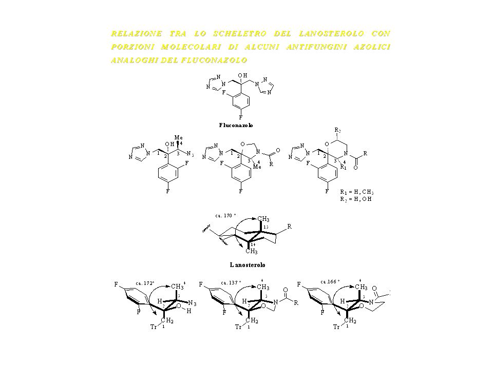
Il lanosterolo si lega al sito attivo dell’enzima ancorandosi con l’ossidrile in C-3 ad un amminoacido del sito attivo e disponendo il C-14 metile,che subirà l’ossidazione, verso l’atomo di Fe del sistema Citocromo P-450. *Machovic R.e Owen W.G.,Biochemistry (1989) Il lanosterolo si lega al sito attivo dell’enzima ancorandosi con l’ossidrile in C-3 ad un amminoacido del sitoe disponendo il C-14 metile,che subirà l’ossidazione,verso l’atomo di Fe del sistema del citocromo P-450
 Il lanosterolo si lega al sito attivo dell’enzima ancorandosi con l’ossidrile in C-3 ad un amminoacido del sitoe disponendo il C-14 metile,che subirà l’ossidazione,verso l’atomo di Fe del sistema del citocromo P-450.")
12
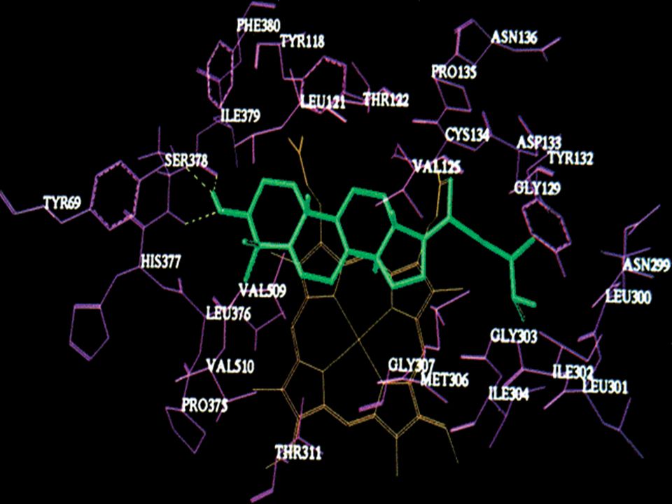
Metilen 24,25 diidrolanosterolo nel sito attivo dell’enzima lanosterolo14α-demetilasi di C.Albicans
*Haitao Ji, Wnnian Zhang J.Med.Chem.,43(13)
")
13
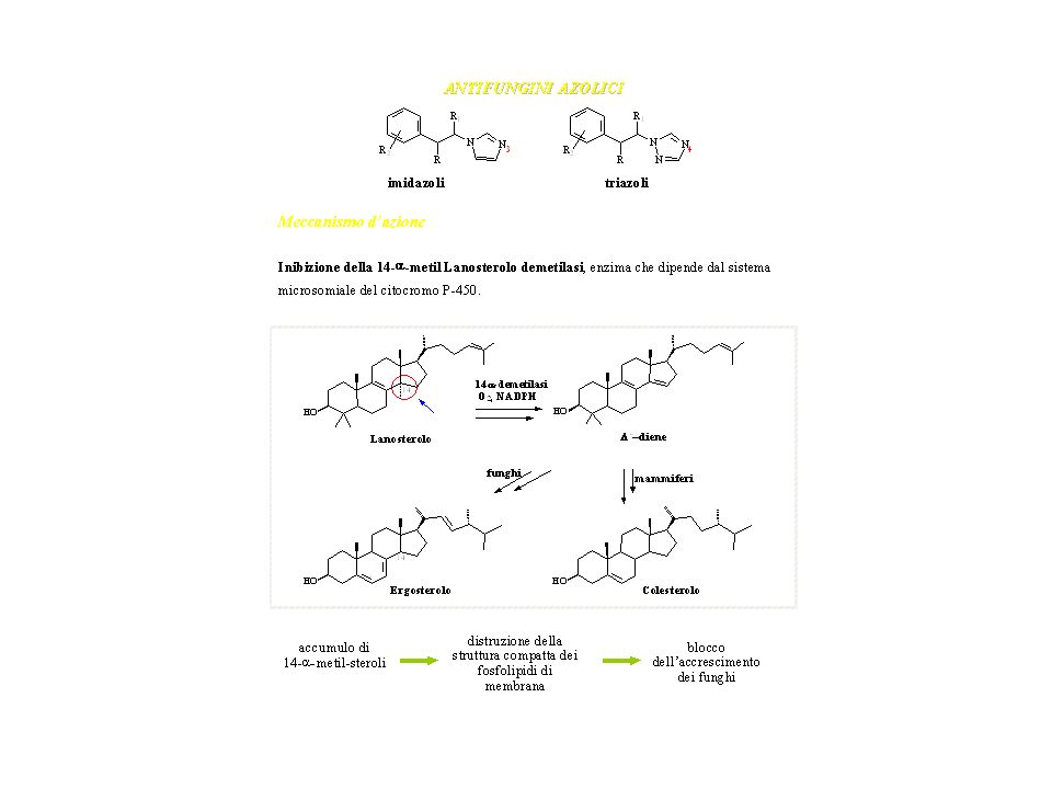
L’atomo di azoto delle strutture azoliche,N-3 per gli imidazoli e N-4 per i triazoli pare sia necessario per la complessazione e la stabilizzazione dello ione ferrico dell’eme.Anche i gruppi R e R-1,presenti sulla struttura N-feniletilica sono importanti per l’azione antifungina

14
Stereoview of active site of lanosterol 14α-demethylase of albicans with bound fluconazole(A) and itraconazole(B) J.Med.Chem.,43(13), ,
, ,")
15
Ribbon representation of the MTCYP51 structures with the inhibitors bound. Front (A) and top (B) views of the of 4-PI- (yellow) and FLU- (blue) bound MTCYP51 superimposed with an rms deviation of Å. Superimpositions for all figures were done by using two-step fitting as implemented in SWISS-PDB VIEWER (33). The first step was performed by using entire structures; for the second step, an rms- deviation cutoff of 1.8 Å was used to select the most structurally homologous regions for subsequent fitting. The second round results in better fitting of the most homologous regions and further divergence of less homologous regions. Heme, red; 4-PI, orange; FLU, light-blue. The I helix is shown also in red. A large cavity of 2,600 Å3, shown in blue, leads from the active site to the molecular surface along the protein domain interface (channel 2). Structural elements significantly deviating among P450 structures are labeled in black, and -sheets that are part of the putative substrate-binding site are labeled in red. All figures, if not otherwise indicated, are generated by using SWISS-PDB VIEWER (33).
 and top (B) views of the of 4-PI- (yellow) and FLU- (blue) bound MTCYP51 superimposed with an rms deviation of 0.45 Å. Superimpositions for all figures were done by using two-step fitting as implemented in SWISS-PDB VIEWER (33). The first step was performed by using entire structures; for the second step, an rms- deviation cutoff of 1.8 Å was used to select the most structurally homologous regions for subsequent fitting. The second round results in better fitting of the most homologous regions and further divergence of less homologous regions. Heme, red; 4-PI, orange; FLU, light-blue. The I helix is shown also in red. A large cavity of 2,600 Å3, shown in blue, leads from the active site to the molecular surface along the protein domain interface (channel 2). Structural elements significantly deviating among P450 structures are labeled in black, and -sheets that are part of the putative substrate-binding site are labeled in red. All figures, if not otherwise indicated, are generated by using SWISS-PDB VIEWER (33)..")
16
A) MTCYP51 active-site chamber
A) MTCYP51 active-site chamber. Structural elements and residues constituting the dome of the active site are indicated. (B and C) Interaction of 4-PI and FLU in the binding site of MTCYP51. Residues located within 4.1 Å of each ligand are shown. Region in C is displaced toward the substrate-binding site as a result of conformational changes in the C helix after FLU binding. Fragments of simulated annealing omit 2Fo Fc map contoured at 1.5 are shown. Podust, Larissa M. et al. (2001) Proc. Natl. Acad. Sci. USA 98,
 MTCYP51 active-site chamber. Structural elements and residues constituting the dome of the active site are indicated. (B and C) Interaction of 4-PI and FLU in the binding site of MTCYP51. Residues located within 4.1 Å of each ligand are shown. Region in C is displaced toward the substrate-binding site as a result of conformational changes in the C helix after FLU binding. Fragments of simulated annealing omit 2Fo Fc map contoured at 1.5 are shown. Podust, Larissa M. et al. (2001) Proc. Natl. Acad. Sci. USA 98,")
17
Antifungini imidazolici

18
antifungini triazolici

Presentazioni simili


You use _______ ________.>")

