Scaricare la presentazione
La presentazione è in caricamento. Aspetta per favore

1
Cellula batterica Parete cellulare

2
STRUTTURE FONDAMENTALI
La cellula batterica STRUTTURE FONDAMENTALI PARETE MEMBRANA STRUTTURE ACCESSORIE PILI FLAGELLI CAPSULA

3
Parete batterica Organizzazione diversa della parete nei Gram positivi
Gram negativi Acido –Alcool resistenti (micobatteri)
")
4
Colorante di contrasto, Safranina
COLORAZIONE DI GRAM Iodio, azione mordenzante Alcool etilico Colorante di contrasto, Safranina

5
COLORAZIONE DI GRAM Batterio Gram positivo Batterio Gram negativo Micrococcus luteus Escherichia coli

6
Funzioni della parete batterica PROTEZIONE
FORMA PROCESSO DI DIVISIONE SOLLECITAZIONI MECCANICHE RIGONFIAMENTO OSMOTICO

7
Figure: 04-33a-b Caption: Protoplasts. (a) In dilute solution breakdown of the cell wall releases the protoplast, but it immediately lyses because the cytoplasmic membrane is very weak. (b) In a solution containing an isotonic concentration of a solute such as sucrose, water does not enter the protoplast and it remains stable. Lysozyme breaks the b-1,4 glycosidic bonds in peptidoglycan (see Figure 4.30).
 In dilute solution breakdown of the cell wall releases the protoplast, but it immediately lyses because the cytoplasmic membrane is very weak. (b) In a solution containing an isotonic concentration of a solute such as sucrose, water does not enter the protoplast and it remains stable. Lysozyme breaks the b-1,4 glycosidic bonds in peptidoglycan (see Figure 4.30).")
8
Forme cellulari dei batteri
bacilli bacilli cocchi spirilli

9
Il monomero del Peptidoglicano
NAM= N-Acetil muramico NAG= N-Acetil glucosoammina, Sono degli AMMINOZUCCHERI

10
Peptidoglicano Scheletro glicanico Catene laterali peptidiche
Acido N-acetilmuramico NAM Acido N-acetilglucosammina NAG Catene laterali peptidiche D- L- aminoacidi Acido diaminopimelico Legami crociati peptidici

11
Struttura del peptidoglicano

12
legami crociati nei batteri G-

13
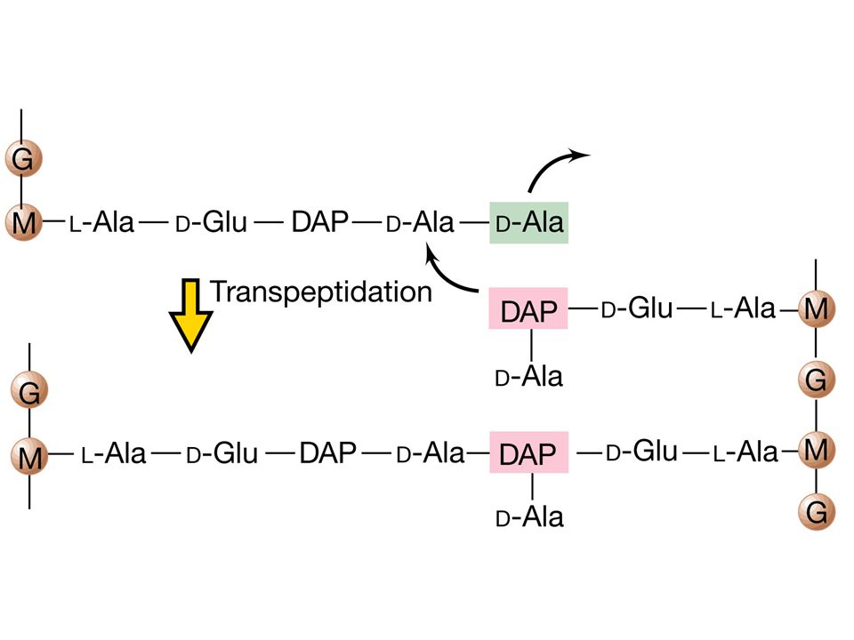
legami crociati nei batteri G+

14
Transpeptidazione

16
Circa il 90% 10-15% Figure: 04-28 Caption:
Cell walls of Bacteria. (a,b) Schematic diagrams of gram-positive and gram-negative cell walls.
 Schematic diagrams of gram-positive and gram-negative cell walls.")
17
Figure: 04-28d Caption: Cell walls of bacteria. Gram-negative bacterium, Leucothrix mucor.

18
ACIDI TEICOICI Figure: 04-32 Caption:
Teichoic acids and the overall structure of the gram-positive cell wall. (a) Structure of the ribitol teichoic acid of Bacillus subtilis. The teichoic acid is a polymer of the repeating ribitol units shown here. (b) Summary diagram of the gram-positive cell wall.
 Structure of the ribitol teichoic acid of Bacillus subtilis. The teichoic acid is a polymer of the repeating ribitol units shown here. (b) Summary diagram of the gram-positive cell wall.")
19
Batteri Gram positivi Figure: 04-32b Caption:
Teichoic acids. (b) Summary diagram of the gram-positive cell wall.
 Summary diagram of the gram-positive cell wall.")
20
Organizzazione della parete dei Gram -negativi
Membrana esterna Figure: 04-36 Caption: The gram-negative cell wall. Note that although the outer membrane is often called the "second lipid bilayer," the chemistry and architecture of this layer differs in many ways from that of the cytoplasmic membrane. (a) Arrangement of lipopolysaccharide, lipid A, phospholipid, porins, and lipoprotein in the outer membrane. See Figure 4.35 for details of the structure of LPS. Lipid A can be toxic in humans, and if so, is referred to as endotoxin (Section 21.12). (b) Molecular model of porin proteins. Note the three pores present, one formed from each of the proteins forming a porin molecule. The view is perpendicular to the plane of the membrane. Model based on X-ray diffraction studies of Rhodobacter blasticus porin.
 Arrangement of lipopolysaccharide, lipid A, phospholipid, porins, and lipoprotein in the outer membrane. See Figure 4.35 for details of the structure of LPS. Lipid A can be toxic in humans, and if so, is referred to as endotoxin (Section 21.12). (b) Molecular model of porin proteins. Note the three pores present, one formed from each of the proteins forming a porin molecule. The view is perpendicular to the plane of the membrane. Model based on X-ray diffraction studies of Rhodobacter blasticus porin.")
21
Struttura del LIPOPOLISACCARIDE (LPS)
Regione variabile CORE POLISACCARIDICO LIPIDE A ANTIGENE O Zuccheri Figure: 04-35 Caption: Structure of the lipopolysaccharide of gram-negative Bacteria. The precise chemistry of lipid A and the polysaccharide components varies among species of gram-negative Bacteria, but the sequence of major components (lipid A–KDO–core–O-specific) is generally uniform. The O-specific polysaccharide varies among species. KDO, ketodeoxyoctonate; Hep, heptose; Glu, glucose; Gal, galactose; GluNac, N-acetylglucosamine; GlcN, glucosamine; P, phosphate. Glucosamine and the lipid A fatty acids are linked by an ester amine bond. The lipid A portion of LPS can be toxic to animals and comprises the endotoxin complex (Section 21.12). Compare this figure with Figures 4.36 and 4.37, and note the color coding of different portions of the LPS in Figures 4.35 and 4.36. Zuccheri insoliti: Hep= Eptosio KDO= Ketodeossioctonico
 is generally uniform. The O-specific polysaccharide varies among species. KDO, ketodeoxyoctonate; Hep, heptose; Glu, glucose; Gal, galactose; GluNac, N-acetylglucosamine; GlcN, glucosamine; P, phosphate. Glucosamine and the lipid A fatty acids are linked by an ester amine bond. The lipid A portion of LPS can be toxic to animals and comprises the endotoxin complex (Section 21.12). Compare this figure with Figures 4.36 and 4.37, and note the color coding of different portions of the LPS in Figures 4.35 and Zuccheri insoliti: Hep= Eptosio. KDO= Ketodeossioctonico.")
22
Struttura del LIPOPOLISACCARIDE (LPS)
ENDOTOSSINA

23
PARETE NEGLI ARCHEA Figure: 04-34 Caption:
Pseudopeptidoglycan and S-layers. (a) Structure of pseudopeptidoglycan, the cell wall polymer of Methanobacterium species. Note the resemblance to the structure of peptidoglycan shown in Figure 4.30, especially the peptide cross-links, in this case between N-acetyltalosaminuronic acid (NAT) residues instead of muramic acid residues. NAG, N-Acetylglucosamine.
 Structure of pseudopeptidoglycan, the cell wall polymer of Methanobacterium species. Note the resemblance to the structure of peptidoglycan shown in Figure 4.30, especially the peptide cross-links, in this case between N-acetyltalosaminuronic acid (NAT) residues instead of muramic acid residues. NAG, N-Acetylglucosamine.")
24
Struttura della parete dei batteri Acido-alcool resistenti
(Micobatteri) Oltre al peptidoglicano, la parete dei batteri acidi-alcool resistenti come Mycobacterium contiene grandi quantità di glicolipidi come acidi micolici, complessi arabinogalactano-lipidi, e lipoarabinomannano.
 Oltre al peptidoglicano, la parete dei batteri acidi-alcool resistenti come Mycobacterium contiene grandi quantità di glicolipidi come acidi micolici, complessi arabinogalactano-lipidi, e lipoarabinomannano.")
25
Membrana citoplasmatica dei batteri
Figure: 04-17 Caption: Diagram of the structure of the cytoplasmic membrane; the inner surface (In) faces the cytoplasm and the outer surface (Out) faces the environment. The matrix of the unit membrane is composed of phospholipids, with the hydrophobic groups directed inward and the hydrophilic groups toward the outside, where they associate with water. Embedded in the matrix are proteins that have considerable hydrophobic character in the region that traverses the fatty acid bilayer. Hydrophilic proteins and other charged substances, such as metal ions, may be attached to the hydrophilic surfaces. Although there are some chemical differences, the overall structure of the cytoplasmic membrane shown is similar in both prokaryotes and eukaryotes (but see an exception to the bilayer design in Figure 4.20d)
 faces the cytoplasm and the outer surface (Out) faces the environment. The matrix of the unit membrane is composed of phospholipids, with the hydrophobic groups directed inward and the hydrophilic groups toward the outside, where they associate with water. Embedded in the matrix are proteins that have considerable hydrophobic character in the region that traverses the fatty acid bilayer. Hydrophilic proteins and other charged substances, such as metal ions, may be attached to the hydrophilic surfaces. Although there are some chemical differences, the overall structure of the cytoplasmic membrane shown is similar in both prokaryotes and eukaryotes (but see an exception to the bilayer design in Figure 4.20d)")
26
FUNZIONE DELLA MEMBRANA CITOPLASMATICA NEI PROCARIOTI
REGOLA IL FLUSSO DEI NUTRIENTI E’ SEDE DI PROCESSI BIOSINTETICI PRODUZIONE DI ENERGIA SITO DI ANCORAGGIO PER STRUTTURE ACCESSORIE FUNZIONE DI SECREZIONE FUNZIONE DI REGOLAZIONE

27
Figure: 04-21 Caption: The major functions of the cytoplasmic membrane.

28
LIPIDI NEI PROCARIOTI LEGAME ESTERE LEGAME ETERE, TIPICO DEGLI ARCHEA ISOPRENILE, TIPICO DEGLI ARCHEA

29
Lipidi negli Archea e organizzazione della membrana
Legame etere fitanile bifitanile

30
Lipidi negli Archea e organizzazione della membrana
Monostrato lipidico

31
Sistemi di trasporto di membrana

32
Figure: 04-25 Caption: Function of the Lac permease (a symporter) of Escherichia coli, and several other well-characterized simple transporters. Although for simplicity the membrane-spanning proteins are drawn here in globular form, note that their structure is actually as depicted in Figure 4.24.
 of Escherichia coli, and several other well-characterized simple transporters. Although for simplicity the membrane-spanning proteins are drawn here in globular form, note that their structure is actually as depicted in Figure")
33
Figure: 04-24 Caption: Structure of membrane-spanning transporters and types of transport events. In prokaryotes, membrane-spanning transporters typically contain 12 alpha helices that align with each other in a circle to form a channel through the membrane. Shown here are three individual transporters, each showing a different type of transport event. For antiporters and symporters, the cotransported molecule is shown in yellow.

34
Traslocazione di gruppo
esterno interno Figure: 04-26 Caption: Mechanism of the phosphotransferase system of Escherichia coli. For glucose uptake, the system consists of five proteins: Enzyme (Enz) I; Enzymes IIa, IIb, and IIc, and HPr. Sequential phosphate transfer occurs from phosphoenolpyruvate (PEP) through the proteins shown to Enzyme IIc. The latter actually transports (and phosphorylates) the sugar.
 I; Enzymes IIa, IIb, and IIc, and HPr. Sequential phosphate transfer occurs from phosphoenolpyruvate (PEP) through the proteins shown to Enzyme IIc. The latter actually transports (and phosphorylates) the sugar.")
35
Spazio periplasmatico
Figure: 04-27 Caption: Mechanism of an ATP-Binding Cassette (ABC-type) transporter. The periplasmic binding protein has high affinity for substrate, the membrane-spanning protein is the transport channel, and the cytoplasmic ATP-hydrolyzing protein supplies the energy for the transport event. In Escherichia coli, the maltose (a disaccharide sugar) transport system is an example of an ABC system.
 transporter. The periplasmic binding protein has high affinity for substrate, the membrane-spanning protein is the transport channel, and the cytoplasmic ATP-hydrolyzing protein supplies the energy for the transport event. In Escherichia coli, the maltose (a disaccharide sugar) transport system is an example of an ABC system.")
36
Strutture accessorie della cellula batterica
Flagelli Pili Capsula, sostanze polimeriche extracellulari

37
Flagelli batterici

38
Flagelli batterici Numero e disposizione dei flagelli in rapporto alla cellula Monotrico Anfitrico Lofotrico Peritrico

39
Flagello batterico uncino Struttura basale

40
Movimento orario Movimento antiorario Movimento antiorario

42
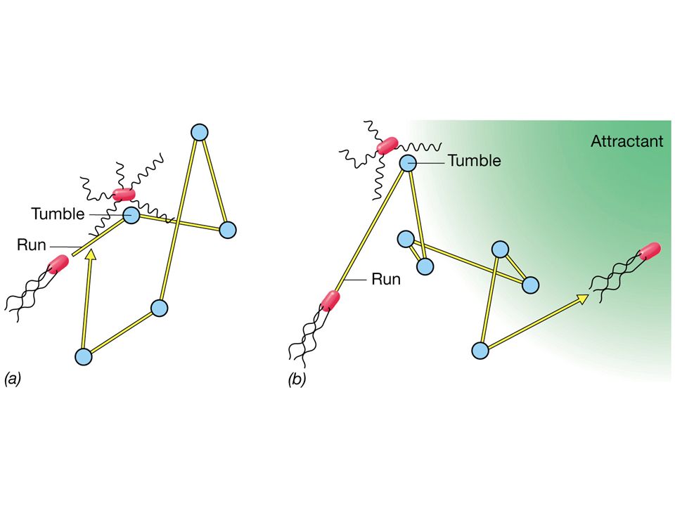
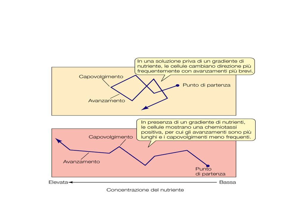
CHEMIOTASSI

44
Velocità relative di alcuni organismi Lunghezza organismo/sec
Gara di velocità tra organismi Velocità relative di alcuni organismi Organismi Km/hr Lunghezza organismo/sec Ghepardo 111 25 UOMO - Michael Johnson 37.5 5.4 BATTERI 10

45
COME SVELARE LA PRESENZA DEI FLAGELLI COLORAZIONE CON FUCSINA BASICA
MOTILITA’ COME SVELARE LA PRESENZA DEI FLAGELLI COLORAZIONE CON FUCSINA BASICA IMMUNOFLUORESCENZA MICROSCOPIO ELETTRONICO COME OSSERVARE LA MOTILITA’ MICROSCOPIO OTTICO USO DI TERRENI DI COLTURA SEMISOLIDI

46
PILI COMUNI,DI TIPO I O FIMBRIE;
PILO F PILI: PILI COMUNI,DI TIPO I O FIMBRIE; PILI DI TIPO IV; PILI SESSUALI DI TIPO F

47
Pili o fimbrie in E. coli

48
SOSTANZE POLIMERICHE EXTRACELLULARI
CAPSULA BATTERICA STRATI MUCOIDI

49
FUNZIONI DELLA CAPSULA
ADERENZA es. Streptococcus mutans VIRULENZA es.Streptococcus pneumoniae RESISTENZA ALL’ESSICCAMENTO RISERVA NUTRIZIONALE DEPOSITO DI SOSTANZE DI RIFIUTO AGGREGAZIONE(BIOFILM) PROTEZIONE(BIOFILM)
 PROTEZIONE(BIOFILM)")
50
Batteri Gram-positivi Batteri Gram-negativi
Capsula batterica Microrganismo Composizione chimica Subunità strutturali Batteri Gram-positivi Bacillus anthracis polipeptide (acido poliglutammico) Acido D-glutammico Bacillus megaterium polipeptide e polisaccaridi Acido D-glutammico, amino zuccheri, zuccheri Streptococcus mutans polisaccaridi (destrano) glucosio Streptococcus pneumoniae amino zuccheri, zuccheri Streptococcus pyogenes polisaccaride (acid ialuronico) N-acetyl-glucosammina e acido glucuronico Batteri Gram-negativi Acetobacter xylinum Polisaccaride (cellulosa) glucosio Escherichia coli polisaccaride (colonic acid) glucosio, galattosio, fucosio, acido glucuronico Pseudomonas aeruginosa polisaccaride Acido mannuronico Azotobacter vinelandii Acido glucuronico Agrobacterium tumefaciens (glucano) glucosio
 Acido D-glutammico. Bacillus megaterium. polipeptide e polisaccaridi. Acido D-glutammico, amino zuccheri, zuccheri. Streptococcus mutans. polisaccaridi. (destrano) glucosio. Streptococcus pneumoniae. amino zuccheri, zuccheri. Streptococcus pyogenes. polisaccaride (acid ialuronico) N-acetyl-glucosammina e acido glucuronico. Batteri Gram-negativi. Acetobacter xylinum. Polisaccaride (cellulosa) glucosio. Escherichia coli. polisaccaride (colonic acid) glucosio, galattosio, fucosio, acido glucuronico. Pseudomonas aeruginosa. polisaccaride. Acido mannuronico. Azotobacter vinelandii. Acido glucuronico. Agrobacterium tumefaciens. (glucano) glucosio.")
Presentazioni simili