Scaricare la presentazione
La presentazione è in caricamento. Aspetta per favore

1
5° CORSO NAZIONALE CONGIUNTO ULTRASONOLOGIA VASCOLARE
BERTINORO, MARZO 2007 5° CORSO NAZIONALE CONGIUNTO ULTRASONOLOGIA VASCOLARE DIAGNOSI E TERAPIA DIAGNOSTICA CON APPARECCHI AD ULTRASUONI ECOCOLORDOPPLER, POWERDOPPLER ED ANGOLO DI INCIDENZA D. Righi

4
PER VALUTARE IL GRADO DI STENOSI DEVO
CONOSCERE LA VELOCITA’ DEL SANGUE PER VALUTARE LA VELOCITA’ DEL SANGUE DEVO CONOSCERE L’ ANGOLO TRA IL FASCIO ULTRASONORO ED IL FLUSSO

5
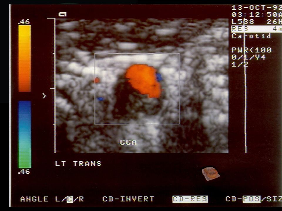
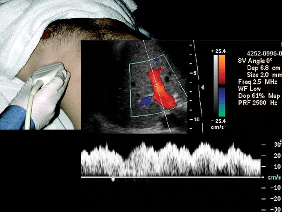
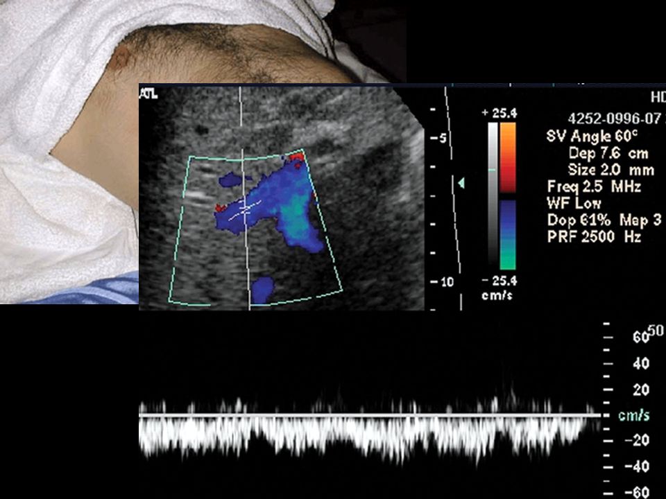
L’ angolo di insonazione è l’ angolo tra il trasduttore (la sonda) e le strutture da esplorare, nel nostro caso i vasi. Influenza la maniera in cui i vasi in esplorazione sono visualizzati. La correzione d’ angolo è necessario per avere una valutazione corretta delle velocità misurate con il doppler pulsato. Non si rende necessaria solo quando il vaso è a zero gradi rispetto al cursore ed alla linea guida.

9
Doppler in Obstetrics Copyright © 2002 by Kypros Nicolaides, Giuseppe Rizzo, Kurt Hecker and Renato Ximenes

11
E’ UNA ED UNA SOLA PER VALUTARE IL GRADO DI STENOSI POSSO
VARIARE LA DIREZIONE DEL FASCIO DI ULTRASUONI UTILIZZANDO I COMANDI DELLA MACCHINA O MUOVENDO LA SONDA MA LA RETTA PARALLELA AL FLUSSO E’ UNA ED UNA SOLA ED UNO ED UNO SOLO E’ QUINDI L’ ANGOLO TRA IL FASCIO ULTRASONORO ED IL FLUSSO

13
Doppler in Obstetrics Copyright © 2002 by Kypros Nicolaides, Giuseppe Rizzo, Kurt Hecker and Renato Ximenes

14
NELL’ ESECUZIONE DI UN ESAME DOPPLER PW MEDIANTE DUPLEX
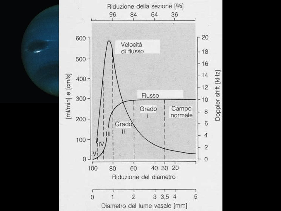
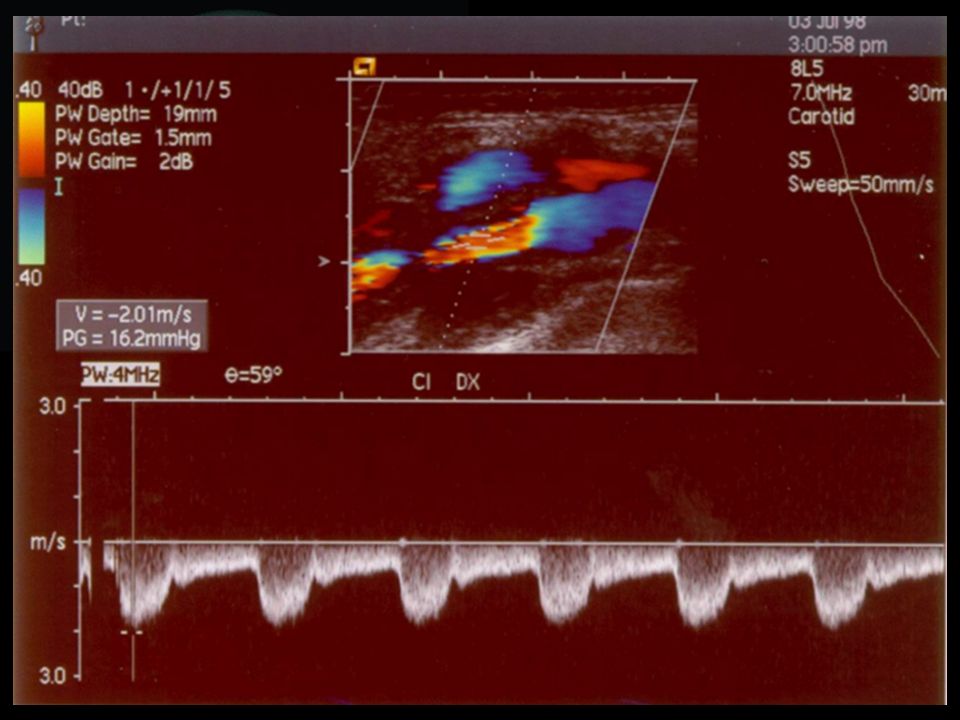
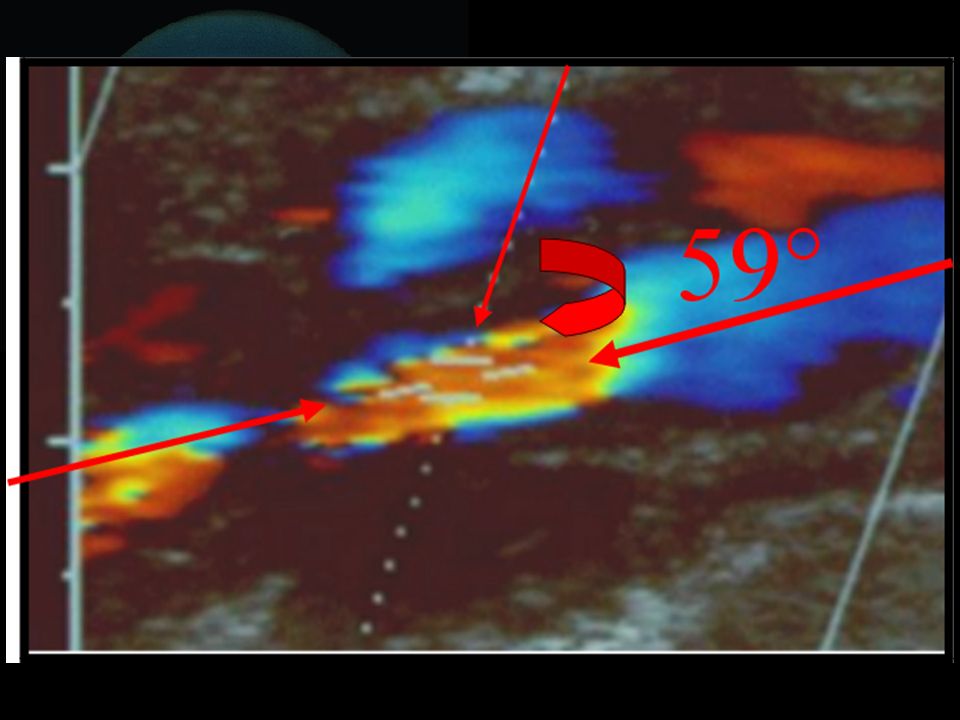
ANGOLO: NELL’ ESECUZIONE DI UN ESAME DOPPLER PW MEDIANTE DUPLEX CONVIENE MANTENERLO TRA 45° E 60° NON SUPERARE MAI I 60° 90° - 61° = ERRATO 60° = OTTIMALE 59° - 1°= GIUSTO

15
The results of the study were published in the Journal of Vascular Surgery (“JVS”).in an article entitled, “Reliability of Extracranial Carotid Artery Duplex Ultrasound Examinations: Value of Vascular Laboratory Accreditation,” authored by O. William Brown, M.D., et.al. The objective of the study reported in this article was to evaluate the reliability of carotid duplex ultrasound testing performed by unaccredited vascular laboratories and to assess the clinical impact on patient management. The study found that of the 174 patients referred for surgical evaluation for carotid endarterectomy, 88 patients (51%) were found to have less than the 60% percent or higher stenosis that had been diagnosed by unaccredited vascular laboratories. The overestimation of stenosis by the unaccredited laboratory was attributed to technical errors (19 arteries), use of B-mode image data alone without the use of velocity criteria for confirmation of the lesion (36 arteries), and use of inappropriate velocity criteria (49 arteries). Because of the study, more than half of all patients who participated in this sub-group were spared the risks of an unnecessary surgery to "correct" a condition that did not exist. Correlating data in all patients with a subsequent evaluation (angiography, MRA, or surgery) supported the findings of the accredited laboratories.
 were found to have less than the 60% percent or higher stenosis that had been diagnosed by unaccredited vascular laboratories. The overestimation of stenosis by the unaccredited laboratory was attributed to technical errors (19 arteries), use of B-mode image data alone without the use of velocity criteria for confirmation of the lesion (36 arteries), and use of inappropriate velocity criteria (49 arteries). Because of the study, more than half of all patients who participated in this sub-group were spared the risks of an unnecessary surgery to correct a condition that did not exist. Correlating data in all patients with a subsequent evaluation (angiography, MRA, or surgery) supported the findings of the accredited laboratories.")
16
国外对于血管狭窄的判断标准报道不一,通常采用收缩峰值流速(PSV)、舒张末期流速(EDV)、颈内动脉/颈总动脉峰值流速比值( PSVica/PSVcca)、PSV1/PSV2(狭窄段1/狭窄远段 2)的比值。Aburahma等提出,ICA狭窄 ≥70%的标准为: (1) ICA的PSV ≥ 195 cm/s,敏感性为98%,特异性为75%,准确率为87%;(2)EDV≥ 97.5cm/s,敏感性为 82%,特异性为 96%,准确率为 89%;(3)PSV为 185 cm/s,EDV为 75 cm/s,PSVica/PSVcca≥2.7 ,敏感性为82%,特异性为96%,准确率为89%。国内对诊断颈动脉狭窄≥70%的标准很少报道,我们的临床检测结果表明,采用综合的判断标准,即 PSV>170 cm/s、EDV>100 cm/s和PSV1 /PSV2 >4:1,并与TCD检测相结合,诊断结果与脑血管造影比较,诊断准确率为99%。 国外对于血管狭窄的判断标准报道不一,通常采用收缩峰值流速(PSV)、舒张末期流速(EDV)、颈内动脉/颈总动脉峰值流速比值( PSVica/PSVcca)、PSV1/PSV2(狭窄段1/狭窄远段 2)的比值。Aburahma等提出,ICA狭窄 ≥70%的标准为: (1) ICA的PSV ≥ 195 cm/s,敏感性为98%,特异性为75%,准确率为87%;(2)EDV≥ 97.5cm/s,敏感性为 82%,特异性为 96%,准确率为 89%;(3)PSV为 185 cm/s,EDV为 75 cm/s,PSVica/PSVcca≥2.7 ,敏感性为82%,特异性为96%,准确率为89%。国内对诊断颈动脉狭窄≥70%的标准很少报道,我们的临床检测结果表明,采用综合的判断标准,即 PSV>170 cm/s、EDV>100 cm/s和PSV1 /PSV2 >4:1,并与TCD检测相结合,诊断结果与脑血管造影比较,诊断准确率为99%。
、舒张末期流速(EDV)、颈内动脉/颈总动脉峰值流速比值( PSVica/PSVcca)、PSV1/PSV2(狭窄段1/狭窄远段 2)的比值。Aburahma等提出,ICA狭窄 ≥70%的标准为: (1) ICA的PSV ≥ 195 cm/s,敏感性为98%,特异性为75%,准确率为87%;(2)EDV≥ 97.5cm/s,敏感性为 82%,特异性为 96%,准确率为 89%;(3)PSV为 185 cm/s,EDV为 75 cm/s,PSVica/PSVcca≥2.7 ,敏感性为82%,特异性为96%,准确率为89%。国内对诊断颈动脉狭窄≥70%的标准很少报道,我们的临床检测结果表明,采用综合的判断标准,即 PSV>170 cm/s、EDV>100 cm/s和PSV1 /PSV2 >4:1,并与TCD检测相结合,诊断结果与脑血管造影比较,诊断准确率为99%。")
17
MEDIA: 210 cm/sec Doppler Velocity Criteria Based on Receiver Operating Characteristic Analysis for the Detection of Threshold Carotid Stenoses Chi-Shin Hwang, MD Wen-Yi Shau, MD, PhD Charles H. Tegeler, MD Journal of Neuroimaging Vol 12 No 2 April 2002

19
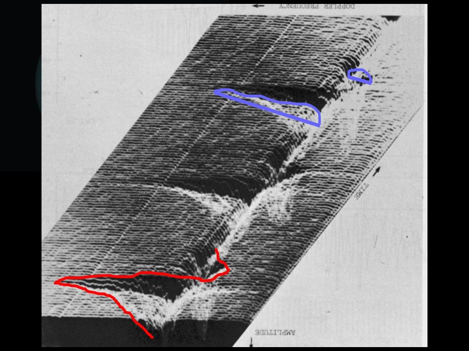
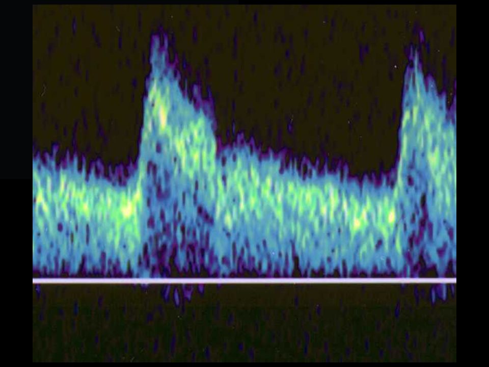
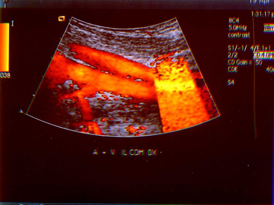
-POWER -POWER DOPPLER -POWER MAP -AMPLITUDE MAP -DOPPLER POWER MODE -COLOR ANGIOGRAPHY

20
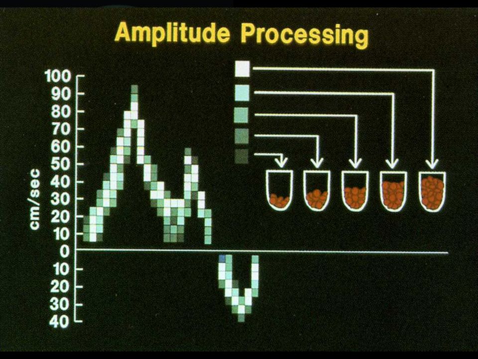
RIFERISCE NON ALLA FREQUENZA MEDIA DEL SEGNALE DOPPLER
IL COLORE DI UN PIXEL SI RIFERISCE NON ALLA FREQUENZA MEDIA DEL SEGNALE DOPPLER OTTENUTO DA QUELLA PARTE DELL’ IMMAGINE, MA ALLA SUA AMPIEZZA O POWER. VIENE RAPPRESENTATO CON UN SOLO COLORE OMOGENEO.

24
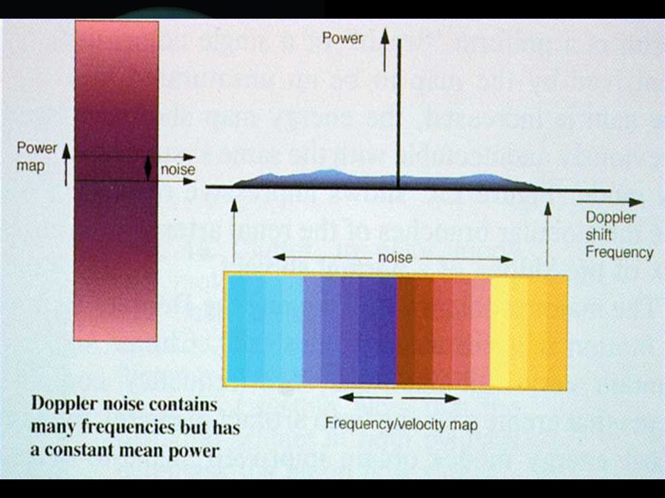
VANTAGGI: - OTTIMO RAPPORTO SEGNALE / RUMORE - SCARSA DIPENDENZA DALL’ ANGOLO - NIENTE ALIASING

26
SVANTAGGI: - MANCA INFORMAZIONE SU DIREZIONE DI FLUSSO - PIU’ SENSIBILE AL MOTO DEI TESSUTI

28
Power and VeloPower As the Power mode is more sensitive than Color mode, it is used to assess the vascular system of tissues and organs, especially in places with slow or weak flow. Directional Power or VeloPower combines the PD sensitivity to slow flows, with an option for detecting the flow direction. As the power of the reflected signal is independent of its direction, Power is less sensitive to Doppler angle and therefore offers an increased sensitivity in the detection of low flow. Power Mode VeloPower

29
Ultraschall Med 1999 Aug;20(4):137-43
[Area reduction in carotid stenosis of the internal carotid artery]. Lyrer P, Bont A, Marugg A, Operschall C, Radu EW Neurologische Universitatsklinik, Abteilung zerebrale Ultraschalldiagnostik,Basel. In 58 patients who suffered from 60 moderate to severe ICA stenoses, B-mode sonography combined with CDE -coded duplex sonography was applied to measure the extent of the stenosis by determining the residual lumen width. CONCLUSIONS: Determination of the degree of stenosis based on CDE alone is not reliable enough to allow orrect diagnosis of severe carotid artery stenosis.

30
Purdue University • Department of Animal Sciences • Lilly Hall

Presentazioni simili


>")
>")


>")