Scaricare la presentazione
La presentazione è in caricamento. Aspetta per favore

1
MIELOFIBROSI Ematologia Adulti Dr. ssa Elena Elli 28/04/2015 1

2
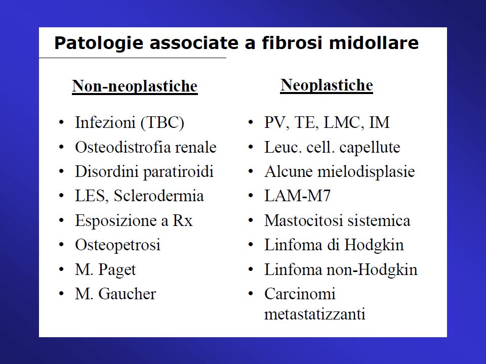
MIELOFIBROSI CRONICA IDIOPATICA (CLASSIFICAZIONE WHO 2001)
")
4
CLASSIFICAZIONE WHO 2008 Neoplasia o disordine clonale mieloproliferativo della cellula staminale emopoietica caratterizzato:da: - fibrosi midollare emopoiesi extramidollare (metaplasia mieloide spleno-epatica) eritropoiesi inefficace iperplasia displastica megacariocitaria aumento forme mieloidi ed eritroidi immature circolanti 4
 eritropoiesi inefficace. iperplasia displastica megacariocitaria. aumento forme mieloidi ed eritroidi immature circolanti. 4.")
5
CARATTERISTICHE MIELOFIBROSI IDIOPATICA
Precursori mieloidi-eritroidi dacriociti CARATTERISTICHE MIELOFIBROSI IDIOPATICA Megacariociti atipici e in clusters densi Osteosclerosi Emopoiesi intrasinusale Fibrosi reticolinica Tefferi, NEJM, 2000 5

6
Patologia clonale MK e monociti Fibrosi midollare reattiva
6

7
EPIDEMIOLOGIA e CLINICA
Incidenza: 0,5-1,5 x abitanti Età mediana: 65 aa (22% pz < 56 aa e 11 % pz < 46 aa) M>F 7
 M>F. 7.")
8
8

9
I sintomi sistemici associati alla MF contribuiscono in modo sostanziale al carico globale di malattia e sono secondari a iperproduzione citochine infiammatorie Sintomi costituzionali Sudorazioni notturne Perdita di peso Febbre Senso di sazietà Splenomegalia Fastidio addominale Tosse Dolore osseo Mieloproliferazione Prurito Stanchezza Sintomi funzionali Inattività Insonnia Prevalenza 1.Scherber R et al, Blood 2011;118(2):401-8 1.Scherber R et al, Blood 2011;118(2):401-8
: Scherber R et al, Blood 2011;118(2):")
10
LABORATORIO - Anemia con anisopoichilocitosi e dacriociti
GB o (15%) con forme immature della serie mieloide ed eritroide circolanti PTL o LDH FAL Ricerca di mutazione JAK2V617F o altri marcatori di clonalità su granulociti sangue periferico (positiva 50-60% casi) Aspirato midollare: spesso punctio sicca. Cariotipo: delezione 13, trisomia 8, delezione 20, più rare alterazioni cromosomi 5 e 7 BOM: valutazione grado di fibrosi, aspetto dei megacariociti e neoangiogenesi 10
 con forme immature della serie mieloide ed eritroide circolanti. PTL o LDH FAL Ricerca di mutazione JAK2V617F o altri marcatori di clonalità su granulociti sangue periferico (positiva 50-60% casi) Aspirato midollare: spesso punctio sicca. Cariotipo: delezione 13, trisomia 8, delezione 20, più rare alterazioni cromosomi 5 e 7. BOM: valutazione grado di fibrosi, aspetto dei megacariociti e neoangiogenesi. 10.")
11
11

12
Circulating hematopoietic progenitor cells in MMM (conta CD34+)
CD34+ hemopoietic stem cells have a constitutional tendency to migrate from bone marrow to blood and to extramedullary organs CD34 + in PB CD34 + in Spleen Barosi et al. Blood 2001 Barosi et al, BJH 2003 12

13
Passamonti et al. Haematologica 2003
15 x 10^6/L CD34+ utile cut-off per distinguere PV ed TE da MMM Passamonti et al. Haematologica 2003 13

14
CD34+ cell number in PB marks disease evolution
CD34+ cells <300x106/L CD34+ cells >300 x106 Barosi et al. Blood 2001 14

15
PATOGENESI 15

16
Alterazione della via JAK/STAT nella MF 6
L’attivazione costitutiva della via JAK/STAT rappresenta l'alterazione biologica principale alla base della MFI La deregolazione della via JAK/STAT può dipendere da diversi meccanismi:2 mutazione JAK2V617F, a carico del gene JAK23 altre mutazioni geniche fra cui MPL e CALR3,4 Essa determina: incontrollata proliferazione Elevata concentrazione di citochine proinfiammatorie3 Alterazione della via JAK/STAT nella MF 6 1. Quintas Cardama A, et al. Clin Cancer Res 2013; 19(8); 1-8; 2. Levine RL, et al. Nat Rev Cancer 2007; 7(9): ; 3. Komrokji RS, et al. Cancer Control 2012; 4. Levine RL. N Engl J Med. 2013; 369(25): ; 5. Savona MR. Leuk Res pii: S (14) doi: /leukres ; 6. Vannucchi AM. N Engl J Med.2010; 363(12):
; 1-8; 2. Levine RL, et al. Nat Rev Cancer 2007; 7(9): ; 3. Komrokji RS, et al. Cancer Control 2012; 4. Levine RL. N Engl J Med. 2013; 369(25): ; 5. Savona MR. Leuk Res pii: S (14) doi: /leukres ; 6. Vannucchi AM. N Engl J Med.2010; 363(12):")
17
L’alterazione del signaling di JAK1 e JAK2 è responsabile delle manifestazioni cliniche della MF
Citochine infiammatorie Mieloproliferazione Fibrosi Sintomi costituzionali Emopoiesi extramidollare (splenomegalia) Eritropoiesi inefficace Verstovsek S, et al. N Engl J Med. 2010; 363:
 Eritropoiesi inefficace. Verstovsek S, et al. N Engl J Med. 2010; 363:")
18
Mutazione JAK2V617F in MFI Presente nel 50-60% dei casi
Forma omozigote più frequente in post-PV-MF Forma eterozigote più frequente in post-ET MF Forme di mielofibrosi iniziale primitiva spesso JAk2V617F negative

19
JAK2 (V617F) nella MIELOFIBROSI
associazione tra mutazione e impronta policitemica della malattia 19

20
JAK2 (V617F) nella MIELOFIBROSI
La mutazione è presente allo stato eterozigote ma anche omozigote, ed è più frequente nelle forme di MFI secondarie a PV. In particolare i pazienti con MMM post-PV presentano la più alta % di alleli mutati (valore mediano 94% in PCR allele-specifica quantitativa) Effetto stimolante mutazione su linea eritroide Associazione tra mutazione e espansione linea mieloide 20
 Effetto stimolante mutazione. su linea eritroide. Associazione tra mutazione e. espansione linea mieloide. 20.")
21
JAK2 (V617F) e conta CD34 in MIELOFIBROSI
Verosimile correlazione tra mobilizzazione di progenitori emopoietici circolanti CD34+ e % di alleli JAK2(V617F). Il processo di mobilizzazione in parte sembra causato dalla attivazione dei neutrofili indotta dalla mutazione JAK2 Relation between JAK2 (V617F) mutation status, granulocyte activation and constitutive mobilization of CD34-positive cells into peripheral blood in myeloproliferative disorders Francesco Passamonti, Blood 2005 21
. Il processo di mobilizzazione in parte sembra causato dalla attivazione dei neutrofili indotta dalla mutazione JAK2. Relation between JAK2 (V617F) mutation status, granulocyte activation and constitutive mobilization of CD34-positive cells into peripheral blood in myeloproliferative disorders. Francesco Passamonti, Blood")
22
JAK2 (V617F) predice evoluzione di malattia in PMF
22

23
23

24
MPL515 mutations in MF JAK2 negative
Sequence analysis of EPOR, TPOR and GCSFR in JAK2-negative PV, ET and MMM led to the discovery of a somatic tryptophan to leucine substitution mutation at the transmembrane- juxtamembrane junction of MPL (MPLW515L). The MPLW515L allele occurs in approximately 10% of pts with JAK2V617F-negative MMM, and in a smaller proportion of pts with ET. A smaller number of pts have an alternate mutation at codon 515, which results in a tryptophan to lysine substitution (MPLW515K). This allele is not observed in PV or other myeloid malignancies, suggesting that activation of JAK- STAT signaling by MPLW515L/K is specific to ET and MMM. 24
. The MPLW515L allele occurs in approximately 10% of pts with JAK2V617F-negative MMM, and in a smaller proportion of pts with ET. A smaller number of pts have an alternate mutation at codon 515, which results in a tryptophan to lysine substitution (MPLW515K). This allele is not observed in PV or other myeloid malignancies, suggesting that activation of JAK- STAT signaling by MPLW515L/K is specific to ET and MMM. 24.")
25
Department of Haematology, University of Florence, Florence, Italy
Anaemia characterises patients with myelofibrosis harbouring Mpl mutation. Guglielmelli P, Pancrazzi A, Bergamaschi G, Rosti V, Villani L, Antonioli E, Bosi A, Barosi G, Vannucchi AM; GIMEMA--Italian Registry of Myelofibrosis; MPD Research Consortium. Department of Haematology, University of Florence, Florence, Italy . The clinical and haematological phenotype of patients with myelofibrosis harbouring MPL(W515L/K) mutation has not been thoroughly investigated. Of 217 myelofibrosis subjects, 18 (8.2%) had an MPL mutation, four of which (22%) co-existed with JAK2(V617F) mutation. When compared with MPL wild-type patients, irrespective of JAK2(V617F) status, those with MPL(W515L/K), were more frequently female, were older (61 years vs. 57 years; P = 0.02), presented with more severe anaemia (haemoglobin, 101 g/l vs. 121 g/l; P = 0.002) and were more likely to require regular transfusional support (P = 0.012). These data indicate that MPL mutation in myelofibrosis characterises patients with more severe anaemic phenotype. Br J Haematol May;137(3):244-7. 25
 mutation has not been thoroughly investigated. Of 217 myelofibrosis subjects, 18 (8.2%) had an MPL mutation, four of which (22%) co-existed with JAK2(V617F) mutation. When compared with MPL wild-type patients, irrespective of JAK2(V617F) status, those with MPL(W515L/K), were more frequently female, were older (61 years vs. 57 years; P = 0.02), presented with more severe anaemia (haemoglobin, 101 g/l vs. 121 g/l; P = 0.002) and were more likely to require regular transfusional support (P = 0.012). These data indicate that MPL mutation in myelofibrosis characterises patients with more severe anaemic phenotype. Br J Haematol May;137(3):")
26
MUTAZIONI GENE CALRETICOLINA IN MF JAK2 E MPL NEGATIVE
Distribuzione delle mutazioni JAK2, MPL e CALR nei pazienti con MF5 Le mutazioni a carico di JAK2 o MPL sono presenti rispettivamente nel 50-60% e nel 5-10% circa dei pazienti con MF o TE2 La mutazione a carico di CALR è la più comune tra i pazienti che non presentano mutazioni a carico di JAK2 o MPL2,3 JAK2, MPL e CALR non mutati Mutazione CALR Mutazione JAK2 Mutazione MPL 1. Rampal R, et al. Blood 2014;123(22): e123-33; 2. Cazzola M, et al. Blood. 2014; 123(24): ; 3. Levine RL. N Engl J Med. 2013; 369(25): ; 4. Komrokji RS, et al. Cancer Control 2012; 5. Klampfl T et al, N Engl J Med 2013;369: 26
: e123-33; 2. Cazzola M, et al. Blood. 2014; 123(24): ; 3. Levine RL. N Engl J Med. 2013; 369(25): ; 4. Komrokji RS, et al. Cancer Control 2012; 5. Klampfl T et al, N Engl J Med 2013;369:")
27
Incidenza cumulativa di anemia (Hb <10 g/dL)1
I pazienti CALR-mutati presentano una malattia meno aggressiva 1.0 0.9 0.8 0.7 0.6 0.5 0.4 0.3 0.2 0.1 0.0 5 10 15 20 25 30 Tempo, anni Incidenza cumulativa Incidenza cumulativa di anemia (Hb <10 g/dL)1 CALR mutato JAK2 mutato MPL mutato Triplo negativo 1.0 0.9 0.8 0.7 0.6 0.5 0.4 0.3 0.2 0.1 0.0 5 10 15 20 25 30 Tempo, anni Incidenza cumulativa Incidenza cumulativa di leucocitosi marcata (Counta leucocitaria >25x109/L)1 1.0 0.9 0.8 0.7 0.6 0.5 0.4 0.3 0.2 0.1 0.0 5 10 15 20 25 30 Tempo, anni Incidenza cumulativa Incidenza cumulativa di trombocitopenia (Conta piastrinica <100x109/L)1 1.Rumi E, et al. Blood. 2014; 124:1062-9
1. CALR mutato JAK2 mutato. MPL mutato Triplo negativo Tempo, anni. Incidenza cumulativa. Incidenza cumulativa di leucocitosi marcata (Counta leucocitaria >25x109/L) Tempo, anni. Incidenza cumulativa. Incidenza cumulativa di trombocitopenia (Conta piastrinica <100x109/L)1. 1.Rumi E, et al. Blood. 2014; 124:")
28
Criteri diagnostici CRITERI RIMM CRITERI ISTOPATOLOGICI (Thiele 2005)
CRITERI WHO 2001 CRITERI WHO 2008 CRITERI PER FORME SECONDARIE A POLICITEMIA VERA E TROMBOCITEMIA ESSENZIALE CRITERI WHO 2014 28

29
The Italian Diagnostic Criteria of MMM
NECESSARY CRITERIA Diffuse bone marrow fibrosis Absence of Ph chromosome or BCR-ABL rearrangement in peripheral blood cells OPTIONAL CRITERIA Splenomegaly of any grade Anisopoikilocytosis with tear-drop erythrocytes Presence of circulating immature myeloid cells Presence of circulating erythroblasts Presence of clusters of megakaryoblasts and anomalous megakaryocytes in bone marrow sections Myeloid metaplasia DIAGNOSIS OF MMM The two necessary criteria plus any other 2 optional criteria when splenomegaly is present, or any other 4 optional criteria when splenomegaly is absent Barosi et al. BJH 1999 29

30
CRITERI ISTOLOGICI Thiele et al. Haematologica 2005; 90: 30

31
GRADING FIBROSI MF-0 sparse fibre reticoliniche senza intersezioni (midollo normale). MF-1 lieve incremento del reticolo con intersezioni . MF-2 diffuso e denso incremento del reticolo con estese intersezioni ed occasionale collagenizzazione e/o osteosclerosi. MF-3 diffusa collagenizzazione e osteosclerosi. EUMNET Consensus Thiele et al. Haematologica 2005 31

32
MF-0 Sparso reticolo lineare senza intersezioni (cross-overs); corrisponde al midollo normale 32

33
MF-1 Network lasso di reticolo con molte intersezioni, specialmente nelle aree perivascolari 33

34
MF-2 Aumento diffuso e denso del reticolo con estese intersezioni, occasionalmente con focali bande di collagene e/o focale osteosclerosi 34

35
MF-3 Aumento diffuso e denso del reticolo con estese intersezioni e bande dense di collagene, spesso associato con osteosclerosi 35

36
Classificazione WHO 2001 MF in stadio prefibrotico (pre- mielofibrosi) MF 0/1 MF in stadio fibrotico MF 2/3 36

37
WHO criteria for MF– prefibrotic stage
Clinical findings No or mild splenomegaly or hepatomegaly Hematologic parameters variable, but often: - Mild anemia Mild to moderate leukocytosis Mild to marked thrombocytosis Morphological findings No or mild leukoerythroblastosis No or minimal red blood cell poikilocytosis; few if any dacrocytes Bone marrow: - Hypercellularity Neutrophilic proliferation Megakaryocytic proliferation and atypia (clustering of megakaryocytes, abnormally lobulated megakaryocytic nuclei, naked megakaryocytic nuclei) - Minimal or absent reticulin fibrosis 37
 - Minimal or absent reticulin fibrosis. 37.")
38
WHO criteria for MF – fibrotic stage
Clinical findings Moderate to marked splenomegaly and hepatomegaly Hematologic parameters: - Moderate to marked anemia - Low, normal or elevated WBC - Platelet count decreased, normal or elevated Morphological findings Leukoerythroblastosis Prominent red blood cell poikilocytosis with dacrocytes Bone marrow: Reticulin and/or collagen fibrosis Decreased cellularity Dilated marrow sinuses with intraluminal hematopoiesis Prominent megakaryocytic proliferation and atypia (clustering of megakaryocytes, abnormally lobulated megakaryocytic nuclei, naked nuclei) New bone marrow formation 38
 New bone marrow formation. 38.")
39
Criteri morfologici sulla BOM per diagnosi differenziale tra ET, IMF, PV
39

40
Nuovi criteri MFI WHO 2008 3 criteri maggiori + 2 minori 40

41
RUOLO CENTRALE DELLA DIAGNOSI ISTOLOGICA NELLA CLASSIFICAZIONE WHO - REVISIONE PROPOSTA 2015
Criteri PV TE MF primaria Criteri maggiori 1.Hb >16,5 g/dL (uomini) o >16 g/dL (donne) o ematocrito >49% (uomini) o >48% (donne) 2. Ipercellularità midollare trilineare (panmielosi) riscontrata alla biopsia ossea con megacariociti pleomorfici 3.Presenza della mutazione JAK2V617F 1.Conta piastrinica maggiore di 450 x109/L 2.Proliferazione della linea megacariocitaria alla biopsia midollare, con aumento del numero di megacariociti ingranditi e maturi 3.Assenza di criteri WHO per LMC, PV, MF, MDS o altre neoplasie mieloidi 4.Presenza di mutazioni a carico di JAK2, CALR o MPL 1.Proliferazione della linea megacariocitaria e atipia associata alla presenza di fibrosi reticolinica o collagene 2.Assenza di criteri WHO per LMC, PV, TE, MDS o altre neoplasie mieloidi 3.Presenza di mutazioni a carico di JAK2, CALR o MPL Criteri minori 1. Valori di eritropoietina sierica inferiori al minimo dell’intervallo di riferimento 1.Presenza di un marker clonale (cariotipo alterato) o assenza di evidenza di trombocitosi reattiva 1.Presenza di marker clonale (cariotipo alterato) o assenza di evidenza di fibrosi midollare reattiva 2.Anemia o splenomegalia palpabile 3. Leucoeritroblastosi o aumento dei livelli sierici di LDH Tefferi A, et al. Leukemia. 2014; 28:
 o >16 g/dL (donne) o ematocrito >49% (uomini) o >48% (donne) 2. Ipercellularità midollare trilineare (panmielosi) riscontrata alla biopsia ossea con megacariociti pleomorfici. 3.Presenza della mutazione JAK2V617F. 1.Conta piastrinica maggiore di 450 x109/L. 2.Proliferazione della linea megacariocitaria alla biopsia midollare, con aumento del numero di megacariociti ingranditi e maturi. 3.Assenza di criteri WHO per LMC, PV, MF, MDS o altre neoplasie mieloidi. 4.Presenza di mutazioni a carico di JAK2, CALR o MPL. 1.Proliferazione della linea megacariocitaria e atipia associata alla presenza di fibrosi reticolinica o collagene. 2.Assenza di criteri WHO per LMC, PV, TE, MDS o altre neoplasie mieloidi. 3.Presenza di mutazioni a carico di JAK2, CALR o MPL. Criteri minori. 1. Valori di eritropoietina sierica inferiori al minimo dell’intervallo di riferimento. 1.Presenza di un marker clonale (cariotipo alterato) o assenza di evidenza di trombocitosi reattiva. 1.Presenza di marker clonale (cariotipo alterato) o assenza di evidenza di fibrosi midollare reattiva. 2.Anemia o splenomegalia palpabile. 3. Leucoeritroblastosi o aumento dei livelli sierici di LDH. Tefferi A, et al. Leukemia. 2014; 28:")
42
Criteri per diagnosi mielofibrosi post-PV/ET
Criteria for post-polycythemia vera (PV) myelofibrosis Criteria for post-essential thrombocythemia (ET) myelofibrosis Proposed criteria for the diagnosis of post-polycythemia vera and post-essential thrombocythemia myelofibrosis: a consensus statement from the International working group for myelofibrosis research and treatment (IWG-MRT) Barosi et al, leukemia 2007 42
 myelofibrosis. Criteria for post-essential thrombocythemia (ET) myelofibrosis. Proposed criteria for the diagnosis of post-polycythemia vera and post-essential thrombocythemia myelofibrosis: a consensus statement from the International working group for myelofibrosis research and treatment (IWG-MRT) Barosi et al, leukemia")
43
Criteria for post-PV myelofibrosis
Required criteria: Documentation of a previous diagnosis of PV as defined by WHO criteria Bone marrow fibrosis grade 2-3 Additional criteria (2 are required): Anemia or sustained loss of requirement of either phlebotomy (in the absence of cytoreductive therapy) or cytoreductive treatment for erythrocytosis A leukoerythroblastic peripheral blood picture Increasing splenomegaly defined as either an increase in palpable splenomegaly of ≥ 5 cm (distance of the tip of the spleen from the left costal margin) or the appearance of a newly palpable splenomegaly Development of ≥ 1 of 3 constitutional symptoms: > 10% weight loss in 6 months, night sweats, unexplained fever (< 37,5 °C) 43
: Anemia or sustained loss of requirement of either phlebotomy (in the absence of cytoreductive therapy) or cytoreductive treatment for erythrocytosis. A leukoerythroblastic peripheral blood picture. Increasing splenomegaly defined as either an increase in palpable splenomegaly of ≥ 5 cm (distance of the tip of the spleen from the left costal margin) or the appearance of a newly palpable splenomegaly. Development of ≥ 1 of 3 constitutional symptoms: > 10% weight loss in 6 months, night sweats, unexplained fever (< 37,5 °C) 43.")
44
Criteria for post-ET myelofibrosis
Required criteria: Documentation of a previous diagnosis of ET as defined by WHO criteria Bone marrow fibrosis grade 2-3 Additional criteria (2 are required): Anemia or ≥ 2 mg/dl decrease from baseline Hb level A leukoerythroblastic peripheral blood picture Increasing splenomegaly defined as either an increase in palpable splenomegaly of ≥ 5 cm (distance of the tip of the spleen from the left costal margin) or the appearance of a newly palpable splenomegaly Increased LDH level Development of ≥ 1 of 3 constitutional symptoms: > 10% weight loss in 6 months, night sweats, unexplained fever (< 37,5 °C) 44
: Anemia or ≥ 2 mg/dl decrease from baseline Hb level. A leukoerythroblastic peripheral blood picture. Increasing splenomegaly defined as either an increase in palpable splenomegaly of ≥ 5 cm (distance of the tip of the spleen from the left costal margin) or the appearance of a newly palpable splenomegaly. Increased LDH level. Development of ≥ 1 of 3 constitutional symptoms: > 10% weight loss in 6 months, night sweats, unexplained fever (< 37,5 °C) 44.")
45
COMPLICANZE Milza Fegato
Ipertensione polmonare, associata a ascite o sanguinamento varici Infarti splenici Emopoiesi extramidollare: - linfoadenopatie (edemi periferici) - sierose (effusione pleurica, ascite) – polmone (IP, infiltrati pneumonia-like) - s. urogenitale (ematuria) - spazi para-epidurali (s. compressiva) Evoluzione in leucemia acuta Milza Fegato 45
 - sierose (effusione pleurica, ascite) – polmone (IP, infiltrati pneumonia-like) - s. urogenitale (ematuria) - spazi para-epidurali (s. compressiva) Evoluzione in leucemia acuta. Milza. Fegato. 45.")
46
MF primaria: riduzione della sopravvivenza e cause di morte
OS mediana 69 mesi (95% CI, 61-76) Altre cause 13% Neoplasia secondaria 4% Ipertensione portale 4% Sanguinamento 5% 33% Infezioni 10% Median OS 5.75 y (cioè meno di 6 anni) Thrombosis: PMF 2.2 x100 p-y (700 pt) PPV-MF 4.2 x100 p-y Nell’ET: x100 p-y Trombosi/CVE* 13% Progressione in assenza di leucemia 19% 50% Leucemia 31% 5 10 15 20 25 30 35 1.Cervantes F et al, Blood 2009;113: Pazienti (%) *CVE: eventi cardio-vascolari 46 46
 Altre cause. 13% Neoplasia secondaria. 4% Ipertensione portale. 4% Sanguinamento. 5% 33% Infezioni. 10% Median OS 5.75 y (cioè meno di 6 anni) Thrombosis: PMF 2.2 x100 p-y (700 pt) PPV-MF 4.2 x100 p-y. Nell’ET: x100 p-y. Trombosi/CVE* 13% Progressione in assenza. di leucemia. 19% 50% Leucemia. 31% Cervantes F et al, Blood 2009;113: Pazienti (%) *CVE: eventi cardio-vascolari")
47
Criteri prognostici 47

48
Variabili alla diagnosi predittive di scarsa sopravvivenza
Età avanzata (> 60 anni) Leucocitosi (GB>30.000/mm3) Leucopenia (GB < 4.000/mm3) Blasti circolanti (> 2%) Hb < 10 g/dl – trasfusione dipendenza Piastrinopenia (PLT < /mm3) Epatomegalia Sintomi costituzionali (febbre, astenia, sudorazione, perdita peso) Anomalie citogenetiche 48
 Leucocitosi (GB>30.000/mm3) Leucopenia (GB < 4.000/mm3) Blasti circolanti (> 2%) Hb < 10 g/dl – trasfusione dipendenza. Piastrinopenia (PLT < /mm3) Epatomegalia. Sintomi costituzionali (febbre, astenia, sudorazione, perdita peso) Anomalie citogenetiche. 48.")
49
Lille scoring system (Dupriez, Blood, 1996)
Fattori prognostici negativi Hb < 10 g/dL WBC < 4.000/mm3 WBC > / mm3 32% Numero fattori Categoria di rischio Mediana sopravvivenza 0 Basso 8 anni 1 Intermedio 2 anni 2 Alto 1 anno 49

50
International Working Group Score (IPSS - Cervantes 2008)
Età > 65 aa Presenza di sintomi costituzionali (peridta di peso, febbre, sudorazioni notturne Marcata anemia (Hb < 10 g/dl) Leucocitosi ( GB > /mm3) Blasti circolanti > 1% 50
 Leucocitosi ( GB > /mm3) Blasti circolanti > 1% 50.")
51
IPSS (International Prognostic Scoring System): classificazione prognostica alla diagnosi di PMF
Fattori di rischio (Ogni fattore di rischio vale 1 punto) 1. Età >65 anni 2. Sintomi costituzionali* 3. Livelli di emoglobina <10 g/dL 4. Conta leucocitaria >25X109/L 5. Blasti periferici circolanti ≥1% * Febbre, perdita di peso e sudorazioni notturne Categorie di rischio Punteggio Rischio basso Rischio intermedio-1 1 Rischio intermedio-2 2 Rischio alto ≥3 % PTS 22% 29% 28% 21% 1,0 Basso Intermedio-1 IC 95% Intermedio-2 Alto IC 95% 0,8 0,6 Sopravvivenza mediana Probabilità 2,2 anni 4 anni 7,9 anni 11,2 anni 0,4 0,2 0,0 2 4 6 8 10 12 14 16 18 20 22 24 Anni 1.Cervantes F et al, Blood 2009;113:
 1. Età >65 anni. 2. Sintomi costituzionali* 3. Livelli di emoglobina <10 g/dL. 4. Conta leucocitaria >25X109/L. 5. Blasti periferici circolanti ≥1% * Febbre, perdita di peso e sudorazioni notturne. Categorie di rischio. Punteggio. Rischio basso. Rischio intermedio Rischio intermedio Rischio alto. ≥3. % PTS. 22% 29% 28% 21% 1,0. Basso. Intermedio-1. IC 95% Intermedio-2. Alto. IC 95% 0,8. 0,6. Sopravvivenza mediana. Probabilità. 2,2 anni. 4 anni. 7,9 anni. 11,2 anni. 0,4. 0,2. 0, Anni. 1.Cervantes F et al, Blood 2009;113:")
52
Dinamic International Working Group Score (DIPSS – Passamonti 2011)
Età > 65 aa Presenza di sintomi costituzionali (peridta di peso, febbre, sudorazioni notturne Marcata anemia (Hb < 10 g/dl) Leucocitosi ( GB > /mm3) Blasti circolanti > 1% 52
 Leucocitosi ( GB > /mm3) Blasti circolanti > 1% 52.")
53
Percentuale cumulativa di sopravvivenza
DIPSS (Dynamic IPSS): classificazione prognostica durante il follow-up Fattori di rischio 1. Età >65 anni (1 punto) 2. Livelli di emoglobina <10 g/dL (2 punti) 3. Conta leucocitaria >25X109/L (1 punto) 4. Blasti periferici circolanti ≥1% (1 punto) 5. Sintomi costituzionali* (1 punto) * Febbre, perdita di peso e sudorazioni notturne Categorie di rischio Punteggio Rischio basso Rischio intermedio-1 1-2 Rischio intermedio-2 3-4 Rischio alto 5-6 1,0 0,0 0,6 5 Tempo (anni) 0,8 Basso Intermedio-2 Percentuale cumulativa di sopravvivenza 0,4 0,2 25 20 15 10 Intermedio-1 Alto Non raggiunto 14,2 anni 4 anni 1,5 anni 1.Passamonti F et al, Blood 2010;115:1703-8
: classificazione prognostica durante il follow-up. Fattori di rischio. 1. Età >65 anni (1 punto) 2. Livelli di emoglobina <10 g/dL (2 punti) 3. Conta leucocitaria >25X109/L (1 punto) 4. Blasti periferici circolanti ≥1% (1 punto) 5. Sintomi costituzionali* (1 punto) * Febbre, perdita di peso e sudorazioni notturne. Categorie di rischio. Punteggio. Rischio basso. Rischio intermedio Rischio intermedio Rischio alto ,0. 0,0. 0,6. 5. Tempo (anni) 0,8. Basso. Intermedio-2. Percentuale cumulativa di sopravvivenza. 0,4. 0, Intermedio-1. Alto. Non raggiunto. 14,2 anni. 4 anni. 1,5 anni. 1.Passamonti F et al, Blood 2010;115:")
54
Sopravvivenza (proporzione)
Stratificazione del rischio nella PMF: il contributo di citogenetica, TD, piastrinopenia (DIPSS-Plus) Fattori di rischio 1. DIPSS rischio int-1 (1 punto) 2. DIPSS rischio int-2 (2 punti) 3. DIPSS alto rischio (3 punti) 4. Citogenetica sfavorevole (1 punto) 5. PLT <100 109/L (1 punto) 6. Trasfusione-dipendenza (1 punto) Categorie di rischio Punteggio Rischio basso Rischio intermedio-1 1 Rischio intermedio-2 2-3 Rischio alto 4-6 15 anni 6,6 anni 2,9 anni 1,3 anni Basso rischio Rischio Int-1 Rischio Int-2 Alto rischio Transfusion-dependent pt: med OS around 2 y Plt <100: med OS around 2 y Sopravvivenza (proporzione) Tempo (anni) 1.Gangat N et al, J Clin Oncol 2011;29(4):392-7 54
 Fattori di rischio. 1. DIPSS rischio int-1 (1 punto) 2. DIPSS rischio int-2 (2 punti) 3. DIPSS alto rischio (3 punti) 4. Citogenetica sfavorevole (1 punto) 5. PLT <100 109/L (1 punto) 6. Trasfusione-dipendenza (1 punto) Categorie di rischio. Punteggio. Rischio basso. Rischio intermedio Rischio intermedio Rischio alto anni. 6,6 anni. 2,9 anni. 1,3 anni. Basso rischio. Rischio Int-1. Rischio Int-2. Alto rischio. Transfusion-dependent pt: med OS around 2 y. Plt <100: med OS around 2 y. Sopravvivenza (proporzione) Tempo (anni) 1.Gangat N et al, J Clin Oncol 2011;29(4):")
55
Frequency of Cytogenetic Abnormalities in PMF
1% 1.4% 3.2% 6% 3.5% 5% 2.8% 2.3 % Caramazza et al., Leukemia Jan;25(1):82-8. 55
:")
56
Impact of Cytogenetics on Survival
Unfavourable Complex Sole or two including +8, -7/7q-, i(17q), inv (3), -5/5q-, 12p-, 11q23 rearrangements Favourable Normal All others Cytogenetic evolutions Patients who acquire over time an unfavourable or very unfavourable karyotype have a worse survival then those who remain with a normal dyploid one Caramazza et al., Leukemia Jan;25(1):82-8. Tam et al. Blood 2009 April; 113 (18) 56
, inv (3), -5/5q-, 12p-, 11q23 rearrangements. Favourable. Normal. All others. Cytogenetic evolutions. Patients who acquire over time an unfavourable or very unfavourable karyotype have a worse survival then those who remain with a normal dyploid one. Caramazza et al., Leukemia Jan;25(1):82-8. Tam et al. Blood 2009 April; 113 (18)")
57
Trasfusione-dipendenza (TD): impatto prognostico alla diagnosi e durante il follow up
1,0 0,1 0,0 0,6 10 Tempo (anni) 0,8 6 4 2 0,3 Non trafusione dipendenti Trafusione dipendenti Probabilità cumulativa di sopravvivenza 0,9 0,7 0,5 0,4 0,2 18 16 14 12 8 p<0,001 OS mediana: 8 anni OS mediana: 2,6 anni 288 pazienti con PMF 14% TD alla diagnosi OS mediana: 2,6 vs 8 anni TD è un FR IPSS-indipendente 220 pazienti regolarmente seguiti 36% diventa TD nel follow up L’insorgenza di TD riduce la probabilità di sopravvivenza (HR: 7,8; 95% CI: 5,1- 11,9; P<0.001) L’insorgenza di TD è un FR DIPSS- indipendente 1,0 0,1 0,0 0,6 10 Tempo (anni) 0,8 6 4 2 0,3 Probabilità cumulativa di sopravvivenza 0,9 0,7 0,5 0,4 0,2 18 16 14 12 8 In 288 consecutive patients with primary myelofibrosis, red blood cell transfusion- dependency at diagnosis affects survival independently of the International Prognostic Scoring System (P<0.001). To evaluate the dynamic impact on survival of red blood cell transfusion-dependency, we performed a Cox’s regression analysis with transfusion status as time-dependent covariate in 220 regularly followed patients with primary myelofibrosis. RBC transfusion-dependency at diagnosis significantly affects survival (P<0.001), with a median survival of 2.6 years (95% CI: ) in patients who received RBC transfusions at diagnosis and eight years (95% CI: ) in those who did not (HR 3.9, 95%CI: ; P<0.001). After adjusting for IPSS categories in multivariate Cox’s proportional hazard regression, transfusion-dependency retained an independent impact on survival (HR 2.4, 95%CI: ; P=0.001). Patients who begin red blood cell transfusions anytime (n=80, 36%) have a significantly worse survival compared to those who continue follow up without transfusions (HR: 7.8, 95%CI: ; P<0.001). Adjusting for Dynamic International Prognostic Scoring System in a multivariate analysis, red blood cell transfusion dependency retained an independent prognostic impact on survival. During follow up, patients may require RBC transfusions. Within the cohort of 220 regularly followed patients (median follow up 3.1 years, range ), RBC transfusion-dependency occurred in 39 (18%). Median time to RBC transfusion- dependency was 2.6 years (range ). Fisher’s exact test demonstrated that the transfusion status was not associated with the presence of the JAK2 (V617F) or MPL mutations. Concerning JAK2 (V617F), our results are in keeping with a study focused on RBC transfusion-dependency at diagnosis while they differ from another study reporting that V617F-positive patients were significantly less likely to require RBC transfusions than V617F-negative. This discrepancy could be explained by the design of the studies, patient accrual, time of follow up and weight of the length bias. The retrospective design of our study didn't allow the exact number of RBC transfusion per month to be assessed. In general, we considered RBC transfusion-dependency as the onset of regular transfusion requirement to correct anemia (at least one unit per month). In multivariable analysis with DIPSS categories and transfusion status as time- dependent covariates, RBC transfusion-dependency retained its prognostic impact on survival (HR 3.7; 95%CI: ; P<0.001). p<0,001 1.Elena C et al, Haematologica 2011;96(1):167-70 57
 0, ,3. Non trafusione dipendenti. Trafusione dipendenti. Probabilità cumulativa di sopravvivenza. 0,9. 0,7. 0,5. 0,4. 0, p<0,001. OS mediana: 8 anni. OS mediana: 2,6 anni. 288 pazienti con PMF. 14% TD alla diagnosi. OS mediana: 2,6 vs 8 anni. TD è un FR IPSS-indipendente. 220 pazienti regolarmente seguiti. 36% diventa TD nel follow up. L’insorgenza di TD riduce la probabilità di sopravvivenza (HR: 7,8; 95% CI: 5,1- 11,9; P<0.001) L’insorgenza di TD è un FR DIPSS- indipendente. 1,0. 0,1. 0,0. 0, Tempo (anni) 0, ,3. Probabilità cumulativa di sopravvivenza. 0,9. 0,7. 0,5. 0,4. 0, In 288 consecutive patients with primary myelofibrosis, red blood cell transfusion- dependency at diagnosis affects survival independently of the International Prognostic Scoring System (P<0.001). To evaluate the dynamic impact on survival of red blood cell transfusion-dependency, we performed a Cox’s regression analysis with transfusion status as time-dependent covariate in 220 regularly followed. patients with primary myelofibrosis. RBC transfusion-dependency at diagnosis significantly affects survival (P<0.001), with a median survival of 2.6 years (95% CI: ) in patients who received RBC transfusions at diagnosis and eight years (95% CI: ) in those who did not (HR 3.9, 95%CI: ; P<0.001). After adjusting for IPSS categories in multivariate Cox’s proportional hazard regression, transfusion-dependency retained. an independent impact on survival (HR 2.4, 95%CI: ; P=0.001). Patients who begin red blood cell transfusions anytime (n=80, 36%) have a significantly worse survival compared to those who continue follow up without transfusions (HR: 7.8, 95%CI: ; P<0.001). Adjusting for Dynamic International Prognostic Scoring System in a multivariate analysis, red blood cell transfusion dependency retained an independent prognostic impact on survival. During follow up, patients may require RBC transfusions. Within the cohort of 220 regularly followed patients (median follow up 3.1 years, range ), RBC transfusion-dependency occurred in 39 (18%). Median time to RBC transfusion- dependency was 2.6 years (range ). Fisher’s exact test demonstrated that the transfusion status was not associated with the presence of the JAK2 (V617F) or MPL mutations. Concerning JAK2 (V617F), our results are in keeping with a study focused on RBC transfusion-dependency at diagnosis while they differ from another study reporting that V617F-positive patients were significantly less likely to require RBC transfusions than V617F-negative. This discrepancy could be explained by the design of the studies, patient accrual, time of follow up and weight of the length bias. The retrospective design of our study didn t allow. the exact number of RBC transfusion per month to be assessed. In. general, we considered RBC transfusion-dependency as the onset. of regular transfusion requirement to correct anemia (at least one. unit per month). In multivariable analysis with DIPSS categories and transfusion status as time- dependent covariates, RBC transfusion-dependency retained its prognostic impact on survival (HR 3.7; 95%CI: ; P<0.001). p<0, Elena C et al, Haematologica 2011;96(1):")
58
Impatto mutazioni driver e trasformanti

59
Significato prognostico delle mutazioni determinanti il fenotipo nella PMF
Probabilità cumulativa di sopravvivenza We studied the impact of driver mutations of JAK2, CALR, (calreticulin gene) or MPL on clinical course, leukemic transformation, and survival of patients with primary myelofibrosis (PMF). Of the 617 subjects studied, 399 (64.7%) carried JAK2 (V617F), 140 (22.7%) had a CALR exon 9 indel, 25 (4.0%) carried an MPL (W515) mutation, and 53 (8.6%) had nonmutated JAK2, CALR, and MPL (so-called triple- negative PMF). Patients with CALR mutation had a lower risk of developing anemia, thrombocytopenia, and marked leukocytosis compared with other subtypes. They also had a lower risk of thrombosis compared with patients carrying JAK2 (V617F). At the opposite, triple-negative patients had higher incidence of leukemic transformation compared with either CALR-mutant or JAK2-mutant patients. Median overall survival was 17.7 years in CALR-mutant, 9.2 years in JAK2-mutant, 9.1 years in MPL-mutant, and 3.2 years in triple-negative patients. In multivariate analysis corrected for age, CALR-mutant patients had better overall survival than either JAK2- mutant or triple-negative patients. The impact of genetic lesions on survival was independent of current prognostic scoring systems. These observations indicate that driver mutations define distinct disease entities within PMF. Accounting for them is not only relevant to clinical decisionmaking, but should also be considered in designing clinical trials. CALR+ were younger, lower DIPSS-plus, less anemia, thrombocytopenia, leukocytosis, less RBC transfusion-dependent. Tempo, anni I pazienti CALR mutati hanno una OS migliore di quelli: - JAK2V617F mutati (HR 2.3, P <0.001) - MPL mutati (HR 2.6, P <0.009) - “Tripli negativi” (HR 6.2, P <0.001) 1.Rumi E et al, Blood 2014;124(7):1062-9 59
 or MPL on clinical course, leukemic transformation, and survival of patients with primary myelofibrosis (PMF). Of the 617 subjects studied, 399 (64.7%) carried JAK2 (V617F), 140 (22.7%) had a CALR exon 9 indel, 25 (4.0%) carried an MPL (W515) mutation, and 53 (8.6%) had nonmutated JAK2, CALR, and MPL (so-called triple- negative PMF). Patients with CALR mutation had a lower risk of developing anemia, thrombocytopenia, and marked leukocytosis compared with other subtypes. They also had a lower risk of thrombosis compared with patients carrying JAK2 (V617F). At the opposite, triple-negative patients had higher incidence of leukemic transformation compared with either CALR-mutant or JAK2-mutant patients. Median overall survival was 17.7 years in CALR-mutant, 9.2 years in JAK2-mutant, 9.1 years in MPL-mutant, and 3.2 years in triple-negative patients. In multivariate analysis corrected for age, CALR-mutant patients had better overall survival than either JAK2- mutant or triple-negative patients. The impact of genetic lesions on survival was independent of current prognostic scoring systems. These observations indicate that driver mutations define distinct disease entities within PMF. Accounting for them is not only relevant to clinical decisionmaking, but should also be considered in designing clinical trials. CALR+ were younger, lower DIPSS-plus, less anemia, thrombocytopenia, leukocytosis, less RBC transfusion-dependent. Tempo, anni. I pazienti CALR mutati hanno una OS migliore di quelli: - JAK2V617F mutati (HR 2.3, P <0.001) - MPL mutati (HR 2.6, P <0.009) - Tripli negativi (HR 6.2, P <0.001) 1.Rumi E et al, Blood 2014;124(7):")
60
Mutazioni di ASXL1, EZH2, SRSF2 o IDH1/2
si associano a OS ridotta e incremento del rischio di trasformazione leucemica HMR (Alto rischio molecolare): ≥1 tra ASXL1, EZH2, SRSF2, IDH1/2 LMR (Basso rischio molecolare): nessuna mutazione nel gruppo di quattro geni LMR HMR P<0.0001 HR = 2.29 (95% CI: ) Trasformazione leucemica Sopravvivenza P<0.0001 LMR HMR MED OS HMR 81 mesi vs LMR 148 mesi In fact, as shown here, in a serie of 382 patients with PMF studied at diagnosis we found that a High Molecular Risk status was associated with reduced OVERALL survival with an hazard ratio of about 2 in multivariate analysis when compared to Low Molecular Risk patients:these patients lacked anyone of the four mutated genes We also found that the High Molecular Risk WAS associated with a significantly greater risk of leukemia transformation using a competite risk analysis WITH AN HAZARD RATIO OF ABOUT 3 HR = (95% CI: ) 1.Vannucchi AM et al, Leukemia 2013;27(9):1861-9 60
: ≥1 tra ASXL1, EZH2, SRSF2, IDH1/2. LMR (Basso rischio molecolare): nessuna mutazione nel gruppo di quattro geni. LMR. HMR. P< HR = 2.29 (95% CI: ) Trasformazione leucemica. Sopravvivenza. P< LMR. HMR. MED OS HMR 81 mesi vs LMR 148 mesi. In fact, as shown here, in a serie of 382 patients with PMF studied at diagnosis we found that a High Molecular Risk status was associated with reduced OVERALL survival with an hazard ratio of about 2 in multivariate analysis when compared to Low Molecular Risk patients:these patients lacked anyone of the four mutated genes. We also found that the High Molecular Risk WAS associated with a significantly greater risk of leukemia transformation using a competite risk analysis WITH AN HAZARD RATIO OF ABOUT 3. HR = 2.96 (95% CI: ) 1.Vannucchi AM et al, Leukemia 2013;27(9):")
61
Sopravvivenza globale
Sopravvivenza globale in base al numero delle mutazioni ad alto rischio molecolare No HMR 1 mutazione 2 mutazioni p < 1,0 0,8 0,6 Nessuna mutazione Mediana 12,2 anni Sopravvivenza globale ≥2 mutazioni Mediana 2,6 anni 1 mutazione Mediana 7,0 anni 0,4 N=370 0,2 N=40 N=127 0,0 5 10 15 20 25 30 Anni HR per l’OS per ≥2 mutazioni : 2,4 (IC 95% 1,6-3,6) 1.Guglielmelli P, et al. Leukemia. 2014; 28(9): 61
 1.Guglielmelli P, et al. Leukemia. 2014; 28(9):")
62
Sopravvivenza libera da leucemia
Sopravvivenza libera da leucemia in base al numero delle mutazioni ad alto rischio molecolare No HMR 1 mutazione 2 mutazioni p < 1,0 0,8 0,6 1 mutazione Mediana 11,1 anni Nessuna mutazione Mediana 26,7 anni Sopravvivenza libera da leucemia ≥2 mutazioni Mediana 6,6 anni 0,4 N=370 N=127 0,2 N=40 0,0 5 10 15 20 25 30 Anni L’impatto negativo della presenza di >2 geni mutati sul tempo alla trasformazione blastica è stato mantenuto nella categoria di rischio basso (HR 1,8; IC 95% 0,8-4,4; p=0,061) e alto (HR 2,5; IC 95% 1,1-5,8, p=0,02) 1.Guglielmelli P, et al. Leukemia. 2014; 28(9): 62
 e alto (HR 2,5; IC 95% 1,1-5,8, p=0,02) 1.Guglielmelli P, et al. Leukemia. 2014; 28(9):")
63
DIPSS-Plus basso e INT-1
Mutazioni di CALR e ASXL1: impatto sull’OS CALR- ASXL1+ CALR+ASXL1 - CALR-ASXL1- / CALR+ASXL1 + P< 10.4 aa 17% 5.8 aa 61% 2.3 aa 22% DIPSS-Plus basso e INT-1 CALR+ASXL1- Med OS 20 aa CALR-ASXL1+ Med OS 4 aa P<0.0001 Frequencies: OVERALL CALR+ASXL1- 46 (17%) CALR+ASXL1+ or CALR-ASXL (61%) CALR-ASXL1+ 62 (22%) LR + Int-1R CALR+ASXL1- 29 CALR+ASXL1+ or CALR-ASXL1- 50 CALR-ASXL1+ 9 Int-2R + HR CALR+ASXL1- 17 CALR+ASXL1+ or CALR-ASXL1- 118 CALR-ASXL1+ 54 Current prognostication in primary myelofibrosis (PMF) is based on the dynamic international prognostic scoring system (DIPSS)-plus, which employs clinical and cytogenetic variables. We recently reported DIPSS-plus independent prognostic significance for calreticulin (CALR) (favorable) and ASXL1 (unfavorable) mutations. In the current study, 570 PMF patients were recruited for derivation (n=277) and validation (n=293) of a molecular prognostic model based on these two mutations. Survival was the longest in CALR(+)ASXL1(-) (median 10.4 years) and shortest in CALR(-)ASXL1(+) patients (median, 2.3 years; hazard ratio (HR), 5.9; 95% confidence interval (CI), ). CALR(+)ASXL1(+) and CALR(-)ASXL1(-) patients had similar survival and were grouped together in an intermediate-risk category (median survival, 5.8 years; HR, 2.5; 95% CI, ). The CALR/ASXL1 mutations-based prognostic model was DIPSS-plus independent (P<0.0001) and effective in identifying low-/intermediate-1-risk patients with shorter (median, 4 years) or longer (median 20 years) survival and high-/intermediate-2-risk patients with shorter (median, 2.3 years) survival. Multivariable analysis distinguished CALR(- )ASXL1(+) mutational status as the most significant risk factor for survival: HR 3.7 vs 2.8 for age >65 years vs 2.7 for unfavorable karyotype. These observations signify immediate clinical relevance and warrant i) CALR and ASXL1 mutation determination in all patients with PMF and ii) molecular revision of DIPSS-plus. CALR-ASXL1- / CALR+ASXL1+ Med OS 9 aa 1.Tefferi A et al, Leukemia 2014; 28(7): 63
 CALR+ASXL1+ or CALR-ASXL (61%) CALR-ASXL1+ 62 (22%) LR + Int-1R. CALR+ASXL CALR+ASXL1+ or CALR-ASXL CALR-ASXL1+ 9. Int-2R + HR. CALR+ASXL CALR+ASXL1+ or CALR-ASXL CALR-ASXL Current prognostication in primary myelofibrosis (PMF) is based on the dynamic international prognostic scoring system (DIPSS)-plus, which employs clinical and cytogenetic variables. We recently reported DIPSS-plus independent prognostic significance for calreticulin (CALR) (favorable) and ASXL1 (unfavorable) mutations. In the current study, 570 PMF patients were recruited for derivation (n=277) and validation (n=293) of a molecular prognostic model based on these two mutations. Survival was the longest in CALR(+)ASXL1(-) (median 10.4 years) and shortest in CALR(-)ASXL1(+) patients (median, 2.3 years; hazard ratio (HR), 5.9; 95% confidence interval (CI), ). CALR(+)ASXL1(+) and CALR(-)ASXL1(-) patients had similar survival and were grouped together in an intermediate-risk category (median survival, 5.8 years; HR, 2.5; 95% CI, ). The CALR/ASXL1 mutations-based prognostic model was DIPSS-plus independent (P<0.0001) and effective in identifying low-/intermediate-1-risk patients with shorter (median, 4 years) or longer (median 20 years) survival and high-/intermediate-2-risk patients with shorter (median, 2.3 years) survival. Multivariable analysis distinguished CALR(- )ASXL1(+) mutational status as the most significant risk factor for survival: HR 3.7 vs 2.8 for age >65 years vs 2.7 for unfavorable karyotype. These observations signify immediate clinical relevance and warrant i) CALR and ASXL1 mutation determination in all patients with PMF and ii) molecular revision of DIPSS-plus. CALR-ASXL1- / CALR+ASXL1+ Med OS 9 aa. 1.Tefferi A et al, Leukemia 2014; 28(7):")
64
588 pazienti (coorte di studio) + 398 pazienti (coorte di validazione)
Score integrato MIPSS (Mutation-enhanced IPSS) nella PMF: fattori di rischio clinici e molecolari 588 pazienti (coorte di studio) pazienti (coorte di validazione) Fattori di rischio Età >60 anni 1.5 Sintomi costituzionali 0.5 Emoglobina <10 g/dL 0.5 Piastrine <200109/L 1.0 Tripla negatività 1.5 JAK2 or MPL mut. 0.5 ASXL1 e SRSF2 mut. 0.5 Categorie di rischio Punteggio Rischio basso 0-0,5 Rischio intermedio-1 1-1,5 Rischio intermedio-2 2-3,5 Rischio alto ≥4 26,4 aa n = 151 To develop the prognostic model, we assigned each factor an integer weight closest to the corresponding HR in 421 patients with all available data: age >65 years (3 points), time to SMF >15 years (2 points), history of thrombosis (2 points), constituonal symptoms (2 points), hemoglobin <10 g/dL (1 point) and circulating blast >= 1% (2 points). To facilitate the use in clinical practice, the MYSEC-PM was recoded into three broader categories: low-risk (score, 0 to 2), intermediate-risk (score, 3 to 6), high-risk (score >6). Survival was significantly different among the risk groups (Figure 1, p< ): median survival and its 95% CI was not reached in low-risk, 7.9 years (95% CI: ) in intermediate and 3.8 (95% CI: 2.2-5) in high-risk. The prediticve value of MYSEC-PM was confirmed in the validation cohort (N=190) with the maximum p value in pairwise comparisons of In the validation cohort, by Akaike information criterion, MYSEC-PM model better predicted survival than the IPSS/DIPSS models (N=190, AIC, vs vs , respectively) and also compared to the DIPSS-plus (N=91, vs , respectively): lower score implies better prediction. 9,7 aa n = 75 Proporzione 6,4 aa n = 259 1,9 aa n = 74 1.Vannucchi AM et al, ASH 2014 # 405 Tempo (anni) 64
 nella PMF: fattori di rischio clinici e molecolari. 588 pazienti (coorte di studio) pazienti (coorte di validazione) Fattori di rischio. Età >60 anni 1.5. Sintomi costituzionali 0.5. Emoglobina <10 g/dL 0.5. Piastrine <200109/L 1.0. Tripla negatività 1.5. JAK2 or MPL mut ASXL1 e SRSF2 mut Categorie di rischio. Punteggio. Rischio basso. 0-0,5. Rischio intermedio ,5. Rischio intermedio ,5. Rischio alto. ≥4. 26,4 aa. n = 151. To develop the prognostic model, we assigned each factor an integer weight closest to the corresponding HR in 421 patients with all available data: age >65 years (3 points), time to SMF >15 years (2 points), history of thrombosis (2 points), constituonal symptoms (2 points), hemoglobin <10 g/dL (1 point) and circulating blast >= 1% (2 points). To facilitate the use in clinical practice, the MYSEC-PM was recoded into three broader categories: low-risk (score, 0 to 2), intermediate-risk (score, 3 to 6), high-risk (score >6). Survival was significantly different among the risk groups (Figure 1, p< ): median survival and its 95% CI was not reached in low-risk, 7.9 years (95% CI: ) in intermediate and 3.8 (95% CI: 2.2-5) in high-risk. The prediticve value of MYSEC-PM was confirmed in the validation cohort (N=190) with the maximum p value in pairwise comparisons of In the validation cohort, by Akaike information criterion, MYSEC-PM model better predicted survival than the IPSS/DIPSS models (N=190, AIC, vs vs , respectively) and also compared to the DIPSS-plus (N=91, vs , respectively): lower score implies better prediction. 9,7 aa. n = 75. Proporzione. 6,4 aa. n = ,9 aa. n = Vannucchi AM et al, ASH 2014 # 405. Tempo (anni) 64.")
65
Cenni di TERAPIA 65

66
(70) Lenalidomide Velcade / mieloproliferation Inibitori di JAK2
Etanercept Zarnestra Anti-VEGF Chem L-R RT L-R Inibitori di JAK2 (IPSS2/ALTO) Barosi, Haematologica reports 2006 66
 Barosi, Haematologica reports")
67
Inibitori JAk2 in fase di studio
* * anti-FLT3 activity 67

68
Ruxolitinib is a potent and selective JAK 1 & JAK 2 inhibitor
In vitro activity Ruxolitinib inhibits JAK2=JAK1>TK2>>>JAK3 Enzymatic and functional potency of ruxolitinib1 Enzyme assays IC50 nM (mean ± SD) N JAK1 3,3 ± 1,2 7 JAK2 2,8 ± 1,2 8 JAK3 428 ± 243 5 TYK2 19 ± 3.2 CHK2 >1,000* cMET >10,000* 1 Whole blsood assay IL-6 stimulation 282 ± 54 6 TPO stimulation 281 ± 62 4 Ruxolitinib ha dimostrato una potente potente attività in vitro 1 Ruxolitinib è una molecola a basso peso molecolare che, agendo da mimetico dell’ATP, inibisce selettivamente JAK2 e JAK1 a concentrazioni pari a 3 nM circa.1 Nessun effetto significativo verso altre chinasi (valutato alla concentrazione >100 volte l’IC50 di JAK1 /2).1 1.Quintás-Cardama A, et al. Blood 2010; 115(15): * Highest concentration evaluated. SD indicates standard deviation; IL, interleukin; and TPO, thrombopoietin. Quintás-Cardama A, et al. Blood 2010; 115(15): 68
 N. JAK1. 3,3 ± 1,2. 7. JAK2. 2,8 ± 1,2. 8. JAK ± TYK2. 19 ± 3.2. CHK2. >1,000* cMET. >10,000* 1. Whole blsood assay. IL-6 stimulation. 282 ± TPO stimulation. 281 ± Ruxolitinib ha dimostrato una potente potente attività in vitro 1. Ruxolitinib è una molecola a basso peso molecolare che, agendo da mimetico dell’ATP, inibisce selettivamente JAK2 e JAK1 a concentrazioni pari a 3 nM circa.1. Nessun effetto significativo verso altre chinasi (valutato alla concentrazione >100 volte l’IC50 di JAK1 /2).1. 1.Quintás-Cardama A, et al. Blood 2010; 115(15): * Highest concentration evaluated. SD indicates standard deviation; IL, interleukin; and TPO, thrombopoietin. Quintás-Cardama A, et al. Blood 2010; 115(15):")
69
Ruxolitinib Inhibits Activity of Wild-Type and Mutated JAK2
James et al. Nature 2005; 434: ; Baxter et al. Lancet 2005; 365: ; Levine et al. Cancer Cell 2005; 7:387-97; Kralovics et al. NEJM. 2005: 352: Ruxolitinib is a small molecule that compete with adenosine triphosphate (ATP) for the ATP-binding catalytic site in the JAK2 kinase domain The V617F mutation is located outside the kinase domain of JAK2, and, therefore, the current JAK2 inhibitors target both normal (wild-type) and mutated JAK2 indiscriminately Myelosuppression is expected side effect due to JAK2 wild-type inhibition
 for the ATP-binding catalytic site in the JAK2 kinase domain. The V617F mutation is located outside the kinase domain of JAK2, and, therefore, the current JAK2 inhibitors target both normal (wild-type) and mutated JAK2 indiscriminately. Myelosuppression is expected side effect due to JAK2 wild-type inhibition.")
70
70

71
Ruxolitinib è in grado di ridurre in maniera sensibile e visibile l’entità della splenomegalia

72
Changes in Select EORTC QLQ-C30 Scores From Baseline at Week 48
COMFORT-II Ruxolitinib improve constitutional symptoms Changes in Select EORTC QLQ-C30 Scores From Baseline at Week 48 Worsening Improvement 9.5 Appetite loss -8.2 6 Insomnia -12.3 4.8 Dyspnea -6.3 3 Pain Ruxolitinib -1.9 BAT 0.4 Fatigue -12.8 15 10 5 -5 -10 -15 Mean Change From Baseline Symptom scale scores related to MF disease burden were significantly improved in patients treated with ruxolitinib compared with the BAT arm Harrison C, et al. N Engl J Med. 2012;366(9): 7272 72
:")
73
COMFORT-II – 3 years follow-up Survival advantage versus BAT
Overall survival by randomized treatment group The probability of survival at 144 weeks was 81% in the ruxolitinib arm and 61% in the BAT arm Median survival time a 144 weeks has not been reached in either arms (Rux = BAT) Cervantes F, et al. Blood. 2013;122(25): 73
 Cervantes F, et al. Blood. 2013;122(25):")
Presentazioni simili


.>")


>")
>")
>")
, Ladetto CANALE A Giorno Ora Docente ARGOMENTO Boccadoro Introduzione.>")


 (ANEMIE REFRATTERIE)>")
