Scaricare la presentazione
La presentazione è in caricamento. Aspetta per favore

1
Cardiopatia Ischemica

2
Ischemia is characterized by an imbalance between myocardial oxygen supply and demand. In some situations this imbalance is caused by a reduction of blood flow and oxygen supply secondary to increased coronary vascular tone, intracoronary platelet aggregation, or thrombus formation. This condition, termed supply ischemia or low-flow ischemia, is responsible for myocardial infarction and most episodes of unstable angina. In other instances, usually in the presence of chronic coronary obstruction, exercise, tachycardia, or emotion leads to an increase in coronary blood flow that is insufficient to meet the rise in myocardial oxygen demand. This condition is termed demand ischemia or high-flow ischemia. It is responsible for many episodes of chronic stable angina. Typically, myocardial ischemia results from both an increase in oxygen demand and a reduction in myocardial oxygen supply. For example, although exercise leads to an overall increase in coronary blood flow, most of the additional flow is distributed toward the subepicardium, whereas subendocardial blood flow may even drop below its resting level. The ischemia of the subendocardium is then caused by both an increase in myocardial oxygen demand and a reduction in regional blood flow.

3
Low-flow ischemia, in contrast to high-flow ischemia or hypoxia, is characterized not only by oxygen deprivation but also by inadequate removal of metabolites consequent to reduced perfusion and by loss of vascular turgor. Coronary flow and coronary perfusion pressure augment left ventricular systolic performance (Gregg effect) and reduce left ventricular diastolic distensibility (Salisbury effect). Buildup of tissue metabolites, especially inorganic phosphate, reduces calcium sensitivity of myofilaments, thereby diminishing contractility. Accordingly, in patients with low-flow ischemia, left ventricular systolic performance is lower and left ventricular diastolic distensibility greater than when the same patients were exposed to high-flow ischemia or hypoxia. Myocardial ischemia may be manifest as anginal discomfort, breathlessness, deviation of the ST segment on the electrocardiogram, reduced uptake of a tracer substance in myocardial perfusion images, or regional or global impairment of ventricular function.

5
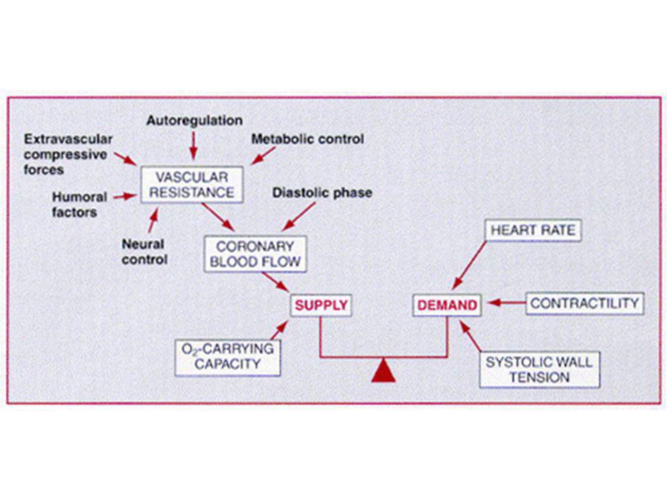
Determinanti del consumo di O2

6
Regolazione del flusso coronarico Concetti generali
Il flusso coronarico è prevalentemente diastolico I determinanti del flusso sono il gradiente pressorio e le resistenze coronariche Il gradiente è dato dalla differenza tra pressione aortica (in diastole) e pressione diastolica in ventricolo sinistro Esiste una pressione critica di chiusura
 e pressione diastolica in ventricolo sinistro. Esiste una pressione critica di chiusura.")
8
Autoregolazione coronarica
Flusso massimo Flusso a riposo Riserva coronarica Flusso coronarico totale (ml/m’) Pressione arteriosa (mm Hg)
 Pressione arteriosa (mm Hg)")
9
Autoregolazione coronarica
Ipertensione Flusso massimo Flusso x 100 g di Ventricolo ipertrofico Flusso x 100 g di Ventricolo normale Riserva coronarica Flusso coronarico totale (ml/m’) Flusso a riposo Pressione arteriosa (mm Hg)
 Flusso a riposo Pressione arteriosa (mm Hg)")
10
MECHANISMS OF AUTOREGULATION
NITRIC OXIDE. Evidence suggests a role for NO in coronary autoregulation. Inhibition of NO raises the lower autoregulatory threshold by about 15 mm Hg. The involvement of NO may be related to the ability of the endothelium to sense changes in perfusion pressure through pressure-sensitive ion channels. MYOGENIC CONTROL. Arteriolar smooth muscle reacts to increased intraluminal pressure by contracting. The consequent augmentation of resistance tends to return blood flow toward normal despite the higher perfusion pressure. This regulatory mechanism, referred to as myogenic control, is an important mechanism in some vascular beds. Their contribution to autoregulation is relatively small.

12
NITRIC OXIDE (NO). This substance increases blood flow during metabolic stimuli. NO production is augmented in response to metabolic stimuli by at least two mechanisms. Hypoxia is a stimulus to release of NO from the endothelium. Furthermore, NO is a principal mediator of flow-mediated dilation. Although hypoxia may initiate hyperemia, flow-mediated dilation sustains and amplifies it. OTHER METABOLIC MEDIATORS. Inhibition of the synthesis of vasodilator prostaglandins and inhibition of K+ATP channels also reduces metabolic vasodilation. A loss or inhibition of one mediator is compensated for by upregulation of others. Although the inhibition of K+ATP channels, adenosine, and NO individually has at most a modest effect on the increase in coronary blood flow during exercise in dogs, inhibition of all three simultaneously nearly abolishes the flow increase.

13
Endotelio: un organo con 2 funzioni
Azione Anticoagulante Azione Antiadesione piastrinica Prostaciclina Adenosina EDRF Glycocalice Repulsione neutrofili Azione dilatante ENDOTHELIAL CELL Eparina Antitrombina III Trombomodulina NO Fattore IX Fattore X Fattore VIII di Von Willebrand Endotelina ICAM-1 Selectina P Azione Procoagulante Azione costrittrice Angiotensina II Adesione Proadesione piastrinica Attrazione neutrofili Cellula endoteliale

14
Fattori di derivazione endoteliale
Fattori di rilasciamento endotelio-derivati: EDRF Ossido d’azoto (NO) Fattore iperpolarizzante endotelio-derivato (EDHF) Prostaciclina Fattori di contrazione endotelio-derivati: EDCF Anione superossido Trombossano Endoperossidi Endotelina
 Fattore iperpolarizzante endotelio-derivato (EDHF) Prostaciclina. Fattori di contrazione endotelio-derivati: EDCF. Anione superossido. Trombossano. Endoperossidi. Endotelina.")
15
Patologia della liberazione di EDRF
Quando le cellule endoteliali scompaiono, vengono rimpiazzate da nuove cellule rigenerate. Tali cellule sono meno capaci di produrre EDRF. Diversi studi hanno dimostrato una tendenza localizzata ad una esagerata vasocostrizione che è una caratteristica precoce della malattia coronarica nell’uomo. Vanhoutte P, Boulanger MC: “L’endotelio: un ruolo fondamentale nella fisiopatologia cardiovascolare” 1994

16
L’Endotelio e il tono vascolare
1980: Furchgott e Zawadski scoprono che l’endotelio è essenziale per la vasodilatazione dell’acetilcolina ACETILCOLINA -5 -6 -9 -9 -7 -8 -8 -7 -6 -5 INTATTO DENUDATO 1986: il gruppo di Ignarro e quello di Moncada identificano nel NO (ossido di azoto) il fattore vasodilatante endotelio-dipendente
 il fattore vasodilatante endotelio-dipendente.")
17
Transmural Distribution of Myocardial Blood Flow
Cross-section of the left ventricular wall in diastole and systole. Factors involved in the susceptibility of the subendocardium to the development of ischemia include the greater dependence of this region on diastolic perfusion and the greater degree of shortening, and therefore of energy expenditure, of this region during systole.

18
Ischemia subendocardica
Il flusso subendocardico è leggermente maggiore del flusso subepicardico a riposo (rapporto =1,16) Tuttavia il gradiente di perfusione è minore, e le forze compressive sono maggiori Ne deriva che questo flusso maggiore è ottenuto per vasodilatazione arteriolare, cioè con riduzione della riserva coronarica
 Tuttavia il gradiente di perfusione è minore, e le forze compressive sono maggiori. Ne deriva che questo flusso maggiore è ottenuto per vasodilatazione arteriolare, cioè con riduzione della riserva coronarica.")
19
Subendocardial ischemia
Epicardial coronary stenoses are associated with reductions in the subendocardial to subepicardial flow ratio. Severe pressure-induced left ventricular hypertrophy, as well as heart failure with elevated left ventricular end-diastolic pressure, may also reduce the endocardial-to-epicardial flow ratio. When the markedly elevated left ventricular end-diastolic pressure in heart failure is corrected, subendocardial coronary flow reserve is restored and the endocardial-to-epicardial flow ratio is normalized. Reduction of myocardial oxygen demand, for example by beta blockers, also decreases epicardial blood flow and increases perfusion pressure and thereby flow to the ischemic subendocardial region.

20
Effects of Coronary Stenoses
As blood traverses a stenosis, pressure (energy) is lost. Principles of fluid dynamics have been applied to estimate this pressure loss and validated in animals models as well as in patients. Although the formulas are complex, they can been simplified as follows: where DP is the pressure drop across a stenosis in millimeters of mercury (mm Hg), Q is the flow across the stenosis in milliliters per second, and dsten is the minimal diameter of the stenosis lumen in millimeters
 is lost. Principles of fluid dynamics have been applied to estimate this pressure loss and validated in animals models as well as in patients. Although the formulas are complex, they can been simplified as follows: where DP is the pressure drop across a stenosis in millimeters of mercury (mm Hg), Q is the flow across the stenosis in milliliters per second, and dsten is the minimal diameter of the stenosis lumen in millimeters.")
21
Energy losses across a stenosis
Energy losses across a stenosis. The pressure gradient due to friction losses within the stenosis (DP) is directly proportional to blood flow (Q), whereas separation losses at the exit to the stenosis due to formation of eddies increase with blood flow squared (Q2). Separation losses predominate at high blood flows.
 is directly proportional to blood flow (Q), whereas separation losses at the exit to the stenosis due to formation of eddies increase with blood flow squared (Q2). Separation losses predominate at high blood flows.")
22
Relation between pressure reduction across a stenosis (DP) and flow through the stenosis (Q)
 and flow through the stenosis (Q)")
23
Relationship between resting (dashed line) and maximal coronary blood flow (solid line) and percentage of diameter stenosis
 and maximal coronary blood flow (solid line) and percentage of diameter stenosis")
24
Riserva coronarica La riserva coronarica è il rapporto tra il flusso massimale (ottenibile con la vasodilatazione massimale) e il flusso a riposo Normalmente questo rapporto è circa uguale a 4 La riserva coronarica comincia a diminuire significativamente per stenosi di circa 60-70%

25
Coronaria destra

26
Coronaria sinistra OAdx

27
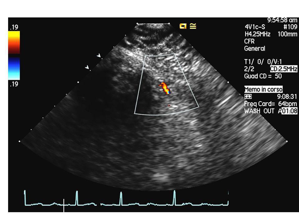
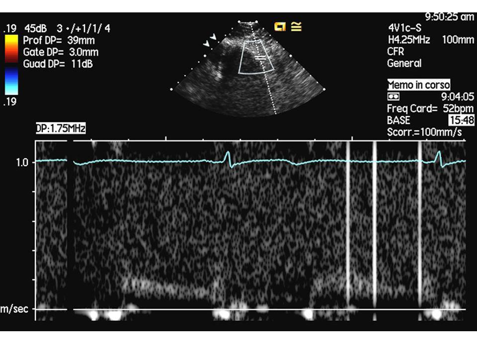
Valutazione della riserva coronarica
Stimolo alla vasodilatazione massimale (adenosina, dipiridamolo) Misurazione del flusso coronarico Diretta: flussimetri intracoronarici Indiretta: Ecodoppler Fractional flow reserve
 Misurazione del flusso coronarico. Diretta: flussimetri intracoronarici. Indiretta: Ecodoppler. Fractional flow reserve.")
28
FFR myo SU DA ADENOSINA i.c 0.84 BASALE 0.86

32
Coronary Collateral Vessels
COLLATERAL FORMATION (ARTERIOGENESIS) Preexisting collaterals are normally closed and nonfunctional, because no pressure gradient exists between the arteries they connect. After coronary occlusion, the distal pressure drops precipitously and preexisting collaterals open virtually instantly. The transformation of preexisting collaterals into mature collaterals is called arteriogenesis is characterized by inflammation and cellular proliferation. MECHANISMS PROMOTING COLLATERAL GROWTH SHEAR STRESS. Pressure gradients across preexisting rudimentary collaterals augment blood flow velocity and shear stress. Shear stress induces widespread functional changes in the endothelium, many of which reflect new gene expression. INFLAMMATION.
 Preexisting collaterals are normally closed and nonfunctional, because no pressure gradient exists between the arteries they connect. After coronary occlusion, the distal pressure drops precipitously and preexisting collaterals open virtually instantly. The transformation of preexisting collaterals into mature collaterals is called arteriogenesis is characterized by inflammation and cellular proliferation. MECHANISMS PROMOTING COLLATERAL GROWTH. SHEAR STRESS. Pressure gradients across preexisting rudimentary collaterals augment blood flow velocity and shear stress. Shear stress induces widespread functional changes in the endothelium, many of which reflect new gene expression. INFLAMMATION.")
33
Coronary Collateral Vessels (2)
ALTRI FATTORI Gravità della stenosi Ipossia Altri fattori di rischio Attività fisica SIGNIFICATO FUNZIONALE La capacità del circolo collaterale non supera il 50% di quella del circolo nativo (corrisponde quindi ad una stenosi del 70-80%)
")
34
Conseguenze metaboliche dell’ischemia
Metabolismo anaerobico: produzione di acido lattico per glicolisi Deplezione di ATP Perdita di potassio

35
Conseguenze emodinamiche dell’ischemia
Alterazioni reversibili: fino a m’ Cascata ischemica: Disfunzione diastolica Disfunzione sistolica Alterazioni ECG Dolore anginoso Tempo di recupero inversamente proporzionale alla profondità dell’ischemia Stunning Hibernation

36
Schematic diagram of stunned myocardium
During coronary occlusion, a wall motion abnormality of the left ventricle is present in the region supplied by the occluded artery. With relief of ischemia and reestablishment of coronary blood flow, there is a persistent wall motion abnormality despite reperfusion and viable myocytes. There is then gradual improvement in function that requires hours to days for recovery.

37
Two possible additive components of postischemic dysfunction: (1) reperfusion-induced pathology, which can be restored through the use of a therapeutic intervention such as an antioxidant or calcium-limiting agent given transiently at the time of reperfusion; and (2) ischemic pathology from which the heart is slowly recovering. These may be additive to each other and to any additional reperfusion-induced component that is not amenable to the chosen intervention.

38
Hibernating myocardium
The term hibernating myocardium refers to the presence of impaired resting left ventricular function, owing to reduced coronary blood flow that can be restored toward normal by revascularization. Hibernation was first noted in patients with coronary artery disease who had no evidence of ongoing ischemia yet whose left ventricular function improved after coronary artery bypass grafting. Even akinetic segments can occasionally regain systolic contraction after revascularization. Hibernating myocardium is present in approximately one third of patients with coronary artery disease and impaired left ventricular function.

39
CHARACTERISTICS OF STUNNING, HIBERNATION, AND ISCHEMIA
PARAMETER STUNNING HIBERNATION TRUE ISCHEMIA Myocardial mechanical function Reduced Coronary blood flow Post-ischemic: normal/high Modestly reduced or low normal; intermittent ischemia-reperfusion Most severely reduced Myocardial energy metabolism Normal or excessive Reduced or low normal; in steady state with intermittent ischemia-reperfusion Reduced; increasingly severe as ischemia proceeds Duration Hours to days; late stunning over weeks Days to hours to months Minutes to hours Outcome Full spontaneous recovery Recovery if revascularized Myocyte necrosis if severe ischemia persists Proposed change in metabolic regulation of calcium Cytosolic overload of calcium in early reperfusion with damage to contractile proteins Possibly just enough glycolytic ATP to prevent contracture Insufficient glycolytic ATP to prevent ischemic contracture and irreversibility ATP = adenosine triphosphate. Modified from Opie LH: The multifarious spectrum of ischemic left ventricular dysfunction: Relevance of new ischemic syndromes. J Mol Cell Cardiol 28:2403–2414, 1966.

40
Forme cliniche della cardiopatia ischemica
Angina da sforzo Sindromi ischemiche acute Con sopraslivellamento del tratto ST (infarto miocardico acuto) Senza sopraslivellamento sel tratto ST Angina instabile Infarto non-Q Cardio(mio)patia ischemica cronica
 Senza sopraslivellamento sel tratto ST. Angina instabile. Infarto non-Q. Cardio(mio)patia ischemica cronica.")
41
Angina da sforzo

42
Netter

43
LE DIVERSE SEDI DEL DOLORE TORACICO
1: posizione del cuore nel torace 2: angina di petto 3: irradiazione nell’attacco card. 4: sede correlata ad emozioni e stati ansiosi Da: M Caccavale. Inervista sul cuore e dintorni, Adn Kronos, Roma

44
ALCUNE POSSIBILI SEDI ED IRRADIAZIONI DEL DOLORE TORACICO DI
ORIGINE ESOFAGEA Sleisenger MH, Fordtran JS: Trattato di gastroenterologia, Piccin Ed, Padova

45
EMANAZIONE SIMPATICA E PARASIMPATICA DEL CUORE ED EMBRICAZIONE DELL’INNERVAZIONE CARDIACA E SPLANCNICA da: S Dalla Volta. La cardiopatia ischemica: dalla teoria alla clinica

46
GRECO LATINO INGLESE ITALIANO agcon: cappio, capestro, laccio
kardia: cuore, bocca dello stomaco Liddel Scott. Le Monnier LATINO angor: stringimento, angoscia, pena cardiacus: di stomaco Castiglioni Mariotti. Loescher INGLESE heart burn: bruciore di stomaco Hazon. Garzanti ITALIANO cardias: sbocco dell’esofago nello stomaco angere: affliggere, angosciare

47
Pazienti % origine esofagea
DOLORE TORACICO DI TIPO ANGINOSO ED ATTRIBUZIONE DELLA SINTOMATOLOGIA ALL’ESOFAGO Pazienti % origine esofagea Delmonico et al., Brand et al., Dart et al., Ferguson et al., Kline et al., De Meester et al., Katz et al., Schofield et al., Nevens et al., Voskuil et al., Frøbert et al., Chauhan et al., Fass et al., Ho et al., Börjesson et al., Romand et al., Netzer et al.,

48
Summary Angina pectoris is a discomfort in the chest or adjacent areas caused by myocardial ischemia. Anginal “equivalents” (i.e., symptoms of myocardial ischemia other than angina), such as dyspnea, faintness, fatigue, and eructations, are common, particularly in the elderly. Nocturnal angina should raise the suspicion of sleep apnea. Chest discomfort while walking in the cold, uphill, or after a meal is suggestive of angina. Features suggesting the absence of angina pectoris include pleuritic pain, pain localized to the tip of one finger, pain reproduced by movement or palpation of the chest wall or arms, and constant pain lasting many hours or, alternatively, very brief episodes of pain lasting seconds. Pain radiating into the lower extremities is also a highly unusual manifestation of angina pectoris. Typical angina pectoris is relieved within minutes by rest or by the use of nitroglycerin.
, such as dyspnea, faintness, fatigue, and eructations, are common, particularly in the elderly. Nocturnal angina should raise the suspicion of sleep apnea. Chest discomfort while walking in the cold, uphill, or after a meal is suggestive of angina. Features suggesting the absence of angina pectoris include pleuritic pain, pain localized to the tip of one finger, pain reproduced by movement or palpation of the chest wall or arms, and constant pain lasting many hours or, alternatively, very brief episodes of pain lasting seconds. Pain radiating into the lower extremities is also a highly unusual manifestation of angina pectoris. Typical angina pectoris is relieved within minutes by rest or by the use of nitroglycerin.")
49
Differential diagnosis of chest pain according to location where pain starts. Serious intrathoracic or subdiaphragmatic diseases are usually associated with pains that begin in the left anterior chest, left shoulder, or upper arm, the interscapular region, or the epigastrium. The scheme is not all inclusive (e.g., intercostal neuralgia occurs in locations other than the left, lower anterior chest area).
..")
50
Fisiopatologia Abitualmente placca aterosclerotica stabile che crea stenosi critica Angina da eccesso di domanda o da riduzione di offerta Angina a soglia fissa/soglia variabile Angina mista Non c’è correlazione tra gravità del sintomo e gravità della malattia L’ischemia può essere silente

51
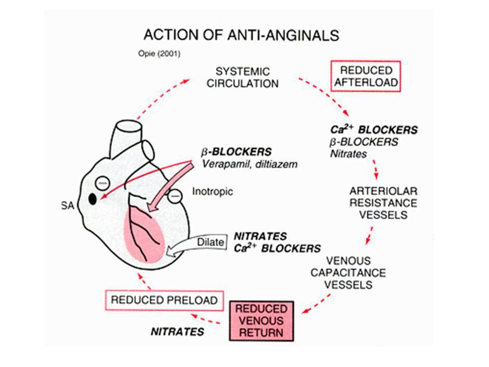
Factors influencing the balance between myocardial O2 requirements (left) and supply (right). Arrows indicate effects of nitrates. In relieving angina pectoris, nitrates exert favorable effects by reducing O2 requirements and increasing supply. Although a reflex increase in heart rate would tend to reduce the time for coronary flow, dilation of collaterals and enhancement of the pressure gradient for flow to occur as the left ventricular end-diastolic pressure (LVEDP) falls tend to increase coronary flow. AoP = aortic pressure; NC = no change. (From Frishman WH: Pharmacology of the nitrates in angina pectoris. Am J Cardiol 56:8I, By permission of Excerpta Medica.)
 falls tend to increase coronary flow. AoP = aortic pressure; NC = no change. (From Frishman WH: Pharmacology of the nitrates in angina pectoris. Am J Cardiol 56:8I, By permission of Excerpta Medica.).")
52
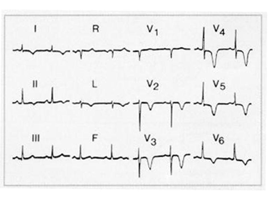
Elettrocardiogramma A riposo e in assenza di sintomi l’elettrocardiogramma può essere normale (spesso è normale) Altre alterazioni possono dipendere dalla malattia di base In presenza di sintomi (angina) il segno fondamentale è la lesione subendocardica, (sottoslivellamento del tratto ST)
 il segno fondamentale è la lesione subendocardica, (sottoslivellamento del tratto ST)")
53
Elettrogenesi della “lesione”
Acute ischemia may alter ventricular action potentials by inducing lower resting membrane potential, decreased amplitude and velocity of phase 0, and an abbreviated action potential duration (pathological early repolarization). These electrophysiological effects create a voltage gradient between ischemic and normal cells during different phases of the cardiac electrical cycle. The resulting currents of injury are reflected on the surface electrocardiogram by deviation of the ST segment
. These electrophysiological effects create a voltage gradient between ischemic and normal cells during different phases of the cardiac electrical cycle. The resulting currents of injury are reflected on the surface electrocardiogram by deviation of the ST segment.")
54
Current-of-injury patterns with acute ischemia
Current-of-injury patterns with acute ischemia. With predominant subendocardial ischemia (A), the resultant ST vector is directed toward the inner layer of the affected ventricle and the ventricular cavity. Overlying leads therefore record ST depression. With ischemia involving the outer ventricular layer (B) (transmural or epicardial injury), the ST vector is directed outward. Overlying leads record ST elevation. Reciprocal ST depression can appear in contralateral leads.
, the resultant ST vector is directed toward the inner layer of the affected ventricle and the ventricular cavity. Overlying leads therefore record ST depression. With ischemia involving the outer ventricular layer (B) (transmural or epicardial injury), the ST vector is directed outward. Overlying leads record ST elevation. Reciprocal ST depression can appear in contralateral leads.")
55
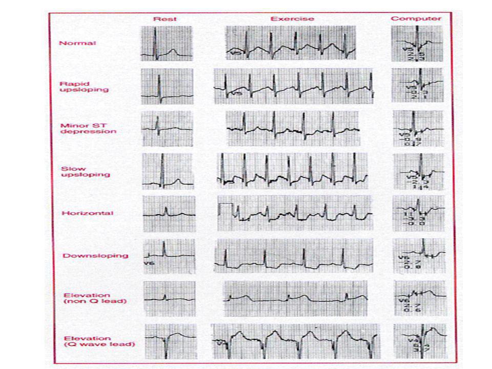
Test ergometrico (prova da sforzo)
Si esegue al cicloergometro o al tapis roulant (treadmill) Esercizio a carichi crescenti monitorizzando frequenza cardiaca e pressione arteriosa Cessazione dello sforzo se compaiono sintomi o alterazioni ECG Positività: alterazioni del tratto ST (più spesso sottoslivellamento)
 Esercizio a carichi crescenti monitorizzando frequenza cardiaca e pressione arteriosa. Cessazione dello sforzo se compaiono sintomi o alterazioni ECG. Positività: alterazioni del tratto ST (più spesso sottoslivellamento)")
57
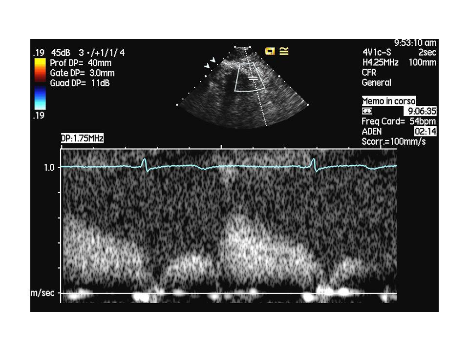
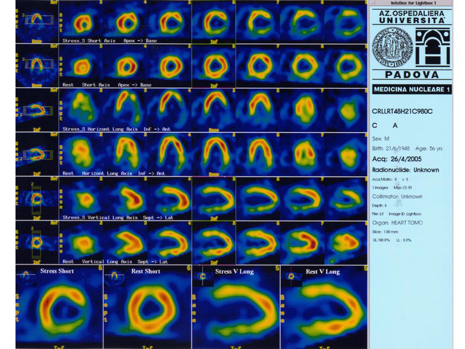
Miocardioscintigrafia da sforzo (o da stress)
")
59
Dobutamina ischemia apice laterale durante stress

60
Treatment 1) identification and treatment of associated diseases that can precipitate or worsen angina; (2) reduction of coronary risk factors; (3) application of general and non-pharmacological methods, with particular attention toward adjustments in life style; (4) pharmacological management; (5) revascularization by percutaneous catheter-based techniques or by coronary bypass surgery
 reduction of coronary risk factors; (3) application of general and non-pharmacological methods, with particular attention toward adjustments in life style; (4) pharmacological management; (5) revascularization by percutaneous catheter-based techniques or by coronary bypass surgery.")
61
Mortalità annua x 100000 maschi
Non fumatore Ex fumatore Fumatore < 15 15-25 >25 RR (>25/NF) BPCO 10 57 127 86 112 225 22,5 (11-42) Cardiopatia ischemica 572 678 892 802 1025 1,8 (1,6-2) Miocardiopatie 61 88 125 122 109 173 2,8 (2,1-3,8) Ipertensione 32 33 44 28 51 60 (1,2-2,9) Cerebrovasculo-patie 152 158 166 121 231 235 1,5 (1,2-1,9) Neoplasia polmonare 14 58 209 105 208 355 25,4 (15-43) BMJ 1994;309: (8 October)
 BPCO ,5. (11-42) Cardiopatia ischemica ,8. (1,6-2) Miocardiopatie ,8. (2,1-3,8) Ipertensione (1,2-2,9) Cerebrovasculo-patie ,5. (1,2-1,9) Neoplasia polmonare ,4. (15-43) BMJ 1994;309: (8 October)")
62
Mortality Risk Reduction Associated With Smoking Cessation in Patients
With Coronary Heart Disease. A Systematic Review Julia A. Critchley, MSc, DPhil; Simon Capewell, MD, FRCPE JAMA. 2003;290:86-97. Objective To conduct a systematic review to determine the magnitude of risk reduction achieved by smoking cessation in patients with CHD. Data Sources Nine electronic databases were searched from start of database to April 2003, supplemented by cross-checking references, contact with experts, and with large international cohort studies (identified by the Prospective Studies Collaboration). Study Selection Prospective cohort studies of patients who were diagnosed with CHD were included if they reported all-cause mortality and had at least 2 years of follow-up. Smoking status had to be measured after CHD diagnosis to ascertain quitting. Data Extraction Two reviewers independently assessed studies to determine eligibility, quality assessment of studies, and results, and independently carried out data extraction using a prepiloted, standardized form. Data Synthesis From the literature search, 665 publications were screened and 20 studies were included. Results showed a 36% reduction in crude relative risk (RR) of mortality for patients with CHD who quit compared with those who continued smoking (RR, 0.64; 95% confidence interval [CI], ). Results from individual studies did not vary greatly despite many differences in patient characteristics, such as age, sex, type of CHD, and the years in which studies took place. Adjusted risk estimates did not differ substantially from crude estimates. Many studies did not adequately address quality issues, such as control of confounding, and misclassification of smoking status. However, restriction to 6 higher-quality studies had little effect on the estimate (RR, 0.71; 95% CI, ). Few studies included large numbers of elderly persons, women, ethnic minorities, or patients from developing countries. Conclusions Quitting smoking is associated with a substantial reduction in risk of all-cause mortality among patients with CHD. This risk reduction appears to be consistent regardless of age, sex, index cardiac event, country, and year of study commencement.
. Study Selection Prospective cohort studies of patients who were diagnosed with CHD were included if they reported all-cause mortality and had at least 2 years of follow-up. Smoking status had to be measured after CHD diagnosis to ascertain quitting. Data Extraction Two reviewers independently assessed studies to determine eligibility, quality assessment of studies, and results, and independently carried out data extraction using a prepiloted, standardized form. Data Synthesis From the literature search, 665 publications were screened and 20 studies were included. Results showed a 36% reduction in crude relative risk (RR) of mortality for patients with CHD who quit compared with those who continued smoking (RR, 0.64; 95% confidence interval [CI], ). Results from individual studies did not vary greatly despite many differences in patient characteristics, such as age, sex, type of CHD, and the years in which studies took place. Adjusted risk estimates did not differ substantially from crude estimates. Many studies did not adequately address quality issues, such as control of confounding, and misclassification of smoking status. However, restriction to 6 higher-quality studies had little effect on the estimate (RR, 0.71; 95% CI, ). Few studies included large numbers of elderly persons, women, ethnic minorities, or patients from developing countries. Conclusions Quitting smoking is associated with a substantial reduction in risk of all-cause mortality among patients with CHD. This risk reduction appears to be consistent regardless of age, sex, index cardiac event, country, and year of study commencement.")
63
Trattamento farmacologico
Nitrati (Trinitroglicerina e altri) In particolare per risolvere l’attacco acuto (via sublinguale) Beta-bloccanti Calcioantagonisti
 In particolare per risolvere l’attacco acuto (via sublinguale) Beta-bloccanti. Calcioantagonisti.")
65
Effects of nitrates in generating NO+ and stimulating guanylate cyclase to cause vasodilation. Note the role of cysteine cascade in stimulating guanylate cyclase. Previously, sulfhydryl (SH) depletion was thought to explain nitrate tolerance. Current emphasis is on the generation of peroxynitrite, which in turn inhibits the conversion of guanosine triphosphate (GTP) to cyclic guanosine monophosphate (GMP). Note that mononitrates bypass hepatic metabolism.
 depletion was thought to explain nitrate tolerance. Current emphasis is on the generation of peroxynitrite, which in turn inhibits the conversion of guanosine triphosphate (GTP) to cyclic guanosine monophosphate (GMP). Note that mononitrates bypass hepatic metabolism..")
66
The mechanisms of action of the nitrates are complex
The mechanisms of action of the nitrates are complex. These drugs decrease cardiac demand and may increase coronary blood supply through a variety of actions. It is likely that the various mechanisms that act to provide relief to angina patients or prevent myocardial ischemia differ among individuals. LV = left ventricular; RV = right ventricular.

68
Mechanisms of nitrate tolerance
Depletion of Sulfhydryl Groups. Neurohormonal Activation. Plasma Volume Expansion. Downregulation of Receptors. Free Radical Generation. MANAGEMENT. The only practical strategy to manage nitrate tolerance is to prevent it by providing a “nitrate-free” interval.

69
INTERACTION WITH SILDENAFIL.
The combination of nitrates and sildenafil may cause serious, prolonged, and potentially life-threatening hypotension. Nitrate therapy is an absolute contraindication to the use of sildenafil and vice versa. Patients who wish to take sildenafil should be aware of the serious nature of this adverse drug interaction and be warned about taking sildenafil within 24 hours of any nitrate preparation, including short-acting sublingual nitroglycerin tablets.

70
Effects of beta blockade on the ischemic heart
Effects of beta blockade on the ischemic heart. Beta blockade has a beneficial effect on ischemic myocardium unless (1) the preload rises substantially as in left-sided heart failure or (2) vasospastic angina is present, in which case spasm may be promoted in some patients. Note the recent proposal that beta blockade diminishes exercise-induced vasoconstriction.
 the preload rises substantially as in left-sided heart failure or (2) vasospastic angina is present, in which case spasm may be promoted in some patients. Note the recent proposal that beta blockade diminishes exercise-induced vasoconstriction.")
71
CANDIDATES FOR USE OF BETA-BLOCKING AGENTS FOR ANGINA
Ideal Candidates Prominent relationship of physical activity to attacks of angina Coexistent hypertension History of supraventricular or ventricular arrhythmias Previous myocardial infarction Left ventricular systolic dysfunction Mild to moderate heart failure symptoms (NYHA functional Class II–III) Prominent anxiety state Poor Candidates Asthma or reversible airway component in chronic lung disease patients Severe left ventricular dysfunction with severe heart failure symptoms (NYHA functional Class IV) History of severe depression Raynaud's phenomenon Symptomatic peripheral vascular disease Severe bradycardia or heart block Brittle diabetes NYHA = New York Heart Association.
 Prominent anxiety state. Poor Candidates Asthma or reversible airway component in chronic lung disease patients. Severe left ventricular dysfunction with severe heart failure symptoms (NYHA functional Class IV) History of severe depression. Raynaud s phenomenon. Symptomatic peripheral vascular disease. Severe bradycardia or heart block. Brittle diabetes. NYHA = New York Heart Association.")
72
CARDIAC ARRHYTHMIA OR CONDUCTION DISTURBANCE Sinus bradycardia
RECOMMENDED DRUG THERAPY (CALCIUM ANTAGONIST VS. BETA BLOCKER) IN PATIENTS WHO HAVE ANGINA IN CONJUNCTION WITH OTHER MEDICAL CONDITIONS CLINICAL CONDITION RECOMMENDED DRUG CARDIAC ARRHYTHMIA OR CONDUCTION DISTURBANCE Sinus bradycardia Nifedipine or amlodipine Sinus tachycardia (not caused by cardiac failure) Beta blocker Supraventricular tachycardia Beta blocker (verapamil) Atrioventricular block Rapid atrial fibrillation (with digitalis) Verapamil or beta blocker Ventricular arrhythmia LEFT VENTRICULAR DYSFUNCTION Heart failure MISCELLANEOUS MEDICAL CONDITIONS Systemic hypertension Beta blocker (calcium antagonist) Severe preexisting headaches Beta blocker (verapamil or diltiazem) COPD with bronchospasm or asthma Nifedipine, amlodipine, verapamil, or diltiazem Hyperthyroidism Raynaud's syndrome Claudication Calcium antagonist Severe depression COPD = chronic obstructive pulmonary disease. (alternatives in parentheses)
 IN PATIENTS WHO HAVE ANGINA IN CONJUNCTION WITH OTHER MEDICAL CONDITIONS. CLINICAL CONDITION. RECOMMENDED DRUG. CARDIAC ARRHYTHMIA OR CONDUCTION DISTURBANCE Sinus bradycardia. Nifedipine or amlodipine. Sinus tachycardia (not caused by cardiac failure) Beta blocker. Supraventricular tachycardia. Beta blocker (verapamil) Atrioventricular block. Rapid atrial fibrillation (with digitalis) Verapamil or beta blocker. Ventricular arrhythmia. LEFT VENTRICULAR DYSFUNCTION Heart failure. MISCELLANEOUS MEDICAL CONDITIONS Systemic hypertension. Beta blocker (calcium antagonist) Severe preexisting headaches. Beta blocker (verapamil or diltiazem) COPD with bronchospasm or asthma. Nifedipine, amlodipine, verapamil, or diltiazem. Hyperthyroidism. Raynaud s syndrome. Claudication. Calcium antagonist. Severe depression. COPD = chronic obstructive pulmonary disease. (alternatives in parentheses)")
73
TABLE 37–3. EFFECTS OF ANTIANGINAL AGENTS ON INDICES OF MYOCARDIAL OXYGEN SUPPLY AND DEMAND*
INDEX NITRATES BETA-ADRENOCEPTOR BLOCKERS CALCIUM ANTAGONISTS ISA Cardioselective No Yes Nifedipine Verapamil Ditiazem SUPPLY Coronary resistance Vascular tone ¯¯ 0 ¯¯¯ Intramyocardial diastolic tension Coronary collateral circulation Duration of diastole 0(¯) 0¯ 0 (¯¯) (¯) (¯) DEMAND Intramyocardial systolic tension Preload ¯0 0¯ Afterload (peripheral vascular resistance) Contractility 0( ) ¯( )† ¯¯( )† ¯( )† Heart rate 0( ) ¯¯( )
 0¯ 0 (¯¯) (¯) (¯) DEMAND Intramyocardial systolic tension. Preload. ¯0. 0¯ Afterload (peripheral vascular resistance) Contractility. 0( ) ¯( )† ¯¯( )† ¯( )† Heart rate. 0( ) ¯¯( )")
74
BETA-ADRENOCEPTOR BLOCKERS CALCIUM ANTAGONISTS ISA Cardioselective No
TABLE 37–3. EFFECTS OF ANTIANGINAL AGENTS ON INDICES OF MYOCARDIAL OXYGEN SUPPLY AND DEMAND* INDEX NITRATES BETA-ADRENOCEPTOR BLOCKERS CALCIUM ANTAGONISTS ISA Cardioselective No Yes Nifedipine Verapamil Ditiazem SUPPLY Coronary resistance Vascular tone ¯¯ 0 ¯¯¯ Intramyocardial diastolic tension Coronary collateral circulation Duration of diastole 0(¯) 0¯ 0 (¯¯) (¯) (¯) DEMAND Intramyocardial systolic tension Preload ¯0 0¯ Afterload (peripheral vascular resistance) Contractility 0( ) ¯( )† ¯¯( )† ¯( )† Heart rate 0( ) ¯¯( ) * = Increase; ¯ = decrease; 0 = little or no definite effect. The number of arrows represents the relative intensity of effect. Symbols in parentheses indicate reflex-mediated effects. †Effect of calcium entry on left ventricular contractility, as assessed in the intact animal model. The net effect on left ventricular performance is variable since it is influenced by alterations in afterload, reflex cardiac stimulation, and the underlying state of the myocardium. ISA = intrinsic sympathomimetic activity. From Shub C, Vlietstra RE, McGoon MD: Selection of optimal drug therapy for the patient with angina pectoris. Mayo Clin Proc 60:539, By permission of the Mayo Foundation.
 0¯ 0 (¯¯) (¯) (¯) DEMAND Intramyocardial systolic tension. Preload. ¯0. 0¯ Afterload (peripheral vascular resistance) Contractility. 0( ) ¯( )† ¯¯( )† ¯( )† Heart rate. 0( ) ¯¯( ) * = Increase; ¯ = decrease; 0 = little or no definite effect. The number of arrows represents the relative intensity of effect. Symbols in parentheses indicate reflex-mediated effects. †Effect of calcium entry on left ventricular contractility, as assessed in the intact animal model. The net effect on left ventricular performance is variable since it is influenced by alterations in afterload, reflex cardiac stimulation, and the underlying state of the myocardium. ISA = intrinsic sympathomimetic activity. From Shub C, Vlietstra RE, McGoon MD: Selection of optimal drug therapy for the patient with angina pectoris. Mayo Clin Proc 60:539, By permission of the Mayo Foundation.")
75
Prognosi in terapia medica
La prognosi è riservata In 15 mesi: Mortalità: 4% Infarto miocardico: 7% Remissione spontanea: 11% Importante il ruolo di: Funzione ventricolare sinistra Estensione dell’ischemia inducibile

76
Graphs showing survival for medically treated CASS patients
Graphs showing survival for medically treated CASS patients. A, Patients with one-, two-, or three-vessel disease and an ejection fraction of 50 to 100 percent stratified by the number of diseased vessels (DISVES). B, Patients with one-, two-, or three-vessel disease and an ejection fraction of 35 to 49 percent stratified by the number of diseased vessels. C, Patients with one-, two-, or three-vessel disease and an ejection fraction of 0 to 34 percent stratified by the number of diseased vessels.
. B, Patients with one-, two-, or three-vessel disease and an ejection fraction of 35 to 49 percent stratified by the number of diseased vessels. C, Patients with one-, two-, or three-vessel disease and an ejection fraction of 0 to 34 percent stratified by the number of diseased vessels.")
77
Indicazioni alla rivascolarizzazione
Mediante angioplastica: presenza di una o più stenosi critiche, preferibilmente senza coinvolgimento della discendente anteriore (indicazioni “quoad valetudinem”) Mediante bypass aortocoronarico (indicazioni “quad vitam”): Malattia del Tronco comune (>50%) Malattia di tre vasi Malattia di due vasi con coinvolgimento della discendente anteriore prossimale
 Mediante bypass aortocoronarico (indicazioni quad vitam ): Malattia del Tronco comune (>50%) Malattia di tre vasi. Malattia di due vasi con coinvolgimento della discendente anteriore prossimale.")
78
Adjusted hazard (mortality) ratios comparing coronary artery bypass grafting (CABG) and medical therapy for nine coronary anatomy severity groups (GR) according to the number of vessels diseased (VD), the presence or absence of a 95 percent proximal stenosis (95 percent), and involvement of the left anterior descending coronary artery (LAD). B, Adjusted hazard (mortality) ratios comparing CABG and percutaneous transluminal angioplasty (PTCA) for nine coronary anatomy groups according to the number of vessels diseased, the presence or absence of a 95 percent proximal stenosis, and LAD involvement.
 ratios comparing CABG and percutaneous transluminal angioplasty (PTCA) for nine coronary anatomy groups according to the number of vessels diseased, the presence or absence of a 95 percent proximal stenosis, and LAD involvement..")
79
Two-year cumulative mortality rates for three treatment strategies
Two-year cumulative mortality rates for three treatment strategies. Significant differences were seen between revascularization and angina-guided strategies (P<.005) and between revascularization and ischemia-guided strategies (P<.05). Angina-guided and ischemia-guided strategies were not significantly different from each other (P=.34). Asymptomatic Cardiac Ischemia Pilot (ACIP) Study Two-Year Follow-up : Outcomes of Patients Randomized to Initial Strategies of Medical Therapy Versus Revascularization. Circulation, Apr 1997; 95:
 and between revascularization and ischemia-guided strategies (P<.05). Angina-guided and ischemia-guided strategies were not significantly different from each other (P=.34). Asymptomatic Cardiac Ischemia Pilot (ACIP) Study Two-Year Follow-up : Outcomes of Patients Randomized to Initial Strategies of Medical Therapy Versus Revascularization. Circulation, Apr 1997; 95:")
80
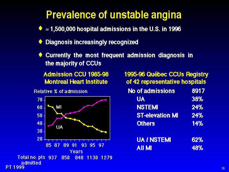
Angina instabile

82
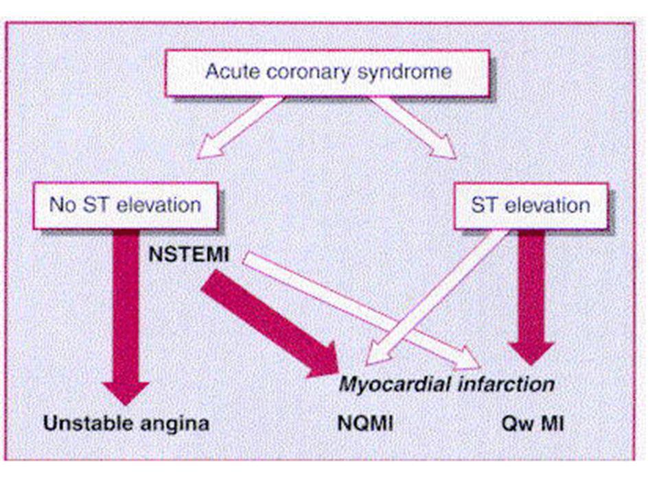
Angina instabile / Nomenclatura
Unstable angina lies in the center of the spectrum of clinical conditions caused by myocardial ischemia. These range from chronic stable angina pectoris to the acute coronary syndromes. The latter, in turn, consist of acute myocardial infarction (MI) associated with electrocardiographic ST segment elevation (STEMI) and unstable angina/non-ST segment elevation MI (UA/NSTEMI). The former is most commonly caused by acute total coronary occlusion, and urgent reperfusion is the mainstay of therapy, UA/NSTEMI is usually associated with severe coronary obstruction but not total occlusion of the culprit coronary artery. If the myocardial ischemia that results from the coronary obstruction is long in duration and/or great in severity, myocardial necrosis occurs, and the patient is classified as having a non-Q-wave MI or, now more aptly termed, NSTEMI
 associated with electrocardiographic ST segment elevation (STEMI) and unstable angina/non-ST segment elevation MI (UA/NSTEMI). The former is most commonly caused by acute total coronary occlusion, and urgent reperfusion is the mainstay of therapy, UA/NSTEMI is usually associated with severe coronary obstruction but not total occlusion of the culprit coronary artery. If the myocardial ischemia that results from the coronary obstruction is long in duration and/or great in severity, myocardial necrosis occurs, and the patient is classified as having a non-Q-wave MI or, now more aptly termed, NSTEMI.")
84
Definizione clinica Unstable angina is defined as angina pectoris (or equivalent type of ischemic discomfort) with at least one of three features: (1) it occurs at rest (or with minimal exertion) usually lasting more than 20 minutes (if not interrupted by nitroglycerin); (2) it is severe and described as frank pain and of new onset (i.e., within 1 month); and (3) it occurs with a crescendo pattern (i.e., more severe, prolonged, or frequent than previously). Some patients with this pattern of ischemic discomfort, especially those with prolonged rest pain, develop evidence of myocardial necrosis on the basis of the release of cardiac markers and thus have a diagnosis of NSTEMI.
 it occurs at rest (or with minimal exertion) usually lasting more than 20 minutes (if not interrupted by nitroglycerin); (2) it is severe and described as frank pain and of new onset (i.e., within 1 month); and. (3) it occurs with a crescendo pattern (i.e., more severe, prolonged, or frequent than previously). Some patients with this pattern of ischemic discomfort, especially those with prolonged rest pain, develop evidence of myocardial necrosis on the basis of the release of cardiac markers and thus have a diagnosis of NSTEMI.")
85
BRAUNWALD CLINICAL CLASSIFICATION OF UNSTABLE ANGINA
DEFINITION DEATH OR MYOCARDIAL INFARCTION TO 1 YEAR* Severity Class I New onset of severe angina or accelerated angina; no rest pain 7.3% Class II Angina at rest within past month but not within preceding 48 hr (angina at rest, subacute) 10.3% Class III Angina at rest within 48 hr (angina at rest, subacute) 10.8%† Clinical Circumstances A (secondary angina) Develops in the presence of extracardiac condition that intensifies myocardial ischemia 14.1% B (primary angina) Develops in the absence of extracardiac condition 8.5% C (postinfarction angina) Develops within 2 weeks after acute myocardial infarction 18.5%‡
 10.3% Class III. Angina at rest within 48 hr (angina at rest, subacute) 10.8%† Clinical Circumstances. A (secondary angina) Develops in the presence of extracardiac condition that intensifies myocardial ischemia. 14.1% B (primary angina) Develops in the absence of extracardiac condition. 8.5% C (postinfarction angina) Develops within 2 weeks after acute myocardial infarction. 18.5%‡")
86
Meccanismi eziopatogenetici Possono agire isolatamente o congiuntamente
Complicanza di placca: trombosi non occlusiva su placca significativa ma non critica (es. stenosi 60%) Trombo piastrinico E’ il meccanismo di gran lunga più frequente Spasmo su stenosi lieve, o in assenza di stenosi (Prinzmetal) Più raro, ma spesso presente anche dopo un trombo piastrinico (trombossano)
 Trombo piastrinico. E’ il meccanismo di gran lunga più frequente. Spasmo su stenosi lieve, o in assenza di stenosi (Prinzmetal) Più raro, ma spesso presente anche dopo un trombo piastrinico (trombossano)")
87
Meccanismi eziopatogenetici Possono agire isolatamente o congiuntamente
Ostruzione meccanica progressiva (crescita di placca) Meccanismo tipico della restenosi dopo angioplastica o intrastent Infiammazione o infezione Aumento della richiesta di flusso da causa extracardiaca Tireotossicosi Anemia
 Meccanismo tipico della restenosi dopo angioplastica o intrastent. Infiammazione o infezione. Aumento della richiesta di flusso da causa extracardiaca. Tireotossicosi. Anemia.")
88
Schematic representation of the causes of unstable angina
Schematic representation of the causes of unstable angina. Each of the five bars represents one of the etiologic mechanisms, and the red portion of the bar represents the extent to which the mechanism is operative. A, Most common form of unstable angina in which atherosclerotic plaque causes moderate (60 percent diameter) obstruction and acute thrombus overlying plaque causes very severe (90 percent diameter) narrowing. B, Mild coronary obstruction, adjacent to which there is intense (90 percent diameter) vasoconstriction.
 obstruction and acute thrombus overlying plaque causes very severe (90 percent diameter) narrowing. B, Mild coronary obstruction, adjacent to which there is intense (90 percent diameter) vasoconstriction.")
89
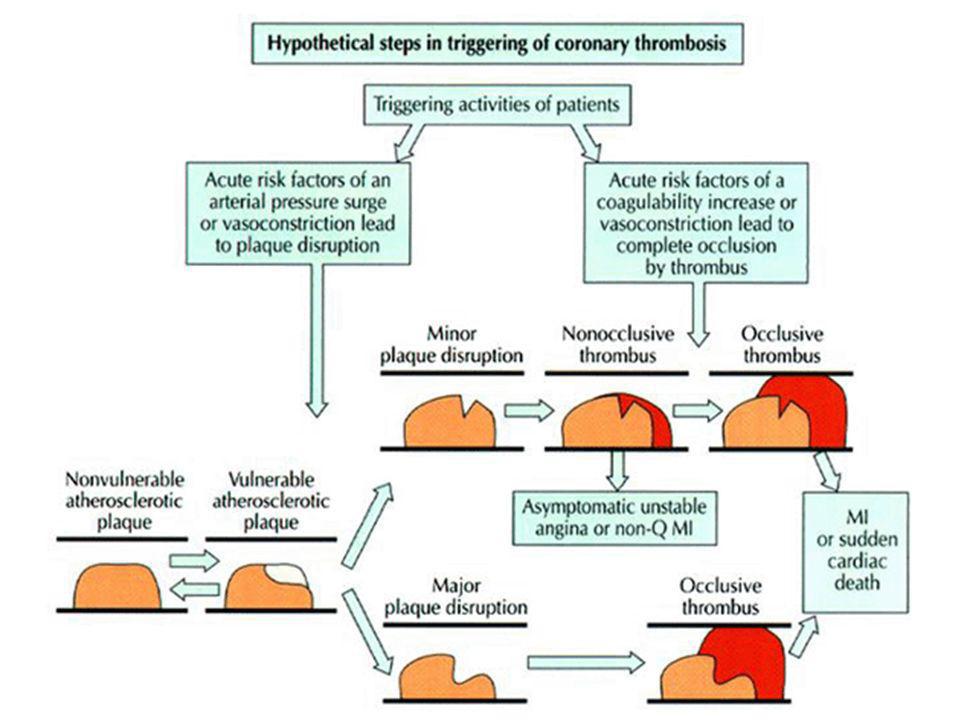
PLAQUE RUPTURE, FISSURE, OR EROSION.
Rupture or erosion of an atherosclerotic plaque with superimposed nonocclusive thrombus is by far the most common cause of UA/NSTEMI. The type of plaque that ruptures, the so-called vulnerable plaques, are usually lesions with less than 50 percent Plaque rupture can be precipitated by multiple factors, including high plaque lipid content, local inflammation causing breakdown of the thin shoulder of the plaque, coronary artery constriction at the site of the plaque, local shear stress forces, platelet activation, and the status of the coagulation system (i.e., a potentially prothrombotic state), all of which culminate in the formation of platelet-rich thrombi at the site of the plaque rupture or erosion and the resultant acute coronary syndrome
, all of which culminate in the formation of platelet-rich thrombi at the site of the plaque rupture or erosion and the resultant acute coronary syndrome.")
90
Platelets initiate thrombosis at the site of a ruptured plaque: the first step is platelet adhesion (1) via the glycoprotein Ib receptor in conjunction with von Willebrand factor. This is followed by platelet activation (2), which leads to a shape change in the platelet, de-granulation of the alpha and dense granules, and expression of glycoprotein IIb/IIIa receptors on the platelet surface with activation of the receptor, such that it can bind fibrinogen. The final step is platelet aggregation (3), in which fibrinogen (or von Willebrand factor) binds to the activated glycoprotein IIb/IIIa receptors of two platelets. Aspirin (ASA) and clopidogrel act to decrease platelet activation, whereas the glycoprotein IIb/IIIa inhibitors inhibit the final step of platelet aggregation.
, in which fibrinogen (or von Willebrand factor) binds to the activated glycoprotein IIb/IIIa receptors of two platelets. Aspirin (ASA) and clopidogrel act to decrease platelet activation, whereas the glycoprotein IIb/IIIa inhibitors inhibit the final step of platelet aggregation.")
91
Vasocostrizione Prinzmetal Vasocostrizione microcircolatoria
Vasocostrizione di coronaria epicardica, focale, anche in assenza di lesione aterosclerotica Vasocostrizione microcircolatoria Vasocostrizione in presenza di placca Serotonina, Trombossano A2

92
Quadro clinico Circa 80% dei pazienti (45% di sesso femminile) hanno già precedenti di cardiopatia ischemica Sintomatologia importante: senso di peso ma spesso vero dolore Insorgenza a riposo o da sforzi lievi Spesso sintomi collaterali neurovegetativi

94
Diagnosi Sintomi e quadro clinico Elettrocardiogramma
Sottoslivellamento ST Onde T Markers cardiospecifici (cTroponina I e T) Livello di rischio Livello di necrosi
 Livello di rischio. Livello di necrosi.")
95
INDICATORS OF INCREASED RISK IN UNSTABLE ANGINA
History Advanced age (>65 years) Diabetes mellitus Post-myocardial infarction angina Prior peripheral vascular disease Prior cerebrovascular disease Clinical Presentation Braunwald Class II or III (acute or subacute rest pain) Braunwald Class B (secondary unstable angina) Heart failure/hypotension Electrocardiogram New/ST segment deviation 0.05 mV New T wave inversion 0.3 mV Left bundle branch block Cardiac Markers Increased troponin T or I or CK-MB Increased C-reactive protein (CRP) Angiogram Thrombus
 Diabetes mellitus Post-myocardial infarction angina Prior peripheral vascular disease Prior cerebrovascular disease. Clinical Presentation Braunwald Class II or III (acute or subacute rest pain) Braunwald Class B (secondary unstable angina) Heart failure/hypotension. Electrocardiogram New/ST segment deviation 0.05 mV New T wave inversion 0.3 mV Left bundle branch block. Cardiac Markers Increased troponin T or I or CK-MB Increased C-reactive protein (CRP) Angiogram Thrombus.")
96
Stratificazione del rischio nelle sindromi coronariche acute

97
Trattamento Stabilizzare (“passivating”) la placca
Aspirina, Clopidogrel, UFH o LMWH Correggere l’ischemia Nitrati, beta-bloccanti, calcioantagonisti Rivascolarizzazione Angioplastica Bypass Prevenzione a lungo termine della progressione della malattia

98
Pooled data from CAPTURE, PRISM-PLUS, and PURSUIT trials of unstable angina, showing benefit of glycoprotein IIb/IIIa inhibition during medical therapy only (left panel), during, and immediately after percutaneous coronary intervention (PCI, right panel).
, during, and immediately after percutaneous coronary intervention (PCI, right panel).")
99
Meccanismi favorevoli degli inibitori della GP IIb-IIIa
Greater resolution of thrombus and improved coronary flow compared with aspirin and heparin Reduced size of an evolving NSTEMI Greater benefit of treatment when administered earlier relative to the onset of pain Warning: No thrombolytics!

100
Infarto miocardico acuto

101
Diagnosi di dimissioni ospedaliere nel triennio 1996 - 1998
1997 1998 Numero Tassi per abitanti N° ricoveri per infarto miocardico 77.033 134,21 80.920 140,7 88.773 154,15 Degenza media IMA 11,62 10,93 10,39 N° ricoveri per angina pectoris 75.966 132,35 75.509 131,29 79.236 137,59 Degenza media AP 7,68 7,19 6,46 N°cateterismi cardiaci 61.378 106,94 72.188 125,52 82.140 142,63 N° By-pass 19.327 33,67 23.318 40,54 25.927 45,02 N° PTCA 14.415 25,11 20.250 35,21 28.104 48,8 IMA = infarto miocardico acuto; AP = angina pectoris Fonte ISTISAN

103
Sopravvivenza post-IMA
100 ! Angina stabile ! 80 + Direct PTCA, statine ! 60 ! Bypass, PTCA, TL, ACE-I 40 ! Bypass 20 ! 2 4 6 8 10 12 FOLLOW-UP (ANNI)
")
104
Infarto miocardico acuto
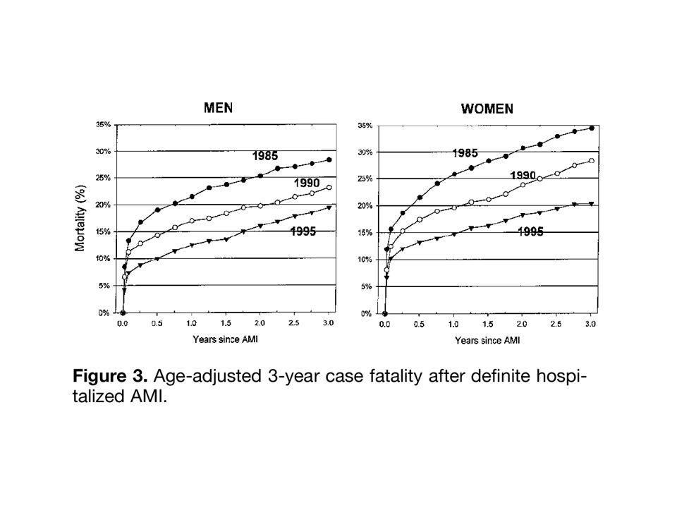
Il 50% della mortalità avviene in fase preospedaliera Mortalità ospedaliera Epoca pre-UCIC: 25% UCIC: 15% Trombolisi: 5-8% PTCA primaria: < 5% (studi randomizzati)
")
105
Patologia Complicanza di placca
Formazione di trombo di piastrine e poi Formazione di trombo di fibrina Il trombo è occlusivo

107
Schematic representation of the progression of myocardial necrosis after coronary artery occlusion. Necrosis begins in a small zone of the myocardium beneath the endocardial surface in the center of the ischemic zone. This entire region of myocardium (dashed outline) depends on the occluded vessel for perfusion and is the area at risk. Note that a very narrow zone of myocardium immediately beneath the endocardium is spared from necrosis because it can be oxygenated by diffusion from the ventricle.
 depends on the occluded vessel for perfusion and is the area at risk. Note that a very narrow zone of myocardium immediately beneath the endocardium is spared from necrosis because it can be oxygenated by diffusion from the ventricle..")
110
Temporal sequence of early biochemical, ultrastructural, histochemical, and histological findings after onset of MI. At the top of the figure are schematically shown the time frames for early and late reperfusion of the myocardium supplied by an occluded coronary artery. For approximately one-half hour after the onset of even the most severe ischemia, myocardial injury is potentially reversible; after that there is progressive loss of viability that is complete by 6 to 12 hours. The benefits of reperfusion (both early and late) are greatest when it is achieved early, with progressively smaller benefits occurring as reperfusion is delayed.
 are greatest when it is achieved early, with progressively smaller benefits occurring as reperfusion is delayed..")
111
Fisiopatologia

112
Fisiopatologia Funzione sistolica Funzione diastolica Aritmie
Precoci Tardive Complicanze

113
Disfunzione ventricolare
Nella zona infartuata A distanza Altre lesioni ischemizzanti Circolo collaterale Disfunzione sistolica Ipocinesia Acinesia Discinesia Disfunzione diastolica Ischemia, Edema, Fibrosi

116
P <<0,001 P = 0,5

119
Aritmie Ipercinetiche precoci: da focus ectopico Ipercinetiche tardive
Extrasistoli e tachicardia ventricolare Fibrillazione ventricolare (morte improvvisa!) Ipercinetiche tardive Come sopra, ma da rientro Ipocinetiche Blocco atrioventricolare, anche completo, particolarmente nell’infarto inferiore (coronaria destra)
 Ipercinetiche tardive. Come sopra, ma da rientro. Ipocinetiche. Blocco atrioventricolare, anche completo, particolarmente nell’infarto inferiore (coronaria destra)")
121
Objective documentation of the circadian pattern in the onset of myocardial infarction (MI).
.")
122
Dolore nell’infarto Insorgenza brusca o graduale, non da sforzo
Durata almeno 20’-30’ Insensibile alla NTG Accompagnato da fenomeni neurovegetativi (nausea, vomito, diarrea) Esprime ischemia continuata: finché c’è dolore, c’è miocardio a rischio! Scompare quasi subito col ripristino del flusso coronarico
 Esprime ischemia continuata: finché c’è dolore, c’è miocardio a rischio! Scompare quasi subito col ripristino del flusso coronarico.")
123
Presentazioni atipiche
“De novo” o peggioramento di insufficienza cardiaca congestizia Semplice angina senza particolare gravità Localizzazione anomala del dolore (solo dorsale, mandibola) Sintomi neurologici centrali (ictus da brusca riduzione del flusso cerebrale) Ansia e agitazione inspiegabile Psicosi acuta Sincope; Astenia invincibile Indigestione acuta Embolizzazione periferica
 Sintomi neurologici centrali (ictus da brusca riduzione del flusso cerebrale) Ansia e agitazione inspiegabile. Psicosi acuta. Sincope; Astenia invincibile. Indigestione acuta. Embolizzazione periferica.")
124
Obiettività clinica Il paziente è sofferente, ansioso, spesso sudato e pallido Pressione arteriosa indifferente: spesso ipotensione, naturalmente sempre presente nei pazienti in shock All’ascoltazione possibili 3. e 4. tono Possibili sfregamenti pericardici a partire dalla terza giornata Ascoltazione toracica: possibili rantoli in presenza di scompenso cardiaco

125
Hyperacute phase of extensive anterior-lateral myocardial infarction
Hyperacute phase of extensive anterior-lateral myocardial infarction. Marked ST elevation melding with prominent T waves is present across the precordium, as well as in leads I and aVl. ST depression, consistent with a reciprocal change, is seen in leads III and aVf. Q waves are present in leads V3 through V6. Marked ST elevations with tall T waves caused by severe ischemia are sometimes referred to as a monophasic current-of-injury pattern. A paradoxical increase in R wave amplitude (V2 and V3 ) may accompany this pattern. This tracing also shows left axis deviation with small or absent inferior R waves, which raises the possibility of a prior inferior infarct.
 may accompany this pattern. This tracing also shows left axis deviation with small or absent inferior R waves, which raises the possibility of a prior inferior infarct.")
126
Sequence of depolarization and repolarization changes with (A) acute anterior-lateral and (B) acute inferior wall Q wave infarctions. With anterior-lateral infarcts, ST elevation in leads I, aVl, and the precordial leads may be accompanied by reciprocal ST depression in leads II, III, and aVf. Conversely, acute inferior (or posterior) infarcts may be associated with reciprocal ST depression in leads V1 to V3.
 infarcts may be associated with reciprocal ST depression in leads V1 to V3..")
127
Variability of electrocardiogram (ECG) patterns with acute myocardial ischemia. The ECG may also be normal or nonspecifically abnormal. Furthermore, these categorizations are not mutually exclusive. For example, a non-Q-wave infarct can evolve into a Q wave infarct, ST elevation can be followed by a non-Q-wave infarct, or ST depression and T wave inversion can be followed by a Q wave infarct.

128
Plot of the appearance of cardiac markers in blood versus time after onset of symptoms. Peak A, early release of myoglobin or CK-MB isoforms after AMI; peak B, cardiac troponin after AMI; peak C, CK-MB after AMI; peak D, cardiac troponin after unstable angina. Data are plotted on a relative scale, where 1.0 is set at the AMI cutoff concentration

129
Markers di danno miocardico
Oggi si usano le troponine (cTnI e cTnT), estremamente specifiche (la concentrazione normale è zero) Essenziale effettuare misurazioni seriate (6-12 ore) almeno fino al picco, poi più distanziate Altri markers sono le CPK, meglio gli isoenzimi (BB, MM, MB) Concomitanti alterazioni di AST e ALT, LDH CPK e Tn sono usate per stimare le dimensioni dell’area necrotica
, estremamente specifiche (la concentrazione normale è zero) Essenziale effettuare misurazioni seriate (6-12 ore) almeno fino al picco, poi più distanziate. Altri markers sono le CPK, meglio gli isoenzimi (BB, MM, MB) Concomitanti alterazioni di AST e ALT, LDH. CPK e Tn sono usate per stimare le dimensioni dell’area necrotica.")
130
Trattamento Il trattamento inizia nella fase preospedaliera
Rapido riconoscimento dei sintomi, ECG a distanza Limitare l’area infartuale Riperfondere al più presto Trombolisi preospedaliera Farmaci antiischemici Proteggere dalle aritmie potenzialmente letali Ricovero in UCIC Prevenire il rimodellamento Prevenire la progressione della malattia

133
Riperfusione miocardica
Va eseguita al più presto, preferibilmente entro tre ore dall’inizio dei sintomi; di scarsa utilità dopo 6 ore Trombolisi controindicata dopo 12 ore Oggi si tende ad ottenerla mediante angioplastica con stent (angioplastica primaria) Trombolisi possibile se il tempo necessario per effettuare un’angioplastica è superiore a 60’-90’ Dipende anche dal tempo già trascorso e dalla gravità della presentazione clinica (shock)
 Trombolisi possibile se il tempo necessario per effettuare un’angioplastica è superiore a 60’-90’ Dipende anche dal tempo già trascorso e dalla gravità della presentazione clinica (shock)")
134
Relationship between coronary blood flow and mortality in AMI

135
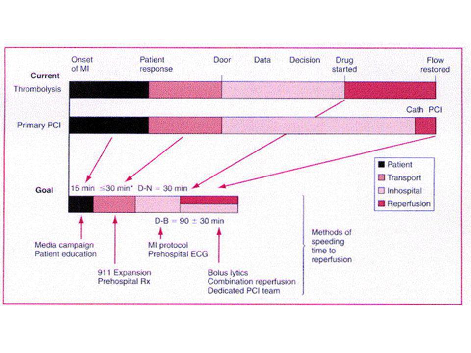
Time-Dependent Benefit of Reperfusion Therapy
Lecture Notes In the GISSI-I trial, patients who received fibrinolytic therapy within 1 hour of symptom onset had a 47% reduction in 21-day mortality compared with placebo; benefit declined as the interval increased. Initiation of treatment between 1 and 3 hours after symptom onset was associated with a mortality reduction of 14%. (GISSI, 1986) The benefit of therapy for patients treated beyond 2 hours is more modest but still significant, and may reflect the ability of the late reperfusion to prevent ventricular enlargement or improve electrophysiologic stability as well as salvage myocardium. The results of other controlled clinical trials of fibrinolytic therapy also support the dramatic relationship between early restoration of patency and myocardial salvage. Using data from the studies by Reimer et al, Bergmann et al, and the GISSI-I clinical trial, Tiefenbrunn and Sobel constructed a reperfusion time-benefit curve that depicts the time-dependent benefit of intervention for AMI. A nonlinear curve provided the best fit for the data. A similar curve was developed by Boersma and colleagues in their analysis of a data set of 22 randomized clinical trials of fibrinolytic therapy. (Reimer et al, 1977; Bergmann et al, 1982; Boersma et al, 1996; Tiefenbrunn and Sobel, 1992) Adapted from Tiefenbrunn AJ, Sobel BE. Circulation. 1992;85:
 The benefit of therapy for patients treated beyond 2 hours is more modest but still significant, and may reflect the ability of the late reperfusion to prevent ventricular enlargement or improve electrophysiologic stability as well as salvage myocardium. The results of other controlled clinical trials of fibrinolytic therapy also support the dramatic relationship between early restoration of patency and myocardial salvage. Using data from the studies by Reimer et al, Bergmann et al, and the GISSI-I clinical trial, Tiefenbrunn and Sobel constructed a reperfusion time-benefit curve that depicts the time-dependent benefit of intervention for AMI. A nonlinear curve provided the best fit for the data. A similar curve was developed by Boersma and colleagues in their analysis of a data set of 22 randomized clinical trials of fibrinolytic therapy. (Reimer et al, 1977; Bergmann et al, 1982; Boersma et al, 1996; Tiefenbrunn and Sobel, 1992) Adapted from Tiefenbrunn AJ, Sobel BE. Circulation. 1992;85:")
138
Trattamento Nitroderivati in infusione Acido acetilsalicilico Eparina
Betabloccanti Evitare i calcioantagonisti Possibilmente inserire al più presto Ace-Inibitori Statine Trattamento del dolore (anche oppioidi) Trattamento dell’insufficienza ventricolare sinistra
 Trattamento dell’insufficienza ventricolare sinistra.")
139
Prevenzione del rimodellamento
Limitare l’estensione della necrosi Rivascolarizzazione precoce! Trattamento dell’ischemia residua Pervietà dell’IRA (?) Trattamento medico ACE-inibitori Bloccanti del recettore dell’angiotensina II Beta-bloccanti Nitrati
 Trattamento medico. ACE-inibitori. Bloccanti del recettore dell’angiotensina II. Beta-bloccanti. Nitrati.")
140
Complicanze Shock cardiogeno Rottura di cuore
Mortalità 80% Rottura di cuore Mortalità 100% Insufficienza mitralica acuta Rottura del setto interventricolare Sindrome di Dressler (a distanza) Non confondere con angina precoce postinfartuale
 Non confondere con angina precoce postinfartuale.")
141
Cause di morte per infarto miocardico acuto
Shock cardiogeno Rottura di cuore Aritmie Oggi raro perché i pazienti sono ricoverati in UCIC Dopo rivascolarizzazione efficace non si verifica rottura di cuore

142
Dimissione Oggi circa in decima giornata
Valutazione del rischio complessivo Valutazione di: Funzione ventricolare Aritmie Ischemia residua Angina precoce postinfartuale

143
TIMI Risk Score for STEMI for predicting 30-day mortality
TIMI Risk Score for STEMI for predicting 30-day mortality. STE = ST elevation; LBBB = left bundle branch block; h/o = history of; HTN = hypertension.

144
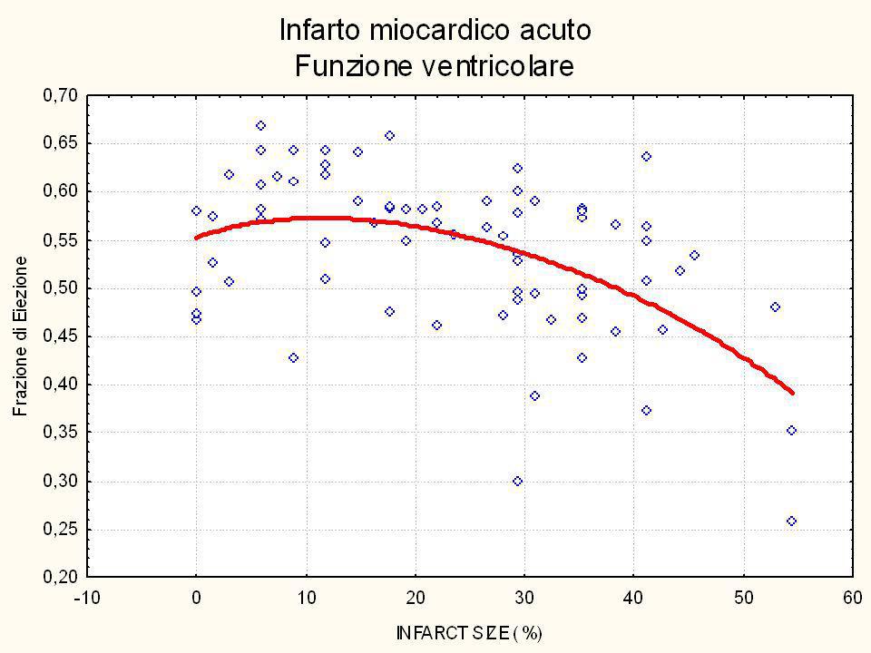
Impact of left ventricular function on survival after myocardial infarction. The curvilinear relationship between left ventricular ejection fraction (LVEF) for patients treated in the thrombolytic era is shown.
 for patients treated in the thrombolytic era is shown..")
Presentazioni simili


>")


>")


