Scaricare la presentazione
La presentazione è in caricamento. Aspetta per favore

1
Sindromi paraneoplastiche
Andrea M.Isidori Dipartimento di Fisiopatologia Medica Dir. Prof. Andrea Lenzi

2
Alterazioni funzionali degli organi interessati dal tumore primitivo
Effetti sistemici delle neoplasie Alterazioni funzionali degli organi interessati dal tumore primitivo Alterazioni funzionali degli organi interessati dalla crescita metastatica Effetti metabolici Effetti tossici Effetti conseguenti all’azione di mediatori noti ed ignoti (fattori di crescita, citokine, ormoni) prodotti dal tumore o derivanti dall’azione di autoAb prodotti dall’organismo contro le cellule tumorali Sindromi paraneoplastiche
 prodotti dal tumore o derivanti dall’azione di autoAb prodotti dall’organismo contro le cellule tumorali. Sindromi paraneoplastiche.")
3
Tumor Host Interactions
Local and Systemic Effects (primary site) Metastases (secondary site)( dedifferentation) Cancer Cachexia Paraneoplastic Syndromes Endocrinopathies Neuromyopathies Gastrointestinal motility syndromes Osteochondral Disorders Vascular Phenomena Fever Dermatological Syndromes
 Metastases (secondary site)( dedifferentation) Cancer Cachexia. Paraneoplastic Syndromes. Endocrinopathies. Neuromyopathies. Gastrointestinal motility syndromes. Osteochondral Disorders. Vascular Phenomena. Fever. Dermatological Syndromes.")
4
Paraneoplastic syndromes
Le Sindromi paraneoplastiche, per definizione, sono fenomeni legati all’interazione neoplasia-ospite, ma NON direttamente riconducibili ad effetti metastatici, compressivi, tossici, infettivi o metabolici tumorali. Sono importanti poiché: Si associano a severa morbilità e mortalità (es. Cushing’s) Sono spesso un “presenting symptom” di una neoplasia misconosciuta (early diagnosis) e un riconoscimento precoce spesso ottimizza le possibilità di intervento Rientrano nella diagnosi differenziale di sindromi comuni (ipercalcemia, iponatriemia, ipopotassiemia)
 Sono spesso un presenting symptom di una neoplasia misconosciuta (early diagnosis) e un riconoscimento precoce spesso ottimizza le possibilità di intervento. Rientrano nella diagnosi differenziale di sindromi comuni (ipercalcemia, iponatriemia, ipopotassiemia)")
5
Paraneoplastic syndromes
Le neoplasie più frequentemente associate a sindromi paraneoplastiche sono: Lung carcinoma (most common) Renal carcinoma Hepatocellular carcinoma Leukemias Lymphomas Breast tumors Ovarian tumors Neural cancers Gastric cancers Pancreatic cancers
 Renal carcinoma. Hepatocellular carcinoma. Leukemias. Lymphomas. Breast tumors. Ovarian tumors. Neural cancers. Gastric cancers. Pancreatic cancers.")
6
Cancer Cachexia Progressive weakness, loss of appetite, anemia and profound weight loss (>20 lbs.) Often correlates with tumor size and extent of metastases Etiology includes a generalized increase in metabolism and central effects of tumor on hypothalamus Probably related to macrophage production of TNF-a and IL-1

7
Endocrine syndromes Hematologic syndromes Cutaneous or dermatologic syndromes Neurologic syndromes Osteoarticular or rheumatologic syndromes Ocular syndromes

8
Sindromi paraneoplastiche
endocrine

9
I primi reports su sindromi endocrine in pazienti affetti da neoplasie maligne non endocrine risalgono agli anni ’20... Lancet 1928 A case of pluriglandular syndrome: diabetes of bearded women. Brown WH Surg Gynecol Obster 1923 Parathyroid hyperplasia and bone destruction in generelized carcinomatosis. Klemperer P

11
Paraneoplastic Endocrine Syndromes
Spectrum of paraneoplastic endocrine HYPERCALCEMIA HYPONATRIEMIA GYNECOMASTIA PTHrP, TGFs, ILs, Vit D, PTH Vasopressin, ANP HCG CUSHING’s ACROMEGALY ACTH, CRH Paraneoplastic Endocrine Syndromes GH, GHRH HYPOGLYCEMIA ONCOGENIC OSTEOMALACIA IGF II HYPERGLYCEMIA FGF23 HPL, PRL, VIP, Renin, LH, GRP Glucagone, GH, DIVERSE SYNTOMS

12
Demonstration of elevated hormone concentrations in the blood
Endocrine Pathology 2003 Paraneoplastic Endocrine Syndromes: A Review DeLellis RA, Xia L. “…A number of criteria have been proposed for the diagnosis of paraneoplastic endocrine syndromes… Demonstration of elevated hormone concentrations in the blood Finding of normal or suppressed endogenous hormone production Demonstration of hormone concentration gradients across the tumor Biochemical or clinical resolution of the syndrome following surgery, radiotherapy or chemotherapy Demonstration of hormone messenger RNA and corresponding hormonal product in tumoral cells

13
Meccanismi patogenetici
Endocrine Pathology 2003 Paraneoplastic Endocrine Syndromes: A Review DeLellis RA, Xia L. Meccanismi patogenetici Theory of randon genetic derepression Attivazione di geni normalmente inattivi per effetto di mutazioni o modificazioni epigenetiche Dedifferentiation Theory Regressione delle cellule tumorali ad uno stato maturativo precoce con produzione di proteine e ormoni fetali ed embrionali (ex. PTHrP)
")
14
Paraneoplastic Syndromes Endocrinopathies
Hypercalcemia (Cancer is the most common cause of hypercalcemia by either humoral or metastatic mechanisms) Squamous cell lung cancer (PTH-like peptide) Multiple myeloma and T-cell lymphoma (IL-1 and perhaps TGF-a) Renal cell carcinoma (prostaglandins) Breast (& Prostate) carcinoma, usually by bone metastasis Parathyroid carcinoma (PTH)
 Squamous cell lung cancer (PTH-like peptide) Multiple myeloma and T-cell lymphoma (IL-1 and perhaps TGF-a) Renal cell carcinoma (prostaglandins) Breast (& Prostate) carcinoma, usually by bone metastasis. Parathyroid carcinoma (PTH)")
15
Paraneoplastic Syndromes Endocrinopathies
Inappropriate ADH syndrome (Hyponatremia) Small cell undifferentiated lung cancer (vassopressin-like hormone. Hypothalamic tumors (vasopressin) Cushing’s Syndrome Small cell undifferentiated lung cancer (ACTH) released through cleavage of pro-opiomelano-cortin gene product. MTC, Thymoma, Ovarian Cancer, Mesothelioma
 Small cell undifferentiated lung cancer (vassopressin-like hormone. Hypothalamic tumors (vasopressin) Cushing’s Syndrome. Small cell undifferentiated lung cancer (ACTH) released through cleavage of pro-opiomelano-cortin gene product. MTC, Thymoma, Ovarian Cancer, Mesothelioma.")
16
Ipercalcemia 0,1% della popolazione generale
fino a 3-5% >50aa Fino al 58% dei pz adulti oncologici ospedalizzati 0,5-1,3% in età pediatrica <5% tumori maligni tratto genitale femminile Mieloma, K squamoso del polmone (quasi nel 100% dei casi), K mammario, k renale, k del tratto genitale femminile, Linfoma HTLV, Am J Med 1997 Hypercalcemia of Malignancy Mundy G, Guise TA
, K mammario, k renale, k del tratto genitale femminile, Linfoma HTLV, Am J Med Hypercalcemia of Malignancy. Mundy G, Guise TA.")
17
Hypercalcemia Primary hyperparathyroidism 23% Paraneoplastic syndrome
Metastasis % (lung, breast cancer, multiple myeloma) Granulomatous desease (tuberculosis, sarcoidosis) Genetic disorders (Familial hypocalciuric hypercalcemia) Long immobilization Medications (lithium, thiazide diuretics, supplements) Dehydration Hyperparathyroidism and cancer are responsible for more than 90% of sustained hypercalcemia.
 Granulomatous desease (tuberculosis, sarcoidosis) Genetic disorders (Familial hypocalciuric hypercalcemia) Long immobilization. Medications (lithium, thiazide diuretics, supplements) Dehydration. Hyperparathyroidism and cancer are responsible for more than 90% of sustained hypercalcemia.")
18
Assenza di metastasi ossee PTH ridotto, PTHrP aumentato
Ipercalcemia osteolitica localizzata (Localized Osteolytic Hypercalcemia -LOH-): da produzione locale di fattori paracrini, quali citochine (IL-6, TGFa e b, TNFa), prostaglandina E e metaboliti della Vitamina D, con effetto stimolatorio sugli osteoclasti Ipercalcemia Maligna (Humoral Hypercalcemia of Malingnancy -HHM-): da produzione di PTH-RP (PTH-related peptide) o più raramente di PTH Ipercalcemia Assenza di metastasi ossee PTH ridotto, PTHrP aumentato
: da produzione locale di fattori paracrini, quali citochine (IL-6, TGFa e b, TNFa), prostaglandina E e metaboliti della Vitamina D, con effetto stimolatorio sugli osteoclasti. Ipercalcemia Maligna (Humoral Hypercalcemia of Malingnancy -HHM-): da produzione di PTH-RP (PTH-related peptide) o più raramente di PTH. Ipercalcemia. Assenza di metastasi ossee. PTH ridotto, PTHrP aumentato.")
19
New Engl J Med 1988 New Engl J Med 2000 Cancer 1991
Humoral Hypercalcemia of cancer. Identification of a novel parathyroid hormone-like peptide Broadus AE, Mangin M, Ikeda K, Insogna KL, Weir EC, Burtis WJ, Stewart AF New Engl J Med 2000 The physiology of parathyroid hormone related protein Strewler GJ Il PTHrP viene individuato nella seconda metà degli anni ’80, espresso in innumerevoli tessuti normali (es. endometrio, placenta, miometrio e decidua durante la gravidanza) 3 ISOFORME La porzione NH2-terminale è simile a quella del PTH e determina simili effetti biologici, mentre il resto della molecola possiede altre funzioni (es. regolazione proliferazione cellulare/apoptosi) Cancer 1991 Immunohistochemical evaluation of PTHrP in human lung cancer and normal tissue with newly developed monoclonal antibody Kitazawa S et al Dosabile con metodo RIA (kit specifici per la porzione C-terminale), PCR, IRMA
 3 ISOFORME. La porzione NH2-terminale è simile a quella del PTH e determina simili effetti biologici, mentre il resto della molecola possiede altre funzioni (es. regolazione proliferazione cellulare/apoptosi) Cancer Immunohistochemical evaluation of PTHrP in human lung cancer and normal tissue with newly developed monoclonal antibody. Kitazawa S et al. Dosabile con metodo RIA (kit specifici per la porzione C-terminale), PCR, IRMA.")
20
PTHrP nelle lesioni tumorali BENIGNE…
Am J Clin Pathol 1996 The Humoral hypercalcemia of benignancy. A newly appreciated syndrome Knecht TP et al PTHrP nelle lesioni tumorali BENIGNE… It has become recently appreciated that the hypercalcemia of malignancy is commonly caused by the increased production of parathyroid hormone-related protein (PTHrP) by the cancer. In fact, the demonstration of increased PTHrP production in a patient with hypercalcemia is regarded as pathognomonic of malignancy. The authors describe a patient with a benign ovarian lesion that produced PTHrP and caused hypercalcemia. They identify other reports of hypercalcemia associated with hypercalcemia and benign tumors, and refer to this syndrome as the humoral hypercalcemia of benignancy. Although apparently rare, a benign PTHrP-producing tumor should be considered in the differential diagnosis of hypercalcemia.
 by the cancer. In fact, the demonstration of increased PTHrP production in a patient with hypercalcemia is regarded as pathognomonic of malignancy. The authors describe a patient with a benign ovarian lesion that produced PTHrP and caused hypercalcemia. They identify other reports of hypercalcemia associated with hypercalcemia and benign tumors, and refer to this syndrome as the humoral hypercalcemia of benignancy. Although apparently rare, a benign PTHrP-producing tumor should be considered in the differential diagnosis of hypercalcemia.")
21
IPERCALCEMIA NEOPLASTICA
Nel 10-20% dei pz oncologici si presenta come emergenza metabolica Emergenza per Ca >14 mg/dl (3.5 mmol/L) Gravità dei sintomi correlata alla velocità di aumento del calcio ionizzato e alla sua concentrazione, alle condizioni generali del e malattie concomitanti LIVELLI CORRETTI DI CALCIO SIERICO Calcio misurato + [ 0.8 x (4.0 – albumina sierica) ] Trattamento Idratazione: NaCl ev 0,25-1 L/h (controllare PVC) Diuretici dell’ansa (furosemide): mg ev ogni 2-4h (controllo elettroliti) Bisfosfonati ev Calcitonina sc/ev 4-8 U/kg ogni 6-12h Corticosteroidi (cortone acetato-prednisone) Emodialisi
 Gravità dei sintomi correlata alla velocità di aumento del calcio ionizzato e alla sua concentrazione, alle condizioni generali del e malattie concomitanti. LIVELLI CORRETTI DI CALCIO SIERICO. Calcio misurato + [ 0.8 x (4.0 – albumina sierica) ] Trattamento. Idratazione: NaCl ev 0,25-1 L/h (controllare PVC) Diuretici dell’ansa (furosemide): mg ev ogni 2-4h (controllo elettroliti) Bisfosfonati ev. Calcitonina sc/ev 4-8 U/kg ogni 6-12h. Corticosteroidi (cortone acetato-prednisone) Emodialisi.")
22
Paraneoplastic SIADH La Seconda più frequente S. Paraneoplastica
Microcitoma polmonare (60%), NET, k tratto urogenitale Iposodiemia (<130 mmol/L) / Sodiuria >20 mEq/L Osmo urin > 100 mOsm/L / Osm plasma < 260 mOsm/L Ann NY Acad Sci 1992 Oxytocin and vasopressin: from genes to peptide Gainer H, Wray S J Intern Med 1995 Syndrome of Inappropriate secretion of antidiuretic hormone (SIADH) in malignant diseases Sorensen JB, Anderson MD, Hansen HH
, NET, k tratto urogenitale. Iposodiemia (<130 mmol/L) / Sodiuria >20 mEq/L. Osmo urin > 100 mOsm/L / Osm plasma < 260 mOsm/L. Ann NY Acad Sci Oxytocin and vasopressin: from genes to peptide. Gainer H, Wray S. J Intern Med Syndrome of Inappropriate secretion of antidiuretic hormone (SIADH) in malignant diseases. Sorensen JB, Anderson MD, Hansen HH.")
23
Causes of hyponatraemia
Urinary Na<20 mmol/l Urinary Na>40 mmol/l GI losses Diuretics Mucosal losses Pancreatitis Sodium depletion post diuretics Diuretics Addison’s disease Cerebral salt wasting Salt wasting nephropathy Hypovolaemic 30-40% Hypothyroidism SIADH with ongoing fluid restriction Primary polydipsia Inappropriate fluid replacement SIADH ACTH deficiency Euvolaemic SIADH rappresenta il 30-40% di tutte le iponatriemie Cardiac failure or cirrhosis on diuretic therapy Cirrhosis Cardiac failure Nephrotic syndrome Hypervolaemic

24
Intracranial pathology
Small cell lung cancer 75% dei casi Nasopharyngeal cancer Mesothelioma GI tract malignancy Pancreatic malignancy GU tract malignancy Lymphoma Sarcoma Pulmonary Malignancy Pneumonia, especially Legionella Mycoplasma Tuberculosis Abscess Vasculitis Positive pressure ventilation Desmopressin Selective serotonin reuptake inhibitors Carbamazepine Prostaglandins Tricyclic antidepressants Phenothiazines Haloperidol 3,4-Methylenedioxymethamphetamine Quinolones Leveteiracetam Cyclophosphamide Vincristine Intracranial pathology Tumour Meningitis Encephalitis Abscess Vasculitis Subarachnoid haemorrhage Subdural haemorrhage Traumatic brain injury Drugs

25
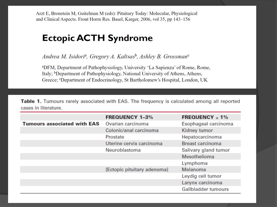
Between 1964 and 2002: 413 patients with CS were investigated 60 had an adrenal adenoma, 30 had an adrenal carcinoma, 5 had macronodular adrenal hyperplasia, 274 of pituitary origin (CD) 44 from an ectopic source of ACTH
 44 from an ectopic source of ACTH.")
26
A COMPARISION BETWEEN THE TWO LARGEST SERIES ON ECTOPIC ACTH SYNDROME
EUROPE (UK) vs. USA (NIH) (n=40) (n=90) Median follow-up m m Are there regional differences in the ectopic ACTH syndrome in different parts of the developed world in tertiary referral centres?
 vs. USA (NIH) (n=40) (n=90) Median. follow-up 60m 26m. Are there regional differences in the ectopic ACTH syndrome in different parts of the developed world in tertiary referral centres")
27
ECTOPIC ACTH SYNDROME 4 patients had markedly fluctuant levels of ACTH – cyclical Cushing’s syndrome 1 pancreatic carcinoid 1 thymic carcinoid 1 bronchial carcinoid 1 occult source

28
St. Bartholomew’s NIH LUNG 47.5% (major organ) CARCINOID 30%
SCLC 17.5% Intrathoracic in general 55% OCCULT 12.5% LUNG 42.2% (major organ) CARCINOID 38% SCLC 3% Tumorlets 0.9% Intrathoracic in general 52% OCCULT 19%
 CARCINOID 38% SCLC 3% Tumorlets 0.9% Intrathoracic in general 52% OCCULT 19%")
29
ECTOPIC ACTH SYNDROME 40 patients 26 revealed on imaging (overt)
14 not apparent 9 became apparent (covert) 5 remained hidden (occult) Barts experience: (Isidori et al., 2005)
 5 remained hidden (occult) Barts experience: (Isidori et al., 2005)")
30
COVERT ECTOPIC ACTH SYNDROME
Of 9 tumours not initially identified: Revealed by CT 4 Revealed by whole-body catheter * 2 Found at surgery/autopsy 3 USING MODERN CROSS-SECTIONAL IMAGING VIRTUALLY ALL ECTOPICS WHICH CAN BE FOUND WILL BE FOUND *before high-quality CT (Isidori et al., 2005)
")
31
St. Bartholomew’s NIH Mean ACTH levels: Mean ACTH levels:
Overt 207 ng/l Covert 125 ng/l Mean Cortisol levels Overt nmol/l Covert nmol/l Mean K+ levels Overt 2.7 mmol/l Covert mmol/l Hypokalaemia in 70% Mean ACTH levels: Overt 205 ng/l Covert 109 ng/l Mean UFC Overt nmol/24h Covert nmol/24h Mean K+ levels Overt 3.4 mmol/l Covert 3.5 mmol/l Hypokalaemia in 74%

32
ECTOPIC ACTH SYNDROME Dynamic Stimulation Tests
High-dose dexamethasone suppression 91% show absent suppression (>50%) CRH stimulation test 94% show absent rise (>20%) One patient showed a response to both tests (1/40=2%)
 CRH stimulation test. 94% show absent rise (>20%) One patient showed a response to both tests (1/40=2%)")
33
ECTOPIC ACTH SYNDROME: NIH experience
Dynamic Stimulation Tests High-dose dexamethasone suppression 86% show absent suppression (UFC) CRH stimulation test 92% show absent rise (>20%)
 CRH stimulation test. 92% show absent rise (>20%)")
34
ECTOPIC ACTH SYNDROME BILATERAL INFERIOR PETROSAL SINUS SAMPLING
1/12 patients showed a central gradient >3 (mesothelioma) At NIH, 1/67 patients showed a central gradient (esthesioneuroblastoma) Therefore, false positive responses in 2/79 (~2%)
 At NIH, 1/67 patients showed a central gradient (esthesioneuroblastoma) Therefore, false positive responses in 2/79 (~2%)")
35
ECTOPIC ACTH SYNDROME TUMOUR MARKERS At NIH 28% show raised gastrin
28% show raised calcitonin 10% show raised urinary 5-HIAA At NIH 31% show raised calcitonin 30% show raised 5-HIAA

36
ECTOPIC ACTH SYNDROME WHOLE BODY VENOUS CATHETER STUDIES 4/22 WERE POSITIVE 2 thymic carcinoids 1 mediastinal lymph node 1 medullary thyroid carcinoma BUT THESE WERE ALL STUDIED BEFORE HIGH-RESOLUTION CT SCANNING

37
ECTOPIC ACTH SYNDROME IMAGING CT LOCALISED TUMOUR IN 82% (NIH=92%)
111In-octreotide localised tumor in 2/8 (25%) At NIH, 21/43 (49%) were positive BUT IT VERY RARELY IDENTIFIES TUMOURS NOT OTHERWISE SEEN!
 At NIH, 21/43 (49%) were positive. BUT IT VERY RARELY IDENTIFIES TUMOURS NOT OTHERWISE SEEN!")
38
ECTOPIC ACTH SYNDROME One patient needed intravenous etomidate
TREATMENT 28/40 treated with steroidogenesis inhibitors for median 9 months Metyrapone Ketoconazole Mitotane One patient needed intravenous etomidate

39
ECTOPIC ACTH SYNDROME 12 patients had primary resection, 10 curative
TREATMENT 12 patients had primary resection, 10 curative 12 patients had bilateral adrenalectomy 14 patients received radiotherapy 11 patients received chemotherapy 2 patients received 131I-MIBG therapy CONCLUSION – Control cortisol excess, remove tumour where possible, consider removing adrenals where not

40
Kaplan-Meier survival curve for ectopic ACTH patients
0.00 0.25 0.50 0.75 1.00 100 200 300 Survival (months) Small cell NET mets NET (Isidori et al., 2005)
 Small cell. NET mets. NET. (Isidori et al., 2005)")
41
Prevalence of Tumours responsible of EAS
60 Percentage (%) Total n=383 Thoracic Tumours 50 40 30 Abdominal Tumours 20 10 GI carcinoids Never-found Thymic tumours Islet Cell Tumours Localized NET Medullary Thyroid K Lung/Mediast. Carcinoids Lung SCLC / Adenok Pheochromocytomas Disseminated NET GI adenocarcinomas Miscellaneous Tumours
 Total n=383. Thoracic Tumours Abdominal Tumours GI carcinoids. Never-found. Thymic tumours. Islet Cell Tumours. Localized NET. Medullary Thyroid K. Lung/Mediast. Carcinoids. Lung SCLC / Adenok. Pheochromocytomas. Disseminated NET. GI adenocarcinomas. Miscellaneous Tumours.")
43
Paraneoplastic Syndromes Endocrinopathies
WDHA syndrome (watery diarrhea, hypokalemia, and achlorhydria) - caused by tumor production of vasoactive intestinal polypeptide (VIP). Islet cell tumors, Intestinal carcinoid tumors Polycythemia - caused by tumor production of erythropoietins Renal cell carcinoma, Cerebellar hemangioma, Hepatocarcinoma
 - caused by tumor production of vasoactive intestinal polypeptide (VIP). Islet cell tumors, Intestinal carcinoid tumors. Polycythemia - caused by tumor production of erythropoietins. Renal cell carcinoma, Cerebellar hemangioma, Hepatocarcinoma.")
44
Sindromi paraneoplastiche
Gastro-motorie

45
Paraneoplastic GI dismotility syndromes
A small proportion of patients with occult or established neoplasms develop a gastrointestinal motility disorder, referred to as paraneoplastic dysmotility. The diagnosis of a paraneoplastic dysmotility requires the onset of gastrointestinal dysmotility associated with the presence of a tumor and presence of specific serum antibodies Kashyap P and Farrugia G, Gastroenterol Clin North Am. 2008

46
Clinical presentation of a para-neoplastic dysmotility syndrome
Pseudoachalasia Gastroparesis Paraneoplastic chronic intestinal pseudoobstruction (CIPO) Chronic constipation The most common neuronal autoantibody associated with a paraneoplastic dysmotility is the type 1 antineuronal nuclear antibody (ANNA-1) [1,2]. ANNA-1 recognizes the nuclear protein Hu which belongs to a family of conserved RNA binding proteins that includes HuC, HuD, HuR and Hel-N1. are expressed in the neurons of the central, peripheral and enteric nervous system, with the exception of HuR which is ubiquitously expressed in proliferating cells [3]. The tumor that most commonly expresses ANNA-1 is small cell lung cancer [4]. Other tumors that may express ANNA-1 include breast, prostate, ovarian carcinomas and lymphomas [5]. Antibodies to ANNA-1 are consequently, most commonly found in patients with small cell lung cancer with associated paraneoplastic gastrointestinal dysmotility. Although there is a very strong association between the presence of ANNA-1 in the setting of a gastrointestinal motility disorder and the presence of an occult or manifest tumor, the exact mechanism by which ANNA-1 antibodies cause enteric neuronal dysfunction is still unclear as the proteins to which the antibody is directed are not expressed on the cell membrane. However, there is some evidence that the antibodies may directly influence motility. A preliminary study in guinea pig ileum suggested that anti-Hu antibodies impair the ascending excitatory reflex and therefore peristalsis. Enteric neuronal degeneration has also been reported in patients with paraneoplastic dysmotility as a possible pathogenetic mechanism Anti HuD positive sera from patients with paraneoplastic gut dysmotility disorder as well as commercial Anti HuD antibodies were shown to induce apoptosis in a human neuroblastoma cell line (SH-Sy5Y) as well as guinea pig cultured myenteric neurons. Pardi et al described a patient with sudden onset of gastroparesis and small bowel dysfunction and the presence of high circulating levels of ANNA-1 [6]. This patient was subsequently found to have decreased and disorganized interstitial cells of Cajal networks and a small cell lung cancer expressing c-Kit, also expressed on interstitial cells of Cajal. Calcium channels were originally classified based on pharmacology as L, N, P/Q, R, and T channels, a classification still used today. This nomenclature corresponds to the current accepted nomenclature that classifies voltage-gated Ca2+ channels into Cav1.1-Cav1.4 (L-type Ca2+ channels), Cav2.1 (P/Q), Cav2.2 (N), Cav2.3 (R), and Cav3.1- Cav3.3 (T) based on the amino acid sequence of the alpha 1 subunit (the pore forming subunit) of the channel. P or Q type calcium ion channels regulate acetylcholine release at the neuromuscular junction as well as central neurotransmission. N type calcium channels are particularly involved in cerebrocortical, cerebellar, spinal and autonomic neurotransmission. Both channel types are expressed in small cell lung cancer and are common targets of autoantibodies in such patients. These antibodies are predominantly seen in patients with Lambert Eaton myasthenic syndrome in association with small cell lung cancer [12]. Lambert-Eaton myasthenic syndrome (LEMS) is a rare disorder of neuromuscular transmission. It is a presynaptic disorder of neuromuscular transmission in which quantal release of acetylcholine (ACh) is impaired, causing a unique set of clinical characteristics, which include proximal muscle weakness, depressed tendon reflexes, posttetanic potentiation, and autonomic changes. The initial presentation can be similar to that of myasthenia gravis, but the progressions of the two diseases have some important differences. Another class of autoantibodies associated with gastrointestinal dysmotility is antibodies against neuronal nicotinic acetylcholine receptors. Antibodies directed towards the extracellular segment of acetylcholine receptor protein in the post synaptic membrane of skeletal muscle are found in patients with myasthenia gravis associated with thymic epithelial tumors [13]. Neuronal nicotinic acetylcholine receptors are also present on neurons in the sympathetic and parasympathetic nervous systems as well as the enteric nervous system. Antibodies targeting this protein can disrupt cholinergic synaptic transmission leading to autonomic failure. These antibodies are seen in both idiopathic and paraneoplastic forms of autonomic neuropathy resulting in autoimmune autonomic neuropathy [14]. Mice injected with rabbit IgG containing ganglionic acetylcholine receptor antibodies develop gastrointestinal dysmotility and autonomic dysfunction. Similar results are obtained by injecting mice with sera from patients with ganglionic acetylcholine receptor antibody. Purkinje Cell Cytoplasmic Autoantibody, type 1 (PCA1) This autoantibody (sometimes called “anti-Yo”) targets a neuronal signal transduction protein Cdr. The antibody was originally defined as a marker of paraneoplastic cerebellar degeneration related to ovarian or breast carcinoma with remarkably limited metastasis [17,18]. In vitro, its Cdr antigen, a prominent cytoplasmic component of large neurons in the central and autonomic/ enteric nervous systems [19], has been shown to promote neuronal apoptosis and degeneration by inhibiting c-myc transcriptional activity [20]. Paraneoplastic gastrointestinal dysmotility has been documented in a minority of PCA-1 seropositive patients (with and without cerebellar ataxia) in association with gynecological or breast carcinoma [21]. Kashyap P and Farrugia G, Gastroenterol Clin North Am. 2008
 Chronic constipation. The most common neuronal autoantibody associated with a paraneoplastic dysmotility is the type 1 antineuronal nuclear antibody (ANNA-1) [1,2]. ANNA-1 recognizes the nuclear protein Hu which belongs to a family of conserved RNA binding proteins that includes HuC, HuD, HuR and Hel-N1. are expressed in the neurons of the central, peripheral and enteric nervous system, with the exception of HuR which is ubiquitously expressed in proliferating cells [3]. The tumor that most commonly expresses ANNA-1 is small cell lung cancer [4]. Other tumors that may. express ANNA-1 include breast, prostate, ovarian carcinomas and lymphomas [5]. Antibodies to ANNA-1 are consequently, most commonly found in patients with small cell lung cancer with associated paraneoplastic gastrointestinal dysmotility. Although there is a very strong association between the presence of ANNA-1 in the setting of a gastrointestinal motility disorder and the presence of an occult or manifest tumor, the exact mechanism by which ANNA-1 antibodies cause enteric neuronal dysfunction is still unclear as the proteins to which the antibody is directed are not expressed on the cell membrane. However, there is some evidence that the antibodies may directly influence motility. A preliminary study in guinea pig ileum suggested that anti-Hu antibodies impair the ascending excitatory reflex and therefore peristalsis. Enteric neuronal degeneration has also been reported in patients with paraneoplastic dysmotility as a possible pathogenetic mechanism Anti HuD positive sera from patients. with paraneoplastic gut dysmotility disorder as well as commercial Anti HuD antibodies were shown to induce apoptosis in a human neuroblastoma cell line (SH-Sy5Y) as well as guinea pig cultured myenteric neurons. Pardi et al described a patient with sudden onset of gastroparesis and small bowel dysfunction and the presence of high circulating levels of ANNA-1 [6]. This patient was subsequently found to have decreased and disorganized interstitial cells of Cajal networks and a small cell lung cancer expressing c-Kit, also expressed on interstitial cells of Cajal. Calcium channels were originally classified based on pharmacology as L, N, P/Q, R, and T channels, a classification still used today. This. nomenclature corresponds to the current accepted nomenclature that classifies voltage-gated Ca2+ channels into Cav1.1-Cav1.4 (L-type Ca2+ channels), Cav2.1 (P/Q), Cav2.2 (N), Cav2.3 (R), and Cav3.1- Cav3.3 (T) based on the amino acid sequence of the alpha 1 subunit (the pore. forming subunit) of the channel. P or Q type calcium ion channels regulate acetylcholine release at the neuromuscular junction as well as central neurotransmission. N type calcium channels are particularly involved in cerebrocortical, cerebellar, spinal and autonomic neurotransmission. Both channel types are expressed in small cell lung cancer and are common targets of autoantibodies in such patients. These antibodies are predominantly seen in patients with Lambert Eaton myasthenic syndrome in association with small cell lung cancer [12]. Lambert-Eaton myasthenic syndrome (LEMS) is a rare disorder of neuromuscular transmission. It is a presynaptic disorder of neuromuscular transmission in which quantal release of acetylcholine (ACh) is impaired, causing a unique set of clinical characteristics, which include proximal muscle weakness, depressed tendon reflexes, posttetanic potentiation, and autonomic changes. The initial presentation can be similar to that of myasthenia gravis, but the progressions of the two diseases have some important differences. Another class of autoantibodies associated with gastrointestinal dysmotility is antibodies against neuronal nicotinic acetylcholine receptors. Antibodies directed towards the extracellular segment of acetylcholine receptor protein in the post synaptic membrane of skeletal muscle are found in patients with myasthenia gravis associated with thymic epithelial tumors [13]. Neuronal nicotinic acetylcholine receptors are also present on neurons in the sympathetic and parasympathetic nervous systems as well as the enteric nervous system. Antibodies targeting this protein can disrupt cholinergic synaptic transmission leading to autonomic failure. These antibodies are seen in both idiopathic and paraneoplastic forms of autonomic neuropathy resulting in autoimmune autonomic neuropathy [14]. Mice injected with rabbit IgG containing ganglionic acetylcholine receptor antibodies develop gastrointestinal dysmotility and autonomic dysfunction. Similar results are obtained by injecting mice with sera from patients with ganglionic acetylcholine receptor antibody. Purkinje Cell Cytoplasmic Autoantibody, type 1 (PCA1) This autoantibody (sometimes called anti-Yo ) targets a neuronal signal transduction protein Cdr. The antibody was originally defined as a marker of paraneoplastic cerebellar degeneration related to ovarian or breast carcinoma with remarkably limited metastasis [17,18]. In vitro, its Cdr antigen, a prominent cytoplasmic component of large neurons in the central and autonomic/ enteric nervous systems [19], has been shown to promote neuronal apoptosis and degeneration by inhibiting c-myc transcriptional activity [20]. Paraneoplastic gastrointestinal dysmotility has been documented in a minority of PCA-1 seropositive patients (with and without cerebellar ataxia) in association with gynecological or breast carcinoma [21]. Kashyap P and Farrugia G, Gastroenterol Clin North Am")
47
autonomic paraneoplastic neurological Hu-related syndromes
A possible autoimmune model for autonomic paraneoplastic neurological Hu-related syndromes. In the lymph nodes, the apoptotic tumour cell is phagocytosed by antigen-presenting dendritic cells, and the tumour antigens are processed and presented to circulating CD8+ T lymphocytes (CTL) via the major histocompatibility complex (MHC) class I pathway. Activated CTL cross the blood brain barrier (BBB) and, together with inflammatory cells, induce neuronal apoptosis through cell-mediated interactions. In addition, plasma cells may cross the BBB or differentiate within the nervous system (activated through cooperation between B cells and T helper CD4+ cells) and produce antibodies (Ab) directed against tumour Hu antigens that crossreact with neuronal antigens with similar epitopes (Hu neuronal proteins). These onconeural antibodies are able to infiltrate neurones by active uptake and bind to intranuclear Hu proteins. Essential processes for cell survival are, thus, compromised and apoptotic neurone death is triggered. Dying or dead neurones themselves can be taken up by neighbouring APCs and a positive feedback process initiated, which contributes to epitope spreading. autonomic paraneoplastic neurological Hu-related syndromes Autoimmunity Reviews 6 (2007) 162–168
 via the. major histocompatibility complex (MHC) class I pathway. Activated CTL cross the blood brain barrier (BBB) and, together with inflammatory cells, induce. neuronal apoptosis through cell-mediated interactions. In addition, plasma cells may cross the BBB or differentiate within the nervous system (activated. through cooperation between B cells and T helper CD4+ cells) and produce antibodies (Ab) directed against tumour Hu antigens that crossreact with. neuronal antigens with similar epitopes (Hu neuronal proteins). These onconeural antibodies are able to infiltrate neurones by active uptake and bind to. intranuclear Hu proteins. Essential processes for cell survival are, thus, compromised and apoptotic neurone death is triggered. Dying or dead neurones. themselves can be taken up by neighbouring APCs and a positive feedback process initiated, which contributes to epitope spreading. autonomic paraneoplastic neurological Hu-related syndromes. Autoimmunity Reviews 6 (2007) 162–168.")
48
Treatment of paraneoplastic dysmotility
No treatments have been convincingly shown to alter outcome (steroids, cyclophosphamide, plasmapheresis, immunoglobulin) Treatment of the underlying primary malignancy Nutritional support either enterally or parenterally Prokinetics, treatment of bacterial overgrowth One additional management strategy is to use high dose IV steroids for 3 days and if there is a clinical response switch to 6-mercatopurine or azathioprine (difficult in the case of chemotherapy) Kashyap P and Farrugia G, Gastroenterol Clin North Am. 2008
 Treatment of the underlying primary malignancy. Nutritional support either enterally or parenterally. Prokinetics, treatment of bacterial overgrowth. One additional management strategy is to use high dose IV steroids for 3 days and if there is a clinical response switch to 6-mercatopurine or azathioprine (difficult in the case of chemotherapy) Kashyap P and Farrugia G, Gastroenterol Clin North Am")
50
Pemfigo Acanthosis nigricans Malattia di Paget
Da autoanticorpi contro la desmoplakina I, proteina dei desmosomi delle cellule epiteliali. Le lesioni bollose pemfigoidi sono conseguenza della perdita della normali adesioni intercellulari a livello dell’epidermide. Linfomi, timoma, sarcomi ed altre neoplasie, soprattutto ematologiche Acanthosis nigricans Iperpigmentazione vellutata, di colore marrone scuro o nero, a livello di ascelle, aree sottomammarie e pieghe inguinali Soprattutto K gastrico. Malattia di Paget Placca eritematosa, simile ad un eczema Quando localizzata a livello delle areole mammarie è quasi sempre associata a K duttale della mammella, mentre la malattia di Paget extramammaria si associa in circa il 50% dei casi a neoplasie genitali.

51
Dermatomiosite Ittiosi Snd di Leser-Trèlat Snd di Sweet
Miopatia infiammatoria associata ad un rash cutaneo violaceo, più evidente nelle aree esposte al sole, edema ed eritema periorbitale, placche eritematose a livello delle articolazioni metacarpofalangee e interfalangee prossimali (papule di Gottron) K polmone, stomaco, utero, ovaio Ittiosi Associata ai linfomi di Hodgkin Placche cutanee a scaglie Snd di Leser-Trèlat Comparsa improvvisa o aumento in numero e dimensioni di cheratosi seborroica Neoplasie gastrointestinali Snd di Sweet Dermatosi neutrofila febbrile acuta (febbre, leucocitosi neutrofila, placche o noduli eritematosi a livello di testa, collo e arti superiori In particolare in corso di leucemia acuta mieloblastica, sindromi mielodisplastiche e malattie mieloproliferative.
 K polmone, stomaco, utero, ovaio. Ittiosi. Associata ai linfomi di Hodgkin. Placche cutanee a scaglie. Snd di Leser-Trèlat. Comparsa improvvisa o aumento in numero e. dimensioni di cheratosi seborroica. Neoplasie gastrointestinali. Snd di Sweet. Dermatosi neutrofila febbrile acuta (febbre, leucocitosi neutrofila, placche o noduli eritematosi a livello di testa, collo e arti superiori. In particolare in corso di leucemia acuta mieloblastica, sindromi mielodisplastiche e malattie mieloproliferative.")
52
Paraneoplastic Syndromes
Think about it…

Presentazioni simili


>")


