Scaricare la presentazione
La presentazione è in caricamento. Aspetta per favore

2
Mechanisms of antisense action on target genes
Mechanisms of antisense action on target genes. Several mechanisms of antisense action have been determined for antisense drugs (shown in green). These include inhibition of transcription (a), inhibition of splicing and inhibition of mRNA maturation by prevention of 5'-cap formation and of polyadenylation (b), and inhibition of ribosomal readthrough (c). Most importantly, antisense drugs hybridized with target RNA have been shown to serve as substrates for RNase H enzymes (shown in red; b,d) that cleave the RNA in DNA–RNA hybrids or RNA–RNA hybrids (as in RNAi) and decrease mRNA levels of the target gene. So, there is a growing repertoire of potential mechanisms of
. These include inhibition of transcription (a), inhibition of splicing and inhibition of mRNA maturation by prevention of 5 -cap formation and of polyadenylation (b), and inhibition of ribosomal readthrough (c). Most importantly, antisense drugs hybridized with target RNA have been shown to serve as substrates for RNase H enzymes (shown in red; b,d) that cleave the RNA in DNA–RNA hybrids or RNA–RNA hybrids (as in RNAi) and decrease mRNA levels of the target gene. So, there is a growing repertoire of potential mechanisms of.")
3
Triple-helix formation at the nucleotide level
Triple-helix formation at the nucleotide level. Shows the formation of Watson–Crick (red) and Hoogsteen bonds (black) between duplex pairs and the third strand (the arrow points to a single base of the third strand). Blue, guanine residue (purine); pink, cytosine residue (pyrimidine).
 and Hoogsteen bonds (black) between duplex pairs and the third strand (the arrow points to a single base of the third strand). Blue, guanine residue (purine); pink, cytosine residue (pyrimidine).")
4
Mechanisms of antisense action on target genes
Mechanisms of antisense action on target genes. Several mechanisms of antisense action have been determined for antisense drugs (shown in green). These include inhibition of transcription (a), inhibition of splicing and inhibition of mRNA maturation by prevention of 5'-cap formation and of polyadenylation (b), and inhibition of ribosomal readthrough (c). Most importantly, antisense drugs hybridized with target RNA have been shown to serve as substrates for RNase H enzymes (shown in red; b,d) that cleave the RNA in DNA–RNA hybrids or RNA–RNA hybrids (as in RNAi) and decrease mRNA levels of the target gene. So, there is a growing repertoire of potential mechanisms of
. These include inhibition of transcription (a), inhibition of splicing and inhibition of mRNA maturation by prevention of 5 -cap formation and of polyadenylation (b), and inhibition of ribosomal readthrough (c). Most importantly, antisense drugs hybridized with target RNA have been shown to serve as substrates for RNase H enzymes (shown in red; b,d) that cleave the RNA in DNA–RNA hybrids or RNA–RNA hybrids (as in RNAi) and decrease mRNA levels of the target gene. So, there is a growing repertoire of potential mechanisms of.")
5
Mechanisms of nucleic-acid-based approaches for gene silencing: antisense compounds. Two mechanisms by which antisense compounds sequence-specifically alter gene expression. Oligodeoxyribonucleic acids (ODNs) can be introduced into the cell through experimental manipulation. The antisense molecules can hybridize to either mRNA or pre-mRNA. The RNA strand of DNA–RNA duplexes is degraded by RNase H. Certain chemically modified antisense molecules complexed with RNA are not recognized by RNase H. These types of compound can be used to inhibit translation of mRNAs or inhibit or alter splicing pathways of pre-mRNAs.
 can be introduced into the cell through experimental manipulation. The antisense molecules can hybridize to either mRNA or pre-mRNA. The RNA strand of DNA–RNA duplexes is degraded by RNase H. Certain chemically modified antisense molecules complexed with RNA are not recognized by RNase H. These types of compound can be used to inhibit translation of mRNAs or inhibit or alter splicing pathways of pre-mRNAs..")
8
ISTRUZIONI PER L’USO DEGLI OLIGONUCLEOTIDI ANTISENSO
Utilizzare controlli appropriati Verificare l’inibizione dell’espressione del bersaglio Utilizzare le minime concentrazioni necessarie a inibire il bersaglio Non utilizzare oligonucleotidi contenenti 4 residui di guanina contigui Non utilizzare oligonucleotidi contenenti un motivo CpG non modificato (negli esperimenti con gli animali)
")
9
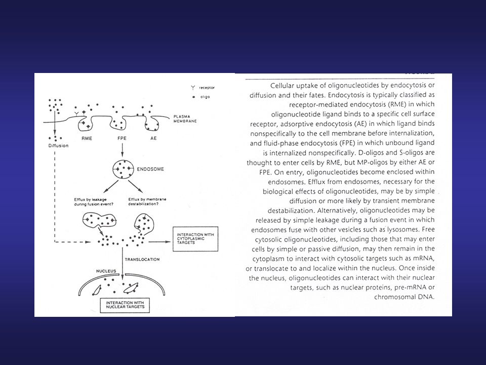
PROBLEMI LEGATI ALL’USO DEGLI OLIGONUCLEOTIDI ANTISENSO
Stabilità biologica Targeting cellulare Uptake cellulare Efflusso dagli endosomi

10
Mechanisms of nucleic-acid-based approaches for gene silencing: ribozymes. General mechanism by which ribozymes silence gene expression. Ribozymes can be produced within the cell through transcription or can be directly introduced into the cell through experimental manipulation. For the hammerhead ribozyme, two arms are used to direct the catalytic centre to target the hydrolysis of the phosphodiester backbone of the mRNA.

11
APTAMERI Sequenze oligonucleotidiche (ssDNA o ssRNA) o peptidiche relativamente brevi (12-30 nucleotidi o aminoacidi) In vivo assumono strutture tridimensionali specifiche in grado di legarsi con elevata affinità (range nM/pM) alle proteine bersaglio
 alle proteine bersaglio.")
12
SELEX systematic evolution of ligands by exponential enrichment
Scheme of systematic evolution of ligands by exponential enrichment (SELEX). A library of DNA or RNA molecules is incubated with the protein target, and those that bind to it are separated from the rest. The sequences with affinity for the target are subsequently amplified to generate a pool of molecules that bind to the protein of interest.
. A library of DNA or RNA molecules is incubated with the protein target, and those that bind to it are separated from the rest. The sequences with affinity for the target are subsequently amplified to generate a pool of molecules that bind to the protein of interest.")
14
APTAMERI vs. ANTICORPI Maggiore stabilità Non immunogenici
Ridotta variabilità da un lotto all’altro Piccole dimensioni migliore penetrazione clearance più rapida possibilità di modificarne la struttura, p.e. per migliorare la stabilità o alterarne le proprietà PK

15
MACUGEN® (pegaptanib sodio)
Primo aptamero approvato, nel dicembre 2004, per l’uso clinico nella degenerazione maculare senile (AMD) ssRNA costituito da 28 nucleotidi, specifico per una delle 6 isoforme di VEGF Modificato a livello dello zucchero per aumentare la resistenza alle nucleasi aggiunta covalente di due molecole di PEG (20 kD ca.) per aumentare t½
 ssRNA costituito da 28 nucleotidi, specifico per una delle 6 isoforme di VEGF. Modificato a livello dello zucchero per aumentare la resistenza alle nucleasi. aggiunta covalente di due molecole di PEG (20 kD ca.) per aumentare t½.")
16
b | Simplified schematic of a process by which RNA aptamers — such as pegaptanib — that bind specifically to protein targets can be prepared. A large library of DNA oligonucleotides are transcribed into RNAs, which are selected for their ability to bind to a target, such as VEGF. Those RNAs that bind best are amplified (by reverse transcription, amplification using the polymerase chain reaction and transcription). Successive selection and amplification cycles result in an exponential increase in the abundance of RNA aptamers that bind to the target.
. Successive selection and amplification cycles result in an exponential increase in the abundance of RNA aptamers that bind to the target..")
17
MACUGEN® (pegaptanib sodio)
a | Sequence and predicted secondary structure of pegaptanib. 2'-O-methylated purines are shown in red, 2'-fluorine-modified pyrimidines are shown in blue and unmodified ribonucleotides are shown in black. The site of attachment of a 40-kDa polyethylene glycol moiety is shown. b | Interaction between the 55-amino-acid heparin-binding domain of vascular endothelial growth factor (VEGF)165 and pegaptanib. Representation of the previously determined NMR solution structure of the free heparin-binding domain of VEGF165 from Fairbrother et al.45 is shown in grey with disulphide bonds in yellow. The aptamer (blue) is shown as a model based on the secondary structure determined by NMR46, with the helical stem regions in teal. The previously reported interaction between cysteine-137 of VEGF165 (cysteine-27 of the heparin-binding domain) and uridine-14 of the aptamer14 is indicated in red.
165 and pegaptanib. Representation of the previously determined NMR solution structure of the free heparin-binding domain of VEGF165 from Fairbrother et al.45 is shown in grey with disulphide bonds in yellow. The aptamer (blue) is shown as a model based on the secondary structure determined by NMR46, with the helical stem regions in teal. The previously reported interaction between cysteine-137 of VEGF165 (cysteine-27 of the heparin-binding domain) and uridine-14 of the aptamer14 is indicated in red.")
18
APTAMERI nella terapia dei tumori
GB10: DNA aptamero che si lega alla tenascina C aptameri peptidici diretti vs. le regioni di dimerizzazione e di legame al DNA di STAT3 AS1411: ssDNA 26-mero ricco di guanosina, forma un complesso con la nucleolina e il fattore modulatore essenziale di NFκB (NEMO)
")
19
Ireson, C. R. et al. Mol Cancer Ther 2006;5:2957-2962
Structure and model for the mechanism of action of AS1411. AS1411 self-anneals into a dimer and adopts a three-dimensional structure that is conferred by eight guanosine-quartets (circles). Squares, thymidine bases. AS1411 is internalized at 1 o'clock following binding to nucleolin, which is expressed on the surface of tumor cells. The mechanism of action involves inhibition of DNA replication and induction of S-phase cell cycle arrest. Copyright ©2006 American Association for Cancer Research
. Squares, thymidine bases. AS1411 is internalized at 1 o clock following binding to nucleolin, which is expressed on the surface of tumor cells. The mechanism of action involves inhibition of DNA replication and induction of S-phase cell cycle arrest. Copyright ©2006 American Association for Cancer Research.")
20
Effetto in vitro di AS1411 IC50 ~1.1
SRB cytotoxicity assay of AS1411 in human lung (A549) and prostate (DU145) cells. AS1411 was incubated with the cells in cell culture medium for 6 d. After 6 d, the medium was removed and replaced with 50% trichloroacetic acid. The cells were washed and incubated with sulforhodamine dye, and absorbance was measured at 564 nm. Points, mean (n = 3). Result is representative of three such experiments. ~1.1
 and prostate (DU145) cells. AS1411 was incubated with the cells in cell culture medium for 6 d. After 6 d, the medium was removed and replaced with 50% trichloroacetic acid. The cells were washed and incubated with sulforhodamine dye, and absorbance was measured at 564 nm. Points, mean (n = 3). Result is representative of three such experiments. ~1.1.")
21
Effetto in vivo di AS1411 In vivo effect of AS1411 on the growth of A549 (A) and SKMES (B) human lung cancer xenografts grown s.c. in nude mice. A, vehicle (PBS) or AS1411 was administered daily (5, 10, and 40 mg/kg, i.v.) for 5 consecutive days. Tumor volumes were measured on day 14 and expressed as tumor volume relative to the volume on the initial day of treatment. Columns, mean (n = 8); bars, SD. B, vehicle ( ), 5 mg/kg ( ), or 10 mg/kg ( ) AS1411 i.p. administration on days 0, 2, and 4 from the time tumors had become established ( 100 mm3).
 and SKMES (B) human lung cancer xenografts grown s.c. in nude mice. A, vehicle (PBS) or AS1411 was administered daily (5, 10, and 40 mg/kg, i.v.) for 5 consecutive days. Tumor volumes were measured on day 14 and expressed as tumor volume relative to the volume on the initial day of treatment. Columns, mean (n = 8); bars, SD. B, vehicle ( ), 5 mg/kg ( ), or 10 mg/kg ( ) AS1411 i.p. administration on days 0, 2, and 4 from the time tumors had become established ( 100 mm3).")
22
AS1411 Studi tossicologici pre-clinici:
nessuna tossicità nel ratto e nel cane Studi clinici (fase I) in NSCLC e carcinomi renali
 in NSCLC e carcinomi renali.")
Presentazioni simili