Scaricare la presentazione
La presentazione è in caricamento. Aspetta per favore

1
Ras

2
The epidermal growth factor receptor signalling pathway
The epidermal growth factor receptor signalling pathway. In response to ligand binding to its extracellular domain, the epidermal growth factor receptor (EGFR) forms homo- or heterodimeric complexes, with either another EGFR or another member of the HER family. This causes structural reorganization within the intracellular portion of the receptor, leading to activation of its kinase activity through autophosphorylation at a tyrosine residue (pY). This, in turn, leads to activation of a range of cell signalling pathways, including the recruitment of the adaptor proteins growth-factor-receptor-bound protein 2 (GRB2) and SOS, leading to activation of the small G proteins RAS and RAF, and signalling through mitogen-activated protein kinase (MAPK) kinase (MEK) and MAPK. EGFR activation also activates the kinase phosphatidylinositol 3-kinase (P13K), which leads to AKT activation, along with the signal transducer and activator of transcription (STAT). Transduction of signals to the nucleus and the activation of gene transcription by factors such as MYC, JUN and FOS leads to the induction of several cellular responses that are required for normal cell growth, including proliferation, survival, differentiation, migration and adhesion. In some tumour cells, EGFR signalling is constitutively active, contributing to the upregulation of many processes that are essential for tumour growth (cell proliferation, survival, angiogenesis, invasion and metastasis) EGFR tyrosine kinase (TK) inhibitors (for example, gefitinib and erlotinib) are small molecules that inhibit ATP binding within the tyrosine-kinase domain of the EGFR, which completely inhibits EGFR autophosphorylation and consequently blocks signal transduction from activated EGFR. As a result, the key mechanisms of tumour growth (blue boxes) are inhibited.
 forms homo- or heterodimeric complexes, with either another EGFR or another member of the HER family. This causes structural reorganization within the intracellular portion of the receptor, leading to activation of its kinase activity through autophosphorylation at a tyrosine residue (pY). This, in turn, leads to activation of a range of cell signalling pathways, including the recruitment of the adaptor proteins growth-factor-receptor-bound protein 2 (GRB2) and SOS, leading to activation of the small G proteins RAS and RAF, and signalling through mitogen-activated protein kinase (MAPK) kinase (MEK) and MAPK. EGFR activation also activates the kinase phosphatidylinositol 3-kinase (P13K), which leads to AKT activation, along with the signal transducer and activator of transcription (STAT). Transduction of signals to the nucleus and the activation of gene transcription by factors such as MYC, JUN and FOS leads to the induction of several cellular responses that are required for normal cell growth, including proliferation, survival, differentiation, migration and adhesion. In some tumour cells, EGFR signalling is constitutively active, contributing to the upregulation of many processes that are essential for tumour growth (cell proliferation, survival, angiogenesis, invasion and metastasis) EGFR tyrosine kinase (TK) inhibitors (for example, gefitinib and erlotinib) are small molecules that inhibit ATP binding within the tyrosine-kinase domain of the EGFR, which completely inhibits EGFR autophosphorylation and consequently blocks signal transduction from activated EGFR. As a result, the key mechanisms of tumour growth (blue boxes) are inhibited.")
3
Signalling upstream of RAS
Signalling upstream of RAS. The activation state of RAS is controlled by the cycle of hydrolysis of bound GTP, which is catalysed by GTPase activating proteins (GAPs), and the replacement of bound GDP with fresh GTP, which is catalysed by guanine nucleotide exchange factors (GEFs). The best-studied activation mechanism involves the assembly of complexes of activated, autophosphorylated growth-factor-receptor tyrosine kinases with the GEF SOS through the adaptor protein GRB2, and possibly SHC, resulting in the recruitment of SOS to the plasma membrane, where RAS is located. Several other GEFs exist that have distinct regulatory mechanisms. In addition, a wide range of GAPs have now been identified for RAS, some of which are also subject to regulation. RAS is also activated through GEFs in response to activation of a wide range of G-protein-coupled receptors.
, and the replacement of bound GDP with fresh GTP, which is catalysed by guanine nucleotide exchange factors (GEFs). The best-studied activation mechanism involves the assembly of complexes of activated, autophosphorylated growth-factor-receptor tyrosine kinases with the GEF SOS through the adaptor protein GRB2, and possibly SHC, resulting in the recruitment of SOS to the plasma membrane, where RAS is located. Several other GEFs exist that have distinct regulatory mechanisms. In addition, a wide range of GAPs have now been identified for RAS, some of which are also subject to regulation. RAS is also activated through GEFs in response to activation of a wide range of G-protein-coupled receptors.")
4
This illustration of the Ras signalling pathway highlights proteins affected by mutations in developmental disorders and cancer. Growth factor binding to cell-surface receptors results in activated receptor complexes, which contain adaptors such as SHC (SH2-containing protein), GRB2 (growth-factor-receptor bound protein 2) and Gab (GRB2-associated binding) proteins. These proteins recruit SHP2 and SOS1, the latter increasing Ras–guanosine triphosphate (Ras–GTP) levels by catalysing nucleotide exchange on Ras. The GTPase-activating protein (GAP) neurofibromin (NF1) binds to Ras–GTP and accelerates the conversion of Ras–GTP to Ras–GDP (guanosine diphosphate), which terminates signalling. Several Ras–GTP effector pathways have been described, and some of the key effectors are depicted here. The BRAF–mitogen-activated and extracellular-signal regulated kinase kinase (MEK)–extracellular signal-regulated kinase (ERK) cascade often determines proliferation and becomes deregulated in certain cancers and in developmental disorders such as cardio-facio-cutaneous syndrome. Ras also activates the phosphatidylinositol 3-kinase (PI3K)– 3-phosphoinositide-dependent protein kinase 1 (PDK1)–Akt pathway that frequently determines cellular survival. RALGDS, RALGDS-like gene (RGL), RGL2 and TIAM1 are exchange factors of Ral and Rac, respectively. Among the effectors of Ral is phospholipase D (PLD) an enzyme that regulates vesicle trafficking. Rac regulates actin dynamics and, therefore, the cytoskeleton. Ras also binds and activates the enzyme phospholipase C (PLC ), the hydrolytic products of which regulate calcium signalling and the protein kinase C (PKC) family. P, phosphate; Y, receptor tyrosine.
, GRB2 (growth-factor-receptor bound protein 2) and Gab (GRB2-associated binding) proteins. These proteins recruit SHP2 and SOS1, the latter increasing Ras–guanosine triphosphate (Ras–GTP) levels by catalysing nucleotide exchange on Ras. The GTPase-activating protein (GAP) neurofibromin (NF1) binds to Ras–GTP and accelerates the conversion of Ras–GTP to Ras–GDP (guanosine diphosphate), which terminates signalling. Several Ras–GTP effector pathways have been described, and some of the key effectors are depicted here. The BRAF–mitogen-activated and extracellular-signal regulated kinase kinase (MEK)–extracellular signal-regulated kinase (ERK) cascade often determines proliferation and becomes deregulated in certain cancers and in developmental disorders such as cardio-facio-cutaneous syndrome. Ras also activates the phosphatidylinositol 3-kinase (PI3K)– 3-phosphoinositide-dependent protein kinase 1 (PDK1)–Akt pathway that frequently determines cellular survival. RALGDS, RALGDS-like gene (RGL), RGL2 and TIAM1 are exchange factors of Ral and Rac, respectively. Among the effectors of Ral is phospholipase D (PLD) an enzyme that regulates vesicle trafficking. Rac regulates actin dynamics and, therefore, the cytoskeleton. Ras also binds and activates the enzyme phospholipase C (PLC ), the hydrolytic products of which regulate calcium signalling and the protein kinase C (PKC) family. P, phosphate; Y, receptor tyrosine..")
5
Regulation of Ras. Ras proteins are GTPases that cycle between an activated GTP-bound and an inactivated GDP-bound form. Guanine nucleotide exchange factors (GEFs) induce dissociation of GDP, which allows binding of GTP. GTPase-activating proteins trigger the hydrolysis of bound GTP. Oncogenic Ras remains in the active GTP-bound form, because the GAP-induced GTP hydrolysis is completely abrogated.
 induce dissociation of GDP, which allows binding of GTP. GTPase-activating proteins trigger the hydrolysis of bound GTP. Oncogenic Ras remains in the active GTP-bound form, because the GAP-induced GTP hydrolysis is completely abrogated..")
6
HRAS, NRAS, KRAS4A and KRAS4B are highly homologous throughout the G domain (amino acids 1–165). The first 85 amino acids are identical in all four proteins and specify binding to guanosine diphosphate (GDP) and guanosine triphosphate (GTP). This includes the P loop (phosphate-binding loop, amino acids 10–16), which binds the -phosphate of GTP, and switch I (amino acids 32–38) and II (amino acids 59–67) which regulate binding to Ras regulators and effectors. The next 80 amino acids (85–165) show 85–90% sequence identity. The C-terminal hypervariable domain (amino acids 165–188/189) specifies membrane localization through post-translational modifications that include the farnesylation of each isoform on the C-terminal CAAX motif (CVLS, CVVM, CIIM and CVIM, respectively) and palmitoylation of key cysteines on HRAS, NRAS and KRAS4A; these cysteines are highlighted below each representation (C). Membrane localization of KRAS4B is facilitated by a stretch of lysines (KKKKKK) proximal to the CVIM motif. To highlight the degree of homology, a box at the bottom of each isoform representation shows the conserved residues in magenta and the variable residues in pink. Somatic RAS mutations found in cancer introduce amino-acid substitutions at positions 12, 13 and 61
 and guanosine triphosphate (GTP). This includes the P loop (phosphate-binding loop, amino acids 10–16), which binds the -phosphate of GTP, and switch I (amino acids 32–38) and II (amino acids 59–67) which regulate binding to Ras regulators and effectors. The next 80 amino acids (85–165) show 85–90% sequence identity. The C-terminal hypervariable domain (amino acids 165–188/189) specifies membrane localization through post-translational modifications that include the farnesylation of each isoform on the C-terminal CAAX motif (CVLS, CVVM, CIIM and CVIM, respectively) and palmitoylation of key cysteines on HRAS, NRAS and KRAS4A; these cysteines are highlighted below each representation (C). Membrane localization of KRAS4B is facilitated by a stretch of lysines (KKKKKK) proximal to the CVIM motif. To highlight the degree of homology, a box at the bottom of each isoform representation shows the conserved residues in magenta and the variable residues in pink. Somatic RAS mutations found in cancer introduce amino-acid substitutions at positions 12, 13 and 61.")
9
REOVIRUS (Respiratory Enteric Orphan Virus)
")
10
Fig. 2. Attenuation of reovirus by viral and cellular adaptation during persistent reovirus infection. Co-adaptation of reovirus and its cellular host resulted in mutation and truncation of the viral S1 gene coding sequence. This in turn caused an attenuation of viral apoptotic potential in healthy cells and tissues (Kim et al., 2007a) while retaining the ability to eliminate tumors in vivo. The mouse on the left was injected in the flank with tumor cells showing massive proliferation, whereas the mouse on the right was injected with tumor cells plus attenuated virus, completely eliminating tumor growth.
 while retaining the ability to eliminate tumors in vivo. The mouse on the left was. injected in the flank with tumor cells showing massive proliferation, whereas the mouse on the right was injected with tumor cells plus. attenuated virus, completely eliminating tumor growth.")
12
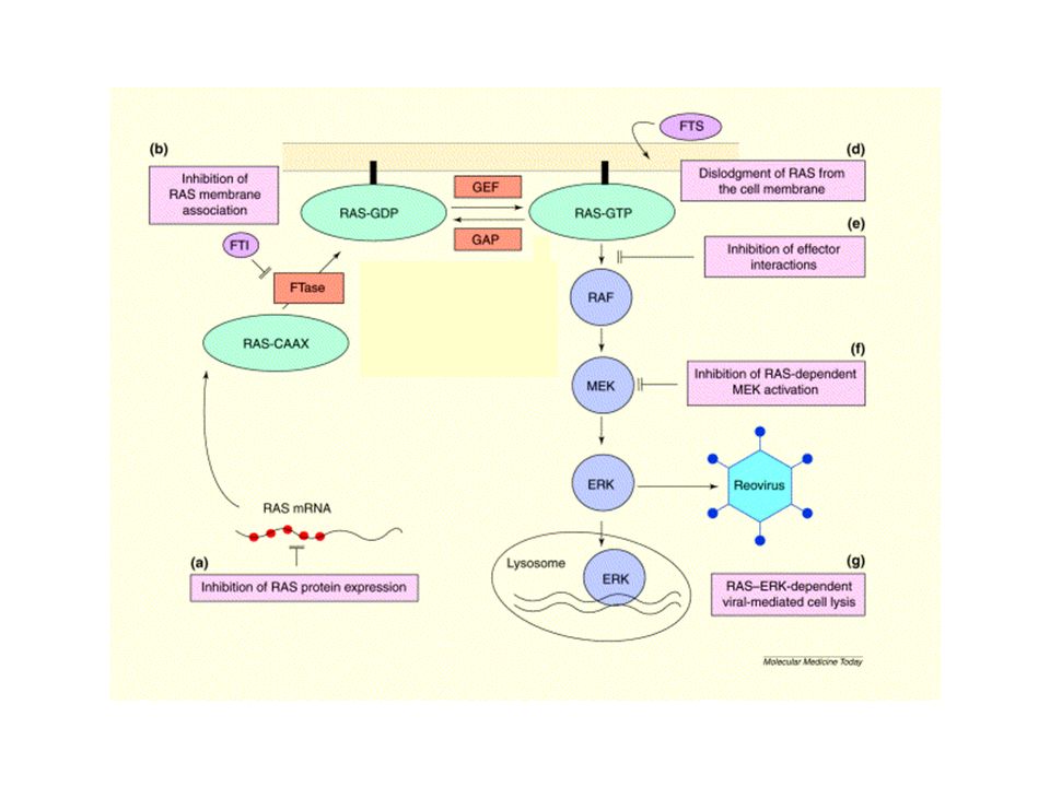
Post-translational processing of RAS proteins
Newly synthesized RAS is a cytosolic protein. Farnesyltransferase (FTase) catalyses the transfer of the 15-carbon isoprenoid chain from farnesyl pyrophosphate (FPP) to a cysteine residue that is close to the carboxyl terminus (C186 in human HRAS) (see a). This results in RAS associating with intracellular membranes via its farnesyl group (F). Farnesyltransferase inhibitors (FTIs) block this farnesylation, so RAS remains in the cytosol and is unable to stimulate its downstream targets. However, when FTase is inhibited, KRAS and NRAS, but not HRAS, can be geranylgeranylated — an alternative 20-carbon isoprenylation is added, and this is catalysed by geranylgeranyltransferase (GGTase) — resulting in rescue of processing of these RAS isoforms. Following isoprenylation, several other processing steps occur. An endopeptidase removes the end three amino acids from the carboxyl terminus of the protein (see b). The new carboxyl terminus is then methylated by a methyltransferase (see c). Following transportation to the plasma membrane, a final processing step (see d) occurs for HRAS and NRAS. Palmitoyltransferase catalyses the addition of two palmitoyl long-chain fatty acid groups (P) to a cysteine residue that is just upstream of the farnesylated carboxy-terminal cysteine. This stabilizes the interaction with the membrane. KRAS does not become palmitoylated, but its interaction with the plasma membrane is promoted by a group of lysine residues near the carboxyl terminus that interact with the negatively charged lipid head groups. The greatest drug discovery effort has gone into developing inhibitors of FTase, but other steps in the pathway might be worth pursuing. The failure of FTIs to block KRAS processing has proved to be a notable problem as KRAS is the most commonly mutated RAS isoform in human tumours. It is apparent that the effects of FTI on KRAS-transformed cells are due to inhibition of other farnesylated proteins, one of which is probably the RAS relative RHOB41.
 catalyses the transfer of the 15-carbon isoprenoid chain from farnesyl pyrophosphate (FPP) to a cysteine residue that is close to the carboxyl terminus (C186 in human HRAS) (see a). This results in RAS associating with intracellular membranes via its farnesyl group (F). Farnesyltransferase inhibitors (FTIs) block this farnesylation, so RAS remains in the cytosol and is unable to stimulate its downstream targets. However, when FTase is inhibited, KRAS and NRAS, but not HRAS, can be geranylgeranylated — an alternative 20-carbon isoprenylation is added, and this is catalysed by geranylgeranyltransferase (GGTase) — resulting in rescue of processing of these RAS isoforms. Following isoprenylation, several other processing steps occur. An endopeptidase removes the end three amino acids from the carboxyl terminus of the protein (see b). The new carboxyl terminus is then methylated by a methyltransferase (see c). Following transportation to the plasma membrane, a final processing step (see d) occurs for HRAS and NRAS. Palmitoyltransferase catalyses the addition of two palmitoyl long-chain fatty acid groups (P) to a cysteine residue that is just upstream of the farnesylated carboxy-terminal cysteine. This stabilizes the interaction with the membrane. KRAS does not become palmitoylated, but its interaction with the plasma membrane is promoted by a group of lysine residues near the carboxyl terminus that interact with the negatively charged lipid head groups. The greatest drug discovery effort has gone into developing inhibitors of FTase, but other steps in the pathway might be worth pursuing. The failure of FTIs to block KRAS processing has proved to be a notable problem as KRAS is the most commonly mutated RAS isoform in human tumours. It is apparent that the effects of FTI on KRAS-transformed cells are due to inhibition of other farnesylated proteins, one of which is probably the RAS relative RHOB41.")
13
FTasi CAAX GGTasi I GGTasi II CX CXC X = Met, Ser, Cys
Zn X = Met, Ser, Cys CAAX Zn GGTasi I X = Leu, Ile GGTasi II Rab CX CXC

14
Fungi, mammals and archaebacteria exclusively use the mevalonate pathway for biosynthesis of isoprenoids. 3-hydroxy 3-methylglutaryl-CoA (HMG-CoA) is converted to mevalonate by HMG-CoA reductase, the rate-limiting enzyme at the apex of the mevalonate pathway. Mevalonate is then converted to isopentenyl pyrophosphate (isopentenyl-PP; the 5-carbon basic isoprene unit), which is subsequently converted to farnesyl pyrophosphate (farnesyl-PP; a 15-carbon isoprenoid) through a series of enzymatic reactions. After addition of isopentenyl-PP, farnesyl-PP can be converted to geranylgeranyl pyrophosphate (geranylgernayl-PP; a 20-carbon isoprenoid), or alternatively farnesyl-PP can be converted to cholesterol or dolichyl phosphate (dolichyl-P), which is used for N-glycosylation of growth factor receptors such as insulin-like growth factor receptor. HMG-CoA reductase is the target of the cholesterol-lowering statins, whereas isopentenyl-PP isomerase and farnesyl-PP synthase are targets of bisphosphonates. Importantly, in normal cells, cholesterol and isoprenoid products suppress HMG-CoA reductase via post-translational downregulation. Conversely, tumour cells are resistant to cholesterol-mediated suppression, although they remain sensitive to isoprenoid-mediated suppression. Addition of isoprenoids to statins may prevent upregulation of HMG-CoA reductase, which might occur as a resistance mechanism to statin therapy. This explains the synergistic activity of statins and isoprenoids. CAAX, C denotes cysteine, A represents any aliphatic amino acid and X may be any amino acid; FTase, farnesyltransferase; GGTase, geranylgeranyltransferase.
 is converted to mevalonate by HMG-CoA reductase, the rate-limiting enzyme at the apex of the mevalonate pathway. Mevalonate is then converted to isopentenyl pyrophosphate (isopentenyl-PP; the 5-carbon basic isoprene unit), which is subsequently converted to farnesyl pyrophosphate (farnesyl-PP; a 15-carbon isoprenoid) through a series of enzymatic reactions. After addition of isopentenyl-PP, farnesyl-PP can be converted to geranylgeranyl pyrophosphate (geranylgernayl-PP; a 20-carbon isoprenoid), or alternatively farnesyl-PP can be converted to cholesterol or dolichyl phosphate (dolichyl-P), which is used for N-glycosylation of growth factor receptors such as insulin-like growth factor receptor. HMG-CoA reductase is the target of the cholesterol-lowering statins, whereas isopentenyl-PP isomerase and farnesyl-PP synthase are targets of bisphosphonates. Importantly, in normal cells, cholesterol and isoprenoid products suppress HMG-CoA reductase via post-translational downregulation. Conversely, tumour cells are resistant to cholesterol-mediated suppression, although they remain sensitive to isoprenoid-mediated suppression. Addition of isoprenoids to statins may prevent upregulation of HMG-CoA reductase, which might occur as a resistance mechanism to statin therapy. This explains the synergistic activity of statins and isoprenoids. CAAX, C denotes cysteine, A represents any aliphatic amino acid and X may be any amino acid; FTase, farnesyltransferase; GGTase, geranylgeranyltransferase..")
15
An overview of CAAX-protein processing
An overview of CAAX-protein processing. a | Proteins that contain a carboxy-terminal CAAX motif are initially modified by the cytosolic CAAX prenyltranferases farnesyltransferase or geranylgeranyltransfersase-I, which add a 15-carbon farnesyl (F) or a 20-carbon geranylgeranyl (GG) group to the cysteine residue. b | Following prenylation, the CAAX protein travels to the endoplasmic reticulum (ER), where the C-terminal three amino acids (AAX) are proteolytically removed by RCE1 — this reaction leaves the prenylcysteine (attached to F or GG) at the C terminus. c | The protein is then carboxyl methylated by isoprenylcysteine carboxyl methyltransferase (ICMT) in a reaction that consumes S-adenosylmethionine (AdoMet) and produces S-adenosylhomocysteine (AdoHcy). d | Following methylation, fully processed CAAX proteins are directed to their appropriate cellular location, which is often the cytoplasmic surface of cell membranes, through mechanisms that are still poorly understood. The insets show detailed structures of the C-terminal processing intermediates.
 or a 20-carbon geranylgeranyl (GG) group to the cysteine residue. b | Following prenylation, the CAAX protein travels to the endoplasmic reticulum (ER), where the C-terminal three amino acids (AAX) are proteolytically removed by RCE1 — this reaction leaves the prenylcysteine (attached to F or GG) at the C terminus. c | The protein is then carboxyl methylated by isoprenylcysteine carboxyl methyltransferase (ICMT) in a reaction that consumes S-adenosylmethionine (AdoMet) and produces S-adenosylhomocysteine (AdoHcy). d | Following methylation, fully processed CAAX proteins are directed to their appropriate cellular location, which is often the cytoplasmic surface of cell membranes, through mechanisms that are still poorly understood. The insets show detailed structures of the C-terminal processing intermediates.")
16
Statins, biphosphonates and isoprenoids, alone or in combinations, target the mevalonate pathway, whereas farnesyltransferase inhibitors (FTIs), geranylgeranyltransferase inhibitors (GGTIs) and dual prenylation inhibitors (DPIs) target the prenylation process. Several strategies have been developed for targeting the functional regulation of the RAS superfamily of GTPases. These focus either on compounds that inhibit the interaction between the RAS superfamily of GTPases and regulatory proteins (for example, interfacial inhibitors), or on drugs that target individual RAS superfamily GTPases (for example, bacterial toxins, GTP analogues, small-interfering RNA (siRNA) inhibitors), or regulatory proteins (siRNA inhibition), thereby blocking GDP/GTP exchange and inhibiting activation of downstream effectors. GAP, GTPase-activating protein; GDI, guanine nucleotide dissociation inhibitor; GEF, guanine nucleotide exchange factor; ICMT, isoprenylcysteine carboxymethyltransferase; RCE1, RAS converting enzyme.
, or on drugs that target individual RAS superfamily GTPases (for example, bacterial toxins, GTP analogues, small-interfering RNA (siRNA) inhibitors), or regulatory proteins (siRNA inhibition), thereby blocking GDP/GTP exchange and inhibiting activation of downstream effectors. GAP, GTPase-activating protein; GDI, guanine nucleotide dissociation inhibitor; GEF, guanine nucleotide exchange factor; ICMT, isoprenylcysteine carboxymethyltransferase; RCE1, RAS converting enzyme..")
17
Zarnestra (Tipifarnib, R115777)
Sarasar (Lonafarnib, SCH66336)
")
18
Three strategies to interfere with Ras signaling in cancer cells: farnesyltransferase inhibitors, antisense oligonucleotides and RNAi.

19
EVIDENZE A FAVORE DI UN EFFETTO Ras-INDIPENDENTE PER GLI FTI
mancanza di correlazione tra suscettibilità e status di Ras discrepanze nella cinetica con cui si sviluppano gli effetti inibizione di della crescita cellulare dipendente da K-Ras e N-Ras

20
RAS processing and association with the plasma membrane
RAS processing and association with the plasma membrane. The four RAS proteins (HRAS, KARAS, KBRAS and NRAS) are synthesized initially as cytosolic, inactive proteins. They undergo a rapid series of post-translational modifications signalled by the carboxy-terminal CAAX tetrapeptide sequence. FTase uses farnesyl diphosphate (FPP) and catalyses the covalent addition of the 15-C farnesyl isoprenoid (F) to the cysteine residue of the CAAX sequence, followed by endoprotease (RCE1) cleavage of the AAX residues and, finally, carboxylmethylation (ICMT) of the now terminal farnesylated cysteine residue. Inhibitors of all three steps have been developed, but only FTase inhibitors (FTIs) have advanced to evaluation in clinical trials. KRAS and NRAS proteins undergo alternative prenylation when FTase activity is blocked by FTIs, resulting in their modification by geranylgeranyl transferase I (GGTase I), which uses geranylgeranyl pyrophosphate (GGPP) and catalyses the addition of the 20-C geranylgeranyl isoprenoid (GG) to the cysteine residue. So, whereas RAS proteins are not normally modified by GG, the GG-modified forms of RAS retain membrane association and transforming activity.
 are synthesized initially as cytosolic, inactive proteins. They undergo a rapid series of post-translational modifications signalled by the carboxy-terminal CAAX tetrapeptide sequence. FTase uses farnesyl diphosphate (FPP) and catalyses the covalent addition of the 15-C farnesyl isoprenoid (F) to the cysteine residue of the CAAX sequence, followed by endoprotease (RCE1) cleavage of the AAX residues and, finally, carboxylmethylation (ICMT) of the now terminal farnesylated cysteine residue. Inhibitors of all three steps have been developed, but only FTase inhibitors (FTIs) have advanced to evaluation in clinical trials. KRAS and NRAS proteins undergo alternative prenylation when FTase activity is blocked by FTIs, resulting in their modification by geranylgeranyl transferase I (GGTase I), which uses geranylgeranyl pyrophosphate (GGPP) and catalyses the addition of the 20-C geranylgeranyl isoprenoid (GG) to the cysteine residue. So, whereas RAS proteins are not normally modified by GG, the GG-modified forms of RAS retain membrane association and transforming activity.")
21
Expected properties of farnesylated proteins involved in FTI-induced growth inhibition. Farnesyltransferase inhibitor (FTI) treatment will result in accumulation of either a nonprenylated or alternatively prenylated protein. If protein function is not dependent on farnesylation, then the protein is not a relevant target. However, if the nonprenylated or geranylgeranyl (GG)-modified protein is functionally distinct from the authentically farnesylated (F) protein — because of altered subcellular localization or interaction with other proteins — then it might be a candidate. If the effect of FTIs is loss of protein function, silencing the expression of the protein should mimic the consequences of FTI treatment. Conversely, if a GG-modified form of the protein is functionally identical to the farnesylated protein, then ectopic expression of a GG-modified variant should prevent FTI-mediated changes. Alternatively, FTIs might induce a gain of function — acquisition of an altered function. The nonprenylated or GG-modified protein might act as a dominant-negative protein, by forming nonproductive, mislocalized complexes with proteins that are important for the function of the farnesylated protein. Ectopic expression of the nonprenylated or GG-modified version of the protein should then cause the same biological consequence as FTI treatment, and ectopic expression of a farnesylated variant of the protein might dampen the consequences of FTI treatment.
 treatment will result in accumulation of either a nonprenylated or alternatively prenylated protein. If protein function is not dependent on farnesylation, then the protein is not a relevant target. However, if the nonprenylated or geranylgeranyl (GG)-modified protein is functionally distinct from the authentically farnesylated (F) protein — because of altered subcellular localization or interaction with other proteins — then it might be a candidate. If the effect of FTIs is loss of protein function, silencing the expression of the protein should mimic the consequences of FTI treatment. Conversely, if a GG-modified form of the protein is functionally identical to the farnesylated protein, then ectopic expression of a GG-modified variant should prevent FTI-mediated changes. Alternatively, FTIs might induce a gain of function — acquisition of an altered function. The nonprenylated or GG-modified protein might act as a dominant-negative protein, by forming nonproductive, mislocalized complexes with proteins that are important for the function of the farnesylated protein. Ectopic expression of the nonprenylated or GG-modified version of the protein should then cause the same biological consequence as FTI treatment, and ectopic expression of a farnesylated variant of the protein might dampen the consequences of FTI treatment..")
22
Farnesyltransferase inhibitors (FTIs) inhibit FTase modification of RAS and other proteins. FTIs are effective inhibitors of HRAS farnesylation and function. FTIs also prevent the farnesylation of KRAS and NRAS; however, these RAS isoforms then undergo alternative prenylation and modification by geranylgeranylation. Experimental evidence supports the possibility that RHOB proteins are important targets for the antitumour activity of FTIs. The increased formation of RHOB-GG, which might have a growth-inhibitory function, is proposed to be the mechanism for this inhibition. However both RHOB-F and RHOB-GG have been shown to inhibit tumour growth in vitro and in vivo51. Other candidate targets for proteins that mediate the antitumour effects of FTIs include other RAS-family GTPases (RHEB and RND), CENP and PRL. Some farnesylated RAS-family proteins (RIG and NOEY2) seem to function as tumour suppressors; so, the loss of function that is caused by FTI treatment might promote, rather than prevent, proliferation in some tissues.
, CENP and PRL. Some farnesylated RAS-family proteins (RIG and NOEY2) seem to function as tumour suppressors; so, the loss of function that is caused by FTI treatment might promote, rather than prevent, proliferation in some tissues..")
24
Structure of the RAF proteins
Structure of the RAF proteins. The RAF-isoforms, A-RAF, B-RAF and C-RAF, share three conserved regions: CR1 (yellow), CR2 (orange) and CR3 (red). The amino acids that are highlighted below the individual isoforms refer to known phosphorylation sites. CR1 contains the RAS-binding domain (RBD) and the cysteine-rich domain (CRD), which are both required for membrane recruitment. Phosphorylation of S43, just N-terminal to this region, blocks C-RAF binding to RAS, probably through steric hindrance. CR2 contains one of the binding sites, which encompasses S259 (the numbering refers to C-RAF). The other two binding sites are centred on S233 and S621. CR3 contains the catalytic domain (the activation segment is highlighted in pink). The negative-charge regulatory region (N-region) is just upstream of CR3 and contains residue Y341, which is conserved in A-RAF (Y302), but is replaced by D448 in B-RAF (shown as a grey box). S338 is conserved in all RAF proteins (S299 in A-RAF and S445 in B-RAF), but it is constitutively phosphorylated in B-RAF (blue star). The catalytic domain contains the two activation-segment phosphorylation sites T491 and S494, which are conserved in A-RAF (T452 and T455) and B-RAF (T598 and S601).
, CR2 (orange) and CR3 (red). The amino acids that are highlighted below the individual isoforms refer to known phosphorylation sites. CR1 contains the RAS-binding domain (RBD) and the cysteine-rich domain (CRD), which are both required for membrane recruitment. Phosphorylation of S43, just N-terminal to this region, blocks C-RAF binding to RAS, probably through steric hindrance. CR2 contains one of the binding sites, which encompasses S259 (the numbering refers to C-RAF). The other two binding sites are centred on S233 and S621. CR3 contains the catalytic domain (the activation segment is highlighted in pink). The negative-charge regulatory region (N-region) is just upstream of CR3 and contains residue Y341, which is conserved in A-RAF (Y302), but is replaced by D448 in B-RAF (shown as a grey box). S338 is conserved in all RAF proteins (S299 in A-RAF and S445 in B-RAF), but it is constitutively phosphorylated in B-RAF (blue star). The catalytic domain contains the two activation-segment phosphorylation sites T491 and S494, which are conserved in A-RAF (T452 and T455) and B-RAF (T598 and S601).")
25
MAPK pathways Stimulus
Proliferation, differentiation, development, survival A-Raf, B-Raf, C-Raf Growth factors, mitogens ERK1/2 MEK1/2 AMLKs, TAK, ASK1 MKK3/6 P38 MAPKa/b MEKK1,4 MLKs ASK1 MKK4/7 SAPK/ JNK1,2,3 Inflammation, apoptosis, proliferation, differentiation Stress, inflammatory cytokines, growth factors MAPKKK MAPKK Human cancers often arise due to mutations in cellular signaling pathways that coordinate and regulate cell proliferation and survival. The mitogen-activated protein kinase (MAPK) pathways are major signal transduction routes that transfer and amplify messages from the cell surface to the nucleus.1,2 There are several distinct MAPK pathways, important in the regulation of cell proliferation, differentiation, development, inflammation, survival and migration.1,2 References 1. Kolch W. Biochem J 2000; 351: 289–305 2. Schaeffer HJ, Weber MJ. Mol Cell Biol 1999; 19: 2435–2444 MAPK Biologic response MAPK, mitogen-activated protein kinase; MEK, MAPK kinase; ERK, extracellular signal-regulated kinase
 pathways are major signal transduction routes that transfer and amplify messages from the cell surface to the nucleus.1,2. There are several distinct MAPK pathways, important in the regulation of cell proliferation, differentiation, development, inflammation, survival and migration.1,2. References. 1. Kolch W. Biochem J 2000; 351: 289– Schaeffer HJ, Weber MJ. Mol Cell Biol 1999; 19: 2435–2444. MAPK. Biologic response. MAPK, mitogen-activated protein kinase; MEK, MAPK kinase; ERK, extracellular signal-regulated kinase.")
26
Growth factors, mitogens
MAPK pathways Stimulus A-Raf, B-Raf, C-Raf Growth factors, mitogens ERK1/2 MEK1/2 Stress, inflammatory cytokines, growth factors AMLKs, TAK, ASK1 MEKK1,4 MLKs ASK1 MAPKKK MAPKK MKK3/6 MKK4/7 The Ras-Raf-MEK-ERK signaling cascade regulates cell proliferation, differentiation and survival.1 In transformed cells, the Ras-Raf-MEK-ERK pathway has been implicated in uncontrolled cell proliferation and survival.1 Reference 1. Kolch W. Biochem J 2000; 351: 289–305 MAPK P38 MAPKa/b SAPK/ JNK1,2,3 Biologic response Proliferation, differentiation, development, survival Inflammation, apoptosis, growth, differentiation MAPK, mitogen-activated protein kinase; MEK, MAPK kinase; ERK, extracellular signal-regulated kinase

27
The role of MEK1/2 SOS1 SHC Grb2 Ras Raf1 MEK1 MEK2 ERK1 ERK2
P SHC SOS1 Ras P Grb2 P Raf1 P MEK1 P The Ras-Raf-MEK-ERK pathway is activated by a range of growth factor receptors, including epidermal growth factor receptor, platelet-derived growth factor receptor, type-1 insulin-like growth factor receptor, and fibroblast growth factor receptors. The pathway can also be activated by cytokines, steroid hormones, and several agonists that act via G-protein-coupled receptors. Growth factor stimulation leads to a signal transduction cascade, which involves sequential activation of a number of cytoplasmic and nuclear targets. In this cascade, the adaptor protein SHC becomes phosphorylated and acts as a binding site for the second adaptor protein, growth factor receptor-bound protein 2 (Grb2).1 Grb2 then recruits the Ras guanine nucleotide exchange factor SOS (son of sevenless) and mediates Ras activation.1 Activated Ras leads to the activation of Raf proteins. Raf serine/threonine kinase activity phosphorylates MEK, which activates MEK catalytic activity.1,2 MEK1/2 is a dual-specific kinase that is essential to the Ras-Raf-MEK-ERK pathway. All three Raf isoforms share Ras as a common upstream activator, whereas MEK is the only commonly accepted downstream substrate.1 References 1. Kolch W. Biochem J 2000; 351: 289–305 2. Harmer SL, DeFranco AL. J Biol Chem 1999; 274: 12183–12191 MEK2 P ERK1 ERK2 Grb2, growth factor receptor-bound protein 2
.1. Grb2 then recruits the Ras guanine nucleotide exchange factor SOS (son of sevenless) and mediates Ras activation.1. Activated Ras leads to the activation of Raf proteins. Raf serine/threonine kinase activity phosphorylates MEK, which activates MEK catalytic activity.1,2. MEK1/2 is a dual-specific kinase that is essential to the Ras-Raf-MEK-ERK pathway. All three Raf isoforms share Ras as a common upstream activator, whereas MEK is the only commonly accepted downstream substrate.1. References. 1. Kolch W. Biochem J 2000; 351: 289– Harmer SL, DeFranco AL. J Biol Chem 1999; 274: 12183– MEK2. P. ERK1. ERK2. Grb2, growth factor receptor-bound protein 2.")
28
The role of MEK1/2 Transcription Transcription factors SOS1 SHC Grb2
Ras P SHC Grb2 P Raf1 P MEK1 P MEK1/2 serves to amplify signals to ERK1/2 (also known as MAPK1/2) through phosphorylation of key tyrosine and threonine residues, thereby activating its catalytic activity. Activation of ERK1/2 via MEK1/2 is a critical step in growth factor signaling, as active ERK mediates a range of cellular effects, eg cell proliferation.1 Active ERK phosphorylates and activates numerous other kinases (eg RSK) and also transcription factors (eg SRF).1 The activation of Ras, therefore, induces a sequential kinase cascade that includes Raf, MEK, and ERK.2 References 1. Kolch W. Biochem J 2000; 351: 289–305 2. Harmer SL, DeFranco AL. J Biol Chem 1999; 274: 12183–12191 MEK2 P ERK1 P Transcription SRF ERK2 P Transcription factors Grb2, growth factor receptor-bound protein 2; SRF, serum response factor
 through phosphorylation of key tyrosine and threonine residues, thereby activating its catalytic activity. Activation of ERK1/2 via MEK1/2 is a critical step in growth factor signaling, as active ERK mediates a range of cellular effects, eg cell proliferation.1. Active ERK phosphorylates and activates numerous other kinases (eg RSK) and also transcription factors (eg SRF).1. The activation of Ras, therefore, induces a sequential kinase cascade that includes Raf, MEK, and ERK.2. References. 1. Kolch W. Biochem J 2000; 351: 289– Harmer SL, DeFranco AL. J Biol Chem 1999; 274: 12183– MEK2. P. ERK1. P. Transcription. SRF. ERK2. P. Transcription factors. Grb2, growth factor receptor-bound protein 2; SRF, serum response factor.")
29
Constitutive Ras-Raf-MEK-ERK activation
P MEK1 P Constitutive activation of the Ras-Raf-MEK-ERK pathway has been implicated in many cancers, including pancreatic, colon, and melanoma. This is caused by cancer-associated, mutational activation of B-Raf and Ras proteins. Mutant Ras and Raf proteins are key activators of the Ras-Raf-MEK-ERK pathway: Oncogenic Ras has been found in ~20-30% of human cancers, including ~25% of lung cancers, 50% of colon cancers, and >90% of pancreatic cancers.1 B-Raf mutations have been identified in >60% of malignant melanomas and ~70% of papillary thyroid cancers.2,3 In addition, uncontrolled growth factor signaling, eg through receptor gene amplification or ligand overexpression, is also a key constitutive activator of this pathway.4 References 1. Oliff A. Biochim Biophys Acta 1999; 1423: C19–C30 2. Davies H et al. Nature 2002; 417: 949–954 3. Cohen Y et al. J Natl Cancer Inst 2003; 95: 625–627 4. Kolch W. Biochem J 2000; 351: 289–305 MEK2 P ERK1 P Transcription SRF ERK2 P

30
MEK1/2 inhibition MEK1/2 inhibition SOS1 SHC Grb2 Ras Raf1 MEK1 MEK2
P SHC SOS1 Ras P Grb2 P Raf1 P MEK1 P MEK1/2 inhibition Inhibition of MEK1/2 is an attractive strategy for therapeutic intervention in cancer because: It has the potential to block inappropriate signal transduction, regardless of the upstream position of any oncogenic aberration ERK1/2 are the only known substrates for MEK1/2 phosphorylation and signaling. MEK2 P ERK2 ERK1

Presentazioni simili


 is a disease state characterized by airflow limitation that is not fully reversible. The.>")


>")