Scaricare la presentazione
La presentazione è in caricamento. Aspetta per favore

1
CLASSIFICAZIONE DELLE NEOPLASIE DEL SISTEMA EMOPOIETICO
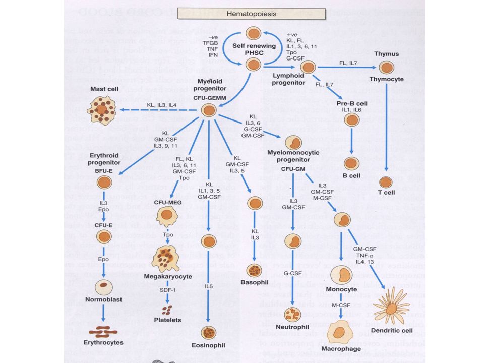
A) DEFINIZIONE Espansione clonale di cellule che riproducono un particolare fenotipo che richiama quello di una normale cellula bloccata ad un determinato stadio differenziativo B) CELLULE D’ORIGINE sindromi mieloproliferative Sindromi mielodisplastiche alcune leucemie acute) cellula staminale pluripotente cellula staminale mieloide multipotente leucemie acute mieloidi cellule staminali committed leucemie acute mieloidi e linfoidi Mieloma, Linfomi Leucemia linfatica cronica cellule già differenziate
 DEFINIZIONE. Espansione clonale di cellule che riproducono un particolare fenotipo che richiama. quello di una normale cellula bloccata ad un determinato stadio differenziativo. B) CELLULE D’ORIGINE. sindromi mieloproliferative. Sindromi mielodisplastiche. alcune leucemie acute) cellula staminale pluripotente. cellula staminale mieloide multipotente. leucemie acute mieloidi. cellule staminali committed. leucemie acute mieloidi e linfoidi. Mieloma, Linfomi. Leucemia linfatica cronica. cellule già differenziate.")
2
SINDROMI MIELOPROLIFERATIVE
Leucemia mieloide cronica Policitemia vera Trombocitemia essenziale Mielofibrosi con metaplasia mieloide Emoglobinuria parossisitica notturna Sindrome ipereosinofila (ALCUNE) IL CLONE NEOPLASTICO MANTIENE LA CAPACITA’ MATURATIVA SINO AGLI STADI TERMINALI croniche IL CLONE NEOPLASTICO PRESENTA UN BLOCCO MATURATIVO INTRAMIDOLLARE AD UNO STADIO IN GENERE PRECOCE acute Leucemie mieloidi acute Classificazione FAB Classificazione WHO
 IL CLONE NEOPLASTICO. MANTIENE LA CAPACITA’ MATURATIVA SINO AGLI. STADI TERMINALI. croniche. IL CLONE NEOPLASTICO. PRESENTA UN BLOCCO. MATURATIVO INTRAMIDOLLARE. AD UNO STADIO IN GENERE. PRECOCE. acute. Leucemie mieloidi acute. Classificazione FAB. Classificazione WHO.")
4
Cellula staminale dell’adulto mutipotente (MAPC)
Keratinocytes Ectoderm stem cell endothelium Mesenchimal stem cell Stroma Adipocyte Muscle Bone Cartilage Angioblast MAPC Mesoderm stem cell Endoderm stem cell Hemopoietic stem cell Neural stem cell Cardiomyocytes Red blood cells White blood cells Platelets Liver Pancreas Brain

5
Erythroid Maturation In the BM RETICULOCYTE POLYCHROMATOPHILIC
POLYCHROMAPHILIC ERYTHROBLAST PROERYTHROBLAST RETICULOCYTE RED BLOOD CELL BASOPHILIC ERYTHROBLAST ORTHOCHROMATIC ERYTHROBLAST RETICULOCYTE POLYCHROMATOPHILIC ERYTHROBLAST RBC PROERYTHROBLAST BASOPHILIC ERYTHROBLAST POLYCHROMATOPHILIC ERYTHROBLAST ORTHOCHROMATIC ERYTHROBLAST

6
Maturation of erythroid precursors in the bone marrow
Note progressive chromatin condensation and progressive reduction of basophilia wich diaseppears totally in the orthochromatic stage Basophilic erythroblast Proerythroblast polychromatophilic erythroblast Orthochromatic erythroblast

7
Normal maturation of megakaryocyets (platelet progenitors)
")
8
Normal stages of maturation
of megakaryocytes in the BM

9
Normal maturation in the BM of granulocyte precursors
MYELOBLAST PROMYELOCYTE NEUTROPHIL MYELOCYTE METAMYELOCYTE

10
3 2 1 Myeloblast With few azurophilic granules Promyelocyte Myelocyte 4 5 Neutrophil Metamyelocyte

11
Morphology of normal human PB lymphocytes
Large granular lymphocyte

12
Normal bone marrow

13
M0 M1 M4 M6 M2 M5 M7 M3

14
PB in CML BM in CML

15
AML-M5

16
AML-M2

17
SINDROMI MELODISPLASTICHE
ESPANSIONE CLOLNALE DI ELEMENTI MIELOIDI AFFETTI DA RALLENTAMENTO MATURATIVO E POSSIBILITA’ DI EVOLVERE IN LEUCEMIA ACUTA ANEMIA REFRATTARIA CON SIDEROBLASTI AD ANELLO preleucemie Anemia refrattaria con eccesso di blasti AREB in trasformazione leucemie oligoblastiche

18
SINDROMI LINFOPROLIFERATIVE
Leucemia linfatica cronica Linfomi non Hodgkin Linfoma di Hodgkin Mieloma IL CLONE NEOPLASTICO RIPRODUCE IL FENOTIPO DELLE CORRISPONDENTI CELLULE D’ORIGINE BLOCCATE AD UN PRECISO STADIO DIFFERENZIATIVO croniche acute Leucemie linfatiche acute IL CLONE NEOPLASTICO PRESENTA UN BLOCCO MATURATIVO AD UNO STADIO IN GENERE PRECOCE Classificazione FAB Classificazione WHO

19
MIDOLLO OSSEO EMOPOIETICO
Schema della differenziazione B-linfocitaria PRECURSORI T-LINFOIDI plasmacellula CELLULA STAMINALE LINFOIDE MIDOLLO OSSEO EMOPOIETICO ANTIGENE Sede infezione Midollo Ricombinazione Ig porzioni VDJ PRECURSORI B-LINFOIDI sIg Centroblasto Cellula pre-B Cellula naive Cellula mantellare Linfocito z. marginale SANGUE LINFONODO centrocicto Blasto follicolare

20
Follicolo linfonodale
MANTELLO FOLLICOLARE Cellula memoria Plasmablasto Plasmacellula BCL6- BCL6- Sopravvivenza Zona apicale chiara Follicolo linfonodale Editing recettoriale Elevata affinità per l’Ag BCL2+ Bassa affinità CENTRO GERMINATIVO Maturazione per affinita’ apoptosi Ipermutazione somatica IgV Switch Ig BCL2- centrocito Zona basale chiara BCL2- BCL6+ blasti B primari centroblasti Zona basale scura

21
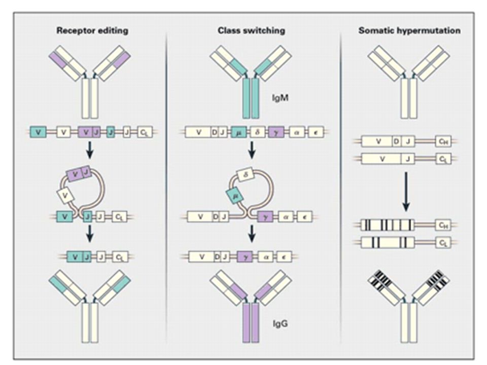
Lymph node Bone Marrow VDJ hypermutation Ig heavy chain switch
MEMORY B-CELL GERMINAL CENTRE VDJ hypermutation Ig heavy chain switch Receptor editing Plasmablast Germinal centre Cell precursor Lymph node Bone Marrow Pre-B cell VDJ rearrangement G, A, E, D Long-lived plasma cell

23
Cell origin of B-lymphoid neoplasia
Pre-germinal centre Post-germinal centre Mantle cell Lymphoma Follicular lymphoma Large cell lymphoma (part) Multiple myeloma B-ALL Lymphoplasmacytic lymphoma Ig gene mutations Pre-plasma cell Plasma cell BM B-precursor cell Naive B-cell (no Ag) Marginal zone Lymphoma CLL Germinal Centre cells Memory B-cell
 Multiple. myeloma. B-ALL. Lymphoplasmacytic. lymphoma. Ig gene. mutations. Pre-plasma cell. Plasma cell. BM. B-precursor. cell. Naive B-cell. (no Ag) Marginal zone. Lymphoma. CLL. Germinal. Centre cells. Memory. B-cell.")
24
Cell origin of T-lymphoid neoplasia
Mycosis fungoides Sezary’s syndrome T-cell CLL/PLL skin Angiocentric NHL Intestinal NHL Mucosae Bowel CD4+ lymphocytes T-ALL THYMUS Hepato-splenic γδ NHL Spleen liver Peripheral blood BM T-precursor cell Lymph node ALCL PTCL CD8+ lymphocytes LGL expansion Germinal centre

25
EPIDEMIOLOGIA NEOPLASIE
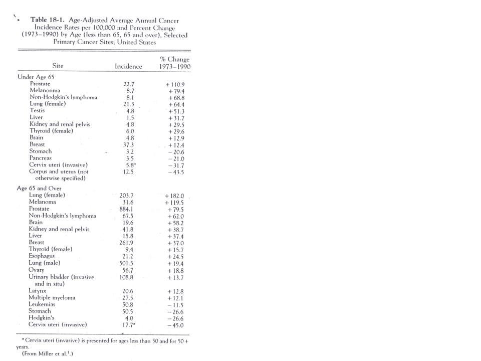
II causa di morte Incremento incidenza e mortalità dell’1% e dello 0.4% per anno negli ultimi 20 anni dal 1973 al 1990: incidenza del 18%, mortalità del 6% mortalità 1% per anno per i casi >65 aa mortalità 0.2% per anno per i casi <65 aa. sopravvivenza a 5 aa: 49% nel 1975 vs 53% nel 1987 L’aumento dell’incidenza non deriva dall’invecchiamento: sono aumentati in molte fasce d’età sono aumentate alcune forme e diminuite altre

27
Non Hodgkin’s lymphoma
The incidence has increased in both sexes (males > females) (Deaths only slightly incresed = earlier diagnosis and better treatment) Rate per
 (Deaths only slightly incresed = earlier diagnosis and better treatment) Rate per")
28
Non Hodgkin’s lymphoma The incidence increases with age in both sexes
(notable exception: Burkitt’s lymphoma which peaked in the first 20 years in pre AIDS era)
")
29
Hematologic malignancies: the magnitude of the problem
Incidence of leukemias and lymphomas compared with other tumors Deaths by leukemias and lymphomas compared with other tumors It is one of the most frequent neoplasia in the young age group

30
Incidenza delle neoplasie nella varie fasce d’età
Età anni Età 0-4 anni Età 5-14 anni leucemie 7,0 3,1 M. Hodgkin 3,7 cervello 4,0 2,9 testicolo 2,5 rene 1,9 ossa 1,5 2,2 Tessuti molli 1,3 M Hodgkin 1,4 2,1 Occhio e orbita Linfomi NH 1,0

31
Incidenza delle neoplasie nella varie fasce d’età
Età anni Età anni Età anni testicolo 8,7 Testicolo 13,5 Polmone 16.7 M Hodgkin 5,2 Linfoma NH 8,9 Melanoma 16,4 2,7 16 4,3 8 SNC 2,5 Tiroide 2,3 5,8

Presentazioni simili


>")


