Scaricare la presentazione
La presentazione è in caricamento. Aspetta per favore

1
MODELLI SPERIMENTALI NELLA RICERCA SUL CANCRO

2
a | Illustration of the rate of testing and throughput of the US National Cancer Institute (NCI) 60 anticancer drug screen (NCI60) and associated prescreens used for synthetic compounds and crude natural product extracts. b | Analysis of the origin of submissions served by the screen in 1999–2005 for the past 40,000 compounds. HT, high throughput

3
When the US National Cancer Institute (NCI) 60 anticancer drug screen (NCI60) tumour cell lines are screened for the activity of test compounds, the resulting data can be displayed and analysed in various ways. For instance, the activity of one test compound on three non-small-cell lung cancer cell lines is shown in part a of the figure. The response parameters GI50 (50% growth inhibition) and LC50 (50% lethal concentration) are extracted from concentration–response curves by linear interpolation. TGI (total growth inhibition) is read as the x-axis intercept. Standard dilutions span a four-log concentration range, here: 1 = 1E-5.2, 2 = 1E-4.2, 3 = 1E-3.2, 4 = 1E-2.2 and 5 = 1E-1.2 (dilutions from a stock solution, expressed in g ml-1). Therefore, in this example for EKVX, the GI50 = 1E-4.05, the TGI = 1E-3.49 and the LC50 level of effect was not reached (>1E-1.2, the maximum concentration tested). Mean Graphs are constructed at each level of effect, with bars depicting the deviation of individual tumour cell lines from the overall mean value for all the cells tested. For purposes of illustration, only the GI50 Mean Graph for leukaemia, lung and colon cancer panels are shown in part b of the figure for two test compounds. Results (shown on the left) are for the immunotoxin transforming growth factor- (TGF)–PE38 (Ref. 42) (NSC ), submitted by Ira Pastan in The profile of cell line response was consistent with expected expression of epidermal growth factor receptors (EGFR) to which TGF binds in lung and colon carcinomas: leukaemic cell lines and small-cell lung cancers were not expected to express the receptor and were relatively unaffected by this chimeric toxin molecule. The Mean Graph based on relative mRNA levels for EGFR, shown on the right, mirrors the relative sensitivity of the panels to TGF–PE38. Relatively sensitive lung cancer cell lines A549, EKVX and NCI-H322 show bars projecting to the right for both drug sensitivity and mRNA level for the EGFR target. The relatively resistant leukaemia cell lines all show mRNA levels below the NCI60 panel mean (bars projecting to the left). Automated COMPARE analysis provides a quantitative method for inter-relating cellular responses with the large database of molecular target information that has been amassed for the NCI60. The program produces rank-ordered lists of compounds or targets based on correlation analysis of the NCI60 profiles. In this example, molecular target information collected long after the original screening provided support for the original targeting strategy.
 and LC50 (50% lethal concentration) are extracted from concentration–response curves by linear interpolation. TGI (total growth inhibition) is read as the x-axis intercept. Standard dilutions span a four-log concentration range, here: 1 = 1E-5.2, 2 = 1E-4.2, 3 = 1E-3.2, 4 = 1E-2.2 and 5 = 1E-1.2 (dilutions from a stock solution, expressed in g ml-1). Therefore, in this example for EKVX, the GI50 = 1E-4.05, the TGI = 1E-3.49 and the LC50 level of effect was not reached (>1E-1.2, the maximum concentration tested). Mean Graphs are constructed at each level of effect, with bars depicting the deviation of individual tumour cell lines from the overall mean value for all the cells tested. For purposes of illustration, only the GI50 Mean Graph for leukaemia, lung and colon cancer panels are shown in part b of the figure for two test compounds. Results (shown on the left) are for the immunotoxin transforming growth factor- (TGF)–PE38 (Ref. 42) (NSC ), submitted by Ira Pastan in The profile of cell line response was consistent with expected expression of epidermal growth factor receptors (EGFR) to which TGF binds in lung and colon carcinomas: leukaemic cell lines and small-cell lung cancers were not expected to express the receptor and were relatively unaffected by this chimeric toxin molecule. The Mean Graph based on relative mRNA levels for EGFR, shown on the right, mirrors the relative sensitivity of the panels to TGF–PE38. Relatively sensitive lung cancer cell lines A549, EKVX and NCI-H322 show bars projecting to the right for both drug sensitivity and mRNA level for the EGFR target. The relatively resistant leukaemia cell lines all show mRNA levels below the NCI60 panel mean (bars projecting to the left). Automated COMPARE analysis provides a quantitative method for inter-relating cellular responses with the large database of molecular target information that has been amassed for the NCI60. The program produces rank-ordered lists of compounds or targets based on correlation analysis of the NCI60 profiles. In this example, molecular target information collected long after the original screening provided support for the original targeting strategy..")
4
When the US National Cancer Institute (NCI) 60 anticancer drug screen (NCI60) tumour cell lines are screened for the activity of test compounds, the resulting data can be displayed and analysed in various ways.
 60 anticancer drug screen (NCI60) tumour cell lines are screened for the activity of test compounds, the resulting data can be displayed and analysed in various ways.")
5
a | Mean Graph that illustrates the pattern of activity at the GI50 (50% growth inhibition) level of effect for bortezomib. b | COMPARE analysis of the synthetic compound screening database identified the three compounds on the right as having the most similar GI50 patterns to bortezomib (top). All three are structurally related boronic acids. For a detailed discussion of COMPARE results, see Holbeck and Sausville81. NCI60, US National Cancer Institute 60 anticancer drug screen.
. All three are structurally related boronic acids. For a detailed discussion of COMPARE results, see Holbeck and Sausville81. NCI60, US National Cancer Institute 60 anticancer drug screen..")
6
MCF7 NCI-H460 SF-268 Compound suppliers provide information about the biological rationale and chemical structures of compounds to be submitted for screening (see figure). Structures are examined and duplicates of previously screened compounds or representatives of well-studied chemical classes are rejected. The accepted samples are sent to a central repository and then to testing, either in a pre-screen model or directly to the US National Cancer Institute (NCI) 60 anticancer drug screen (NCI60). The results are then examined by a committee of scientists, and compounds that show a pattern of interest are retested. The Biological Evaluation Committee then considers the activity in the context of chemical and other available information. Compounds that have reproducible patterns of interest, usually those with a relatively unique, COMPARE-negative, profile of activity are initially tested in an in vivo hollow-fibre assay. Compounds that show significant activity in this model are then tested further in xenograft assays. Compounds with significant activity, and usually a lead as to their mechanism of action, can be presented to the Drug Development Group as candidates for NCI clinical development. Data returned to the compound supplier can be used for extramural development or licensing. DTP, Developmental Therapeutics Program.
. Structures are examined and duplicates of previously screened compounds or representatives of well-studied chemical classes are rejected. The accepted samples are sent to a central repository and then to testing, either in a pre-screen model or directly to the US National Cancer Institute (NCI) 60 anticancer drug screen (NCI60). The results are then examined by a committee of scientists, and compounds that show a pattern of interest are retested. The Biological Evaluation Committee then considers the activity in the context of chemical and other available information. Compounds that have reproducible patterns of interest, usually those with a relatively unique, COMPARE-negative, profile of activity are initially tested in an in vivo hollow-fibre assay. Compounds that show significant activity in this model are then tested further in xenograft assays. Compounds with significant activity, and usually a lead as to their mechanism of action, can be presented to the Drug Development Group as candidates for NCI clinical development. Data returned to the compound supplier can be used for extramural development or licensing. DTP, Developmental Therapeutics Program.")
7
Fundamental differences in the cell culture environment compared with the breast microenvironment affect cell behaviour136. a | Two-dimensional (2D) cultures grown on plastic lack exposure to components of the extracellular matrix that are present in vivo, are exposed to a non-physiological substratum, lack heterotypic cell–cell interactions and do not recapitulate three-dimensional (3D) tissue architecture136, 137, 138. b | The morphology and behaviour of non-transformed breast epithelial cells in 3D reconstituted basement membrane (rBM) cultures more accurately mimic breast structure and function136. Non-transformed breast epithelial cells grown in these conditions undergo a well-characterized morphogenesis process139. However, when breast cancer cells are grown in 3D cultures they fail to become growth arrested, lack polarity, display aberrant architectures and may become invasive39, 136. Protocols are available to make the implementation of these methods achievable139.
 cultures grown on plastic lack exposure to components of the extracellular matrix that are present in vivo, are exposed to a non-physiological substratum, lack heterotypic cell–cell interactions and do not recapitulate three-dimensional (3D) tissue architecture136, 137, 138. b | The morphology and behaviour of non-transformed breast epithelial cells in 3D reconstituted basement membrane (rBM) cultures more accurately mimic breast structure and function136. Non-transformed breast epithelial cells grown in these conditions undergo a well-characterized morphogenesis process139. However, when breast cancer cells are grown in 3D cultures they fail to become growth arrested, lack polarity, display aberrant architectures and may become invasive39, 136. Protocols are available to make the implementation of these methods achievable139..")
8
Fundamental differences in the cell culture environment compared with the breast microenvironment affect cell behaviour136c | Mammosphere cultures, which are distinct from the morphogenesis assays that are performed on 3D rBM cultures, provide an assay for self-renewal of cells. A specific density and number of cells are plated in low-adherence plates within a defined medium. These conditions prevent differentiation and allow self-renewal of stem or progenitor cells. d | Xenograft tumours are produced by injecting tumour cells (0.5–5 106) into the flank (subcutaneous) or into the mammary fat pad (orthotopic) of immunocompromised mice. Tumour formation usually occurs rapidly and reproducibly, and large cohorts of tumour-bearing mice can be generated. Xenografts of hormone-dependent cell lines allow the examination of hormone regulation in breast cancer. For these reasons, xenografts are widely used as preclinical models (for a review see Ref. 69). Xenografts have also been used extensively to investigate metastasis. In spontaneous metastasis assays, metastases arise from orthotopic tumours, and tumour cells must be competent in all steps of metastasis. In experimental metastasis assays, tumour cells are injected into the vasculature via intracardiac or intravenous injection140, bypassing some crucial steps of metastasis141. Not surprisingly, the relevance of experimental metastasis assays has been controversial142.
 into the flank (subcutaneous) or into the mammary fat pad (orthotopic) of immunocompromised mice. Tumour formation usually occurs rapidly and reproducibly, and large cohorts of tumour-bearing mice can be generated. Xenografts of hormone-dependent cell lines allow the examination of hormone regulation in breast cancer. For these reasons, xenografts are widely used as preclinical models (for a review see Ref. 69). Xenografts have also been used extensively to investigate metastasis. In spontaneous metastasis assays, metastases arise from orthotopic tumours, and tumour cells must be competent in all steps of metastasis. In experimental metastasis assays, tumour cells are injected into the vasculature via intracardiac or intravenous injection140, bypassing some crucial steps of metastasis141. Not surprisingly, the relevance of experimental metastasis assays has been controversial142..")
9
MODELLI ANIMALI NELLA RICERCA SUL CANCRO
CANCEROGENESI CHIMICA O FISICA

10
MODELLI ANIMALI NELLA RICERCA SUL CANCRO
CANCEROGENESI EPATICA nel RATTO DIETILNITROSAMINA (DEN) mg/Kgi.p. EPATECTOMIA PARZIALE (70%) COMPARSA DI FOCI GST-P POSITIVI 3-5 settimane 5-7 settimane
 mg/Kgi.p. EPATECTOMIA PARZIALE. (70%) COMPARSA DI. FOCI. GST-P POSITIVI. 3-5 settimane. 5-7 settimane.")
11
MODELLI ANIMALI NELLA RICERCA SUL CANCRO
CANCEROGENESI CUTANEA NEL TOPO (I) UVB, 1,3 kJ/m2 COMPARSA DI TUMORI NEL 50% DEGLI ANIMALI giornalmente x 11 settimane
 UVB, 1,3 kJ/m2. COMPARSA DI TUMORI. NEL 50% DEGLI ANIMALI. giornalmente x 11 settimane.")
12
MODELLI ANIMALI NELLA RICERCA SUL CANCRO
CANCEROGENESI CUTANEA NEL TOPO (II) 2 settimane 20-40 settimane PAPILLOMI TPA APPLICAZIONE TOPICA 3,4 nmol 2X SETTIMANA DMBA APPLICAZIONE TOPICA 10 nmol
 2 settimane settimane. PAPILLOMI. TPA. APPLICAZIONE TOPICA. 3,4 nmol 2X SETTIMANA. DMBA. APPLICAZIONE TOPICA. 10 nmol.")
13
CANCEROGENESI MAMMARIA NEL TOPO

14
CANCEROGENESI MAMMARIA NEL RATTO
Modello di HUGGINS Modello MNU

15
MODELLI ANIMALI PER LO STUDIO DEI TUMORI COLORETTALI
APCmin mouse: Topi ottenuti mediante trattamento con etilnitrosourea, seguito da selezione per trasmissione di mutazioni nella linea germinale. Missense mutation nel gene APC (Adenomatous Polyposis Coli) APC716 mouse modello simile al precedente, ma in questo caso il gene APC è troncato AOM-treated rat ratti trattati con azossimetano sviluppano lesioni preneoplastiche (aberrant crypt foci)
 APC716 mouse. modello simile al precedente, ma in questo caso il gene APC è troncato. AOM-treated rat. ratti trattati con azossimetano sviluppano lesioni preneoplastiche (aberrant crypt foci)")
16
MODELLI ANIMALI NELLA RICERCA SUL CANCRO
CANCEROGENESI CHIMICA O FISICA TRAPIANTO DI CELLULE TUMORALI O FRAMMENTI DI TUMORE (generalmente nel topo) ALLOTRAPIANTO (allograft) XENOTRAPIANTO (xenograft)
 ALLOTRAPIANTO (allograft) XENOTRAPIANTO (xenograft)")
19
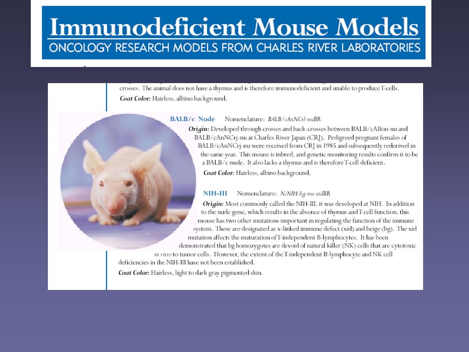
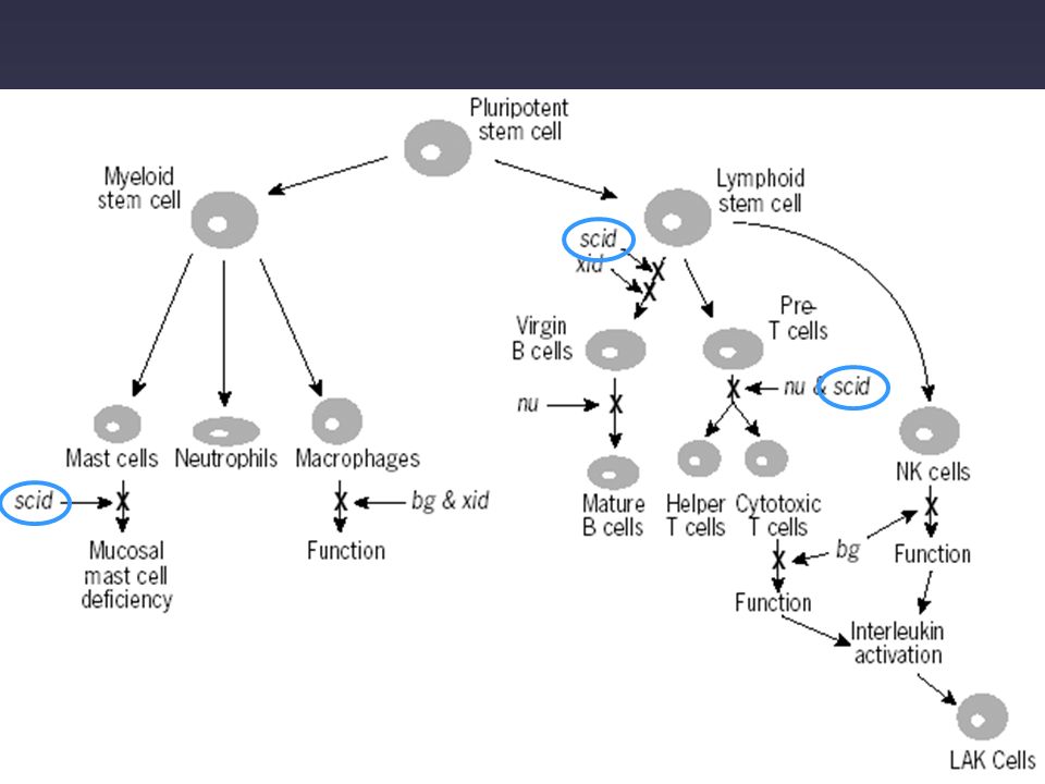
Topi nudi (nu/nu) atimici: presentano una mutazione omozigote a livello del gene nu (cromosoma 11)
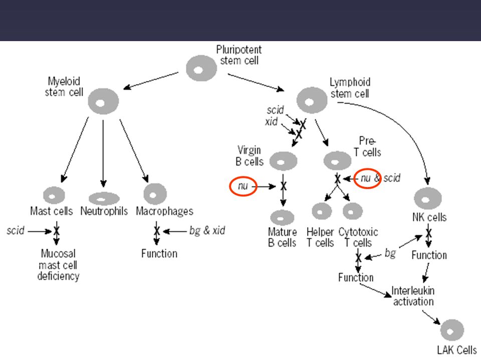
T linfociti B linfociti Cellule NK Macrofagi Cellule LAK scarsissimi difetti nella maturazione attività significativamente elevata normali

21
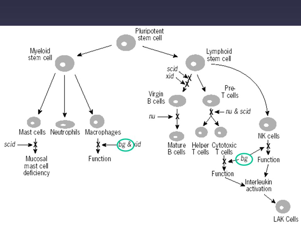
Mutazioni combinate bg (beige)/nu
Mutazioni combinate bg (beige)/nu. La mutazione bg (cromosoma 13) comporta pigmentazione della cute e blocco nella funzionalità delle cellule NK B linfociti T linfociti Cellule NK Macrofagi Cellule LAK = attività Difetto della coagulazione
/nu. La mutazione bg (cromosoma 13) comporta pigmentazione della cute e blocco nella funzionalità delle cellule NK. B linfociti. T linfociti. Cellule NK. Macrofagi. Cellule LAK. = attività. Difetto della coagulazione.")
23
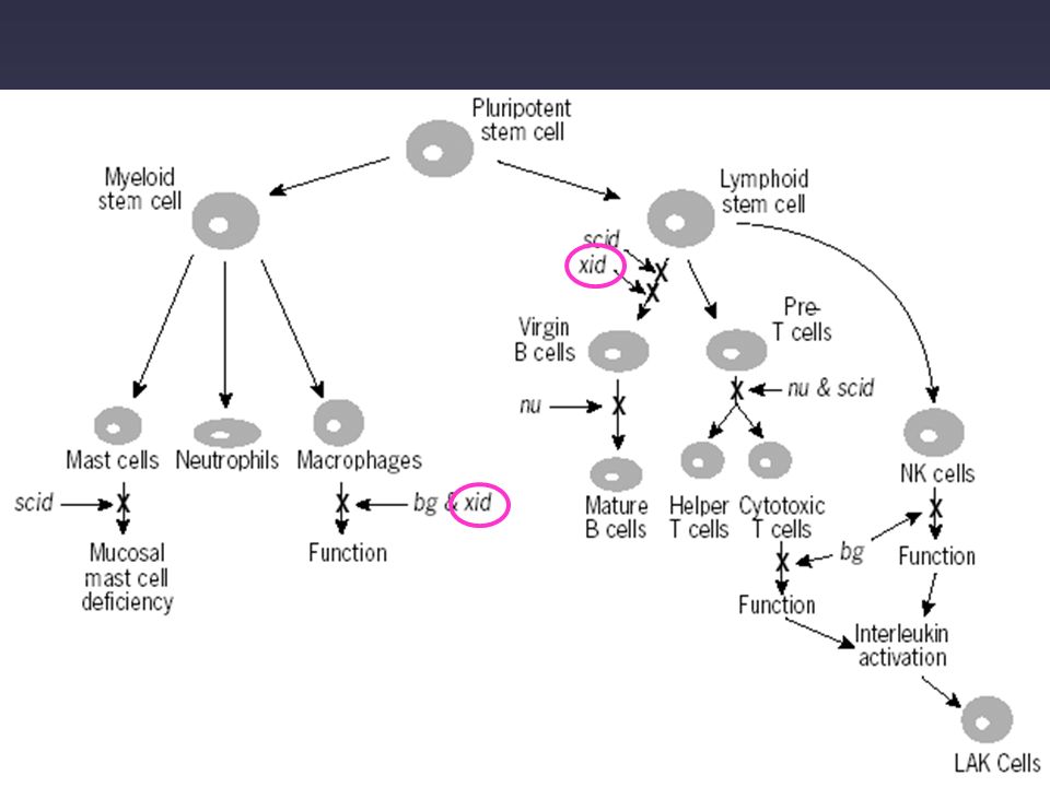
Mutazioni combinate bg/nu/xid (X-linked immunodeficiency)
B linfociti T linfociti Cellule NK Macrofagi Cellule LAK Difetto della coagulazione

24
difetti nella maturazione scarsissimi normali
Topi SCID (Severe Combined ImmunoDeficiency): possiedono una mutazione recessiva a livello del cromosoma 16 B linfociti T linfociti Cellule NK Macrofagi Cellule LAK difetti nella maturazione scarsissimi normali C.B-17 scid/scid (scid mice) have few circulating lymphocytes and little or no serum immunoglobin (usually below 0.02 mg/ml). Lymph nodes and thymus are approximately one-tenth or less of normal size. Natural killer cell differentiation is unaffected by the mutation, however T cell receptor genes are not expressed in natural killer cells. Macrophage activation and antigen presenting function is unimpaired, as is NK cell activity. No specific functional immune system is present in the scid mouse. The scid mutation has its main impact on the rearrangement of the genes during the maturation of T and B cells. During the development of lymphocytes the genes coding for antigen receptor segments are rearranged in order to create the diversity of immune globulins and T cell receptors. It has been shown that scid lymphocytes express a defective recombinase, so that no functional antigen receptor gene rearrangement can occur and it appears that this is the explanation for the lack of T and B lymphocytes in scid mice. In the ‘leaky’ scid mice (see the following section) , A variable percentage of young adult mice (2-20 %) appear "leaky" in that they develop low numbers of functional B and T cells. The occurrence of immunoglobulins and T-cell receptor positive lymphocytes could be explained by a residual activity of the recombinase system, arise through mutation of the genes encoding the proteins involved in the recombinase system. "Leaky" scid mice are recognised through analysis of the IgG serum level. By month of age, virtually all scid mice are leaky. Scid-hu mice: the SCID-hu system involve surgical transplantation of human fetal or adult organs into SCID mice. This in vivo model allows interaction between tumor cells and a human organ environment. Several in vivo models based on xenografts of human tumors in immunodeficient mice do not reproduce the incidence and/or pattern of metastatization observed in man. In all xenograft models, transplanted human tumor cells need to metastasize to and grow in mouse organs; apparently, some human tumors require a specific human tissue microenvironment to reproduce their clinical growth and invasion patterns in experimental in vivo models.
: possiedono una mutazione recessiva a livello del cromosoma 16. B linfociti. T linfociti. Cellule NK. Macrofagi. Cellule LAK. difetti nella maturazione. scarsissimi. normali. C.B-17 scid/scid (scid mice) have few circulating lymphocytes and little or no serum immunoglobin (usually below 0.02 mg/ml). Lymph nodes and thymus are approximately one-tenth or less of normal size. Natural killer cell differentiation is unaffected by the mutation, however T cell receptor genes are not expressed in natural killer cells. Macrophage activation and antigen presenting function is unimpaired, as is NK cell activity. No specific functional immune system is present in the scid mouse. The scid mutation has its main impact on the rearrangement of the genes during the maturation of T and B cells. During the development of lymphocytes the genes coding for antigen receptor segments are rearranged in order to create the diversity of immune globulins and T cell receptors. It has been shown that scid lymphocytes express a defective recombinase, so that no functional antigen receptor gene rearrangement can occur and it appears that this is the explanation for the lack of T and B lymphocytes in scid mice. In the ‘leaky’ scid mice (see the following section) , A variable percentage of young adult mice (2-20 %) appear leaky in that they develop low numbers of functional B and T cells. The occurrence of immunoglobulins and T-cell receptor positive lymphocytes could be explained by a residual activity of the recombinase system, arise through mutation of the genes encoding the proteins involved in the recombinase system. Leaky scid mice are recognised through analysis of the IgG serum level. By month of age, virtually all scid mice are leaky. Scid-hu mice: the SCID-hu system involve surgical transplantation of human fetal or adult organs into SCID mice. This in vivo model allows interaction between tumor cells and a human organ environment. Several in vivo models based on xenografts of human tumors in immunodeficient mice do not reproduce the incidence and/or pattern of metastatization observed in man. In all xenograft models, transplanted human tumor cells need to metastasize to and grow in mouse organs; apparently, some human tumors require a specific human tissue microenvironment to reproduce their clinical growth and invasion patterns in experimental in vivo models.")
26
LIMITI DEGLI XENOTRAPIANTI COME MODELLI DI TUMORI SOLIDI
Non si sviluppano attraverso un processo multistep Manca l’intervento di cellule infiammatorie e sistema immunitario Raramente sono in grado di dare luogo a metastasi Non riproducono fedelmente le interazioni tra cellule tumorali e ambiente stromale

27
XENOGEN TECHNOLOGY

28
XENOGEN TECHNOLOGY

29
The Nobel Prize in Chemistry 2008
"for the discovery and development of the green fluorescent protein, GFP" Osamu Shimomura Martin Chalfie Roger Y. Tsien

30
a | The image shows HT-1080 human fibrosarcoma cells migrating in a skin capillary 14 hours after injection into the heart. Histone H2B–green fluorescent protein (GFP) is evident in the nucleus, and retrovirally expressed red fluorescent protein (RFP) is seen in the cytoplasm. Note the high degree of cell and nuclear deformation of tumour cells. Image courtesy of K. Yamauchi, N. Yamamoto, P. Jiang, and R.M.H. b | This image shows an HT-1080 human fibrosarcoma cell, expressing GFP in the nucleus and RFP in the cytoplasm, extravasating from a blood vessel in the skin (indicated by the arrow). Note that the blood vessel contains numerous tumour cells. The extravasating cell was visualized 2 hours after cell injection of the labelled tumour cells into the heart. Image courtesy of K. Yamauchi, N. Yamamoto, P. Jiang and R.M.H. c | This image shows extravasated Lewis lung carcinoma cells growing within a blood vessel. These cells were labelled in the nucleus with histone H2B–GFP and in the cytoplasm with a retrovirally-expressed RFP. The dual-colour cells were injected into the epigastric cranialis vein and the image from a live mouse with a skin flap was taken 120 hours post-injection. Image courtesy of K. Yamauchi and R.M.H.
 is evident in the nucleus, and retrovirally expressed red fluorescent protein (RFP) is seen in the cytoplasm. Note the high degree of cell and nuclear deformation of tumour cells. Image courtesy of K. Yamauchi, N. Yamamoto, P. Jiang, and R.M.H. b | This image shows an HT-1080 human fibrosarcoma cell, expressing GFP in the nucleus and RFP in the cytoplasm, extravasating from a blood vessel in the skin (indicated by the arrow). Note that the blood vessel contains numerous tumour cells. The extravasating cell was visualized 2 hours after cell injection of the labelled tumour cells into the heart. Image courtesy of K. Yamauchi, N. Yamamoto, P. Jiang and R.M.H. c | This image shows extravasated Lewis lung carcinoma cells growing within a blood vessel. These cells were labelled in the nucleus with histone H2B–GFP and in the cytoplasm with a retrovirally-expressed RFP. The dual-colour cells were injected into the epigastric cranialis vein and the image from a live mouse with a skin flap was taken 120 hours post-injection. Image courtesy of K. Yamauchi and R.M.H..")
31
After mixed implantation in severe combined immunodeficient mice green fluorescent protein (GFP)-labelled or red fluorescent protein (RFP)-labelled HT-1080 human fibrosarcoma cells were used to determine the clonality of metastatic colonies. a | Two clonal lung colonies, one GFP-expressing, the other RFP-expressing, growing next to one another. b | A non-clonal mixed red and green fluorescent RFP–GFP colony.

32
The images show extensive locoregional and metastatic growth of red fluorescent protein-expressing Mia-PaCa-2 human pancreatic cancer cells. The images show either the whole-body image (a) or the image gathered after autopsy (b). The tumour load, as determined by whole-body imaging, significantly correlates with traditional measurements obtained after autopsy as shown in the graph (c; r = 0.89). The presence of abdominal ascites only slightly reduces the accuracy of such measurements (r = 0.83) when compared with mice without ascites (r = 0.95). Reproduced with permission from Ref. 54 © (2003) Elsevier Science.
 or the image gathered after autopsy (b). The tumour load, as determined by whole-body imaging, significantly correlates with traditional measurements obtained after autopsy as shown in the graph (c; r = 0.89). The presence of abdominal ascites only slightly reduces the accuracy of such measurements (r = 0.83) when compared with mice without ascites (r = 0.95). Reproduced with permission from Ref. 54 © (2003) Elsevier Science..")
33
This series of images show Mia-PaCa-2 pancreatic cancer (expressing red fluorescent protein) progression and evaluation of therapeutic efficacy over time. The images were obtained using real-time whole-body imaging of representative mice from each treatment group on days 10, 17, 24, 48 and 56 after tumour implantation. Thick arrows show the primary tumour and thin arrows indicate metastases. Treatment with irinotecan suppressed primary and metastatic tumour growth compared with controls. Gemcitabine successfully induced temporary regression of disease over the first month, after which growth and distant metastasis of the tumour accelerated despite continued treatment. Reproduced with permission from

34
The image shows dendritic-cell-like, green fluorescent protein (GFP)-expressing host cells directly contacting B16F10–red fluorescent protein (RFP)-expressing melanoma cells (interactions between cells indicated by arrows). This image was taken from a GFP-expressing transgenic mouse injected with RFP-expressing tumour cells. Reproduced with permission from Ref. 39 © (2003) National Academy of Sciences of the USA.
 National Academy of Sciences of the USA..")
35
MODELLI ANIMALI NELLA RICERCA SUL CANCRO
CANCEROGENESI CHIMICA O FISICA TRAPIANTO DI CELLULE TUMORALI O FRAMMENTI DI TUMORE (generalmente nel topo) ANIMALI GENETICAMENTE MODIFICATI (generalmente topi)
 ANIMALI GENETICAMENTE MODIFICATI (generalmente topi)")
36
Cancer arises through the sequential accumulation of genetic and epigenetic changes that drive the conversion of normal cells into highly malignant derivatives. a | In sporadic cancer, the initiating mutation affects a single cell in an otherwise normal microenvironment. b | Conventional tumour-suppressor gene knockouts or oncogene-bearing transgenic mice do not mimic sporadic tumour development because the initiating mutation is present throughout the body or a particular tissue. Consequently, the functions of the microenvironment in either stimulating or inhibiting tumour growth can be impaired. Other unwanted side effects include tumorigenesis outside the tissue of interest, or the induction of mechanisms that balance the effects of the mutation.

37
Cancer arises through the sequential accumulation of genetic and epigenetic changes that drive the conversion of normal cells into highly malignant derivatives c | Conditional gene-mutation strategies allow the induction of somatic mutations in a tissue-restricted and time-specific manner, and so can faithfully mimic sporadic tumour onset and progression under well-controlled conditions. d | Simultaneous induction of multiple mutations will result in the development of specific tumours with high penetrance and short latency.

38
IL SISTEMA Cre-LoxP

39
Schematic representation of the strategy for in vivo tissue specific Cre-loxP-mediated recombination. (c) Generation of tissue specific knockout mice. Transgenic mice expressing Cre in a tissue specific manner are intercrossed with homozygous mice from step (b). In the resulting double mutants Cre will excise exons 1 and 2, leading to the inactivation of the geen of interest only in tissues and cells where Cre is expressed.
 Generation of tissue specific knockout mice. Transgenic mice expressing Cre in a tissue specific manner are intercrossed with homozygous mice from step (b). In the resulting double mutants Cre will excise exons 1 and 2, leading to the inactivation of the geen of interest only in tissues and cells where Cre is expressed..")
Presentazioni simili


 Dominanza incompleta>")


>")


>")

 to different types of stress (blue boxes). Activation of p53 can result in a number.>")
 aspetti energetici>")