Scaricare la presentazione
La presentazione è in caricamento. Aspetta per favore

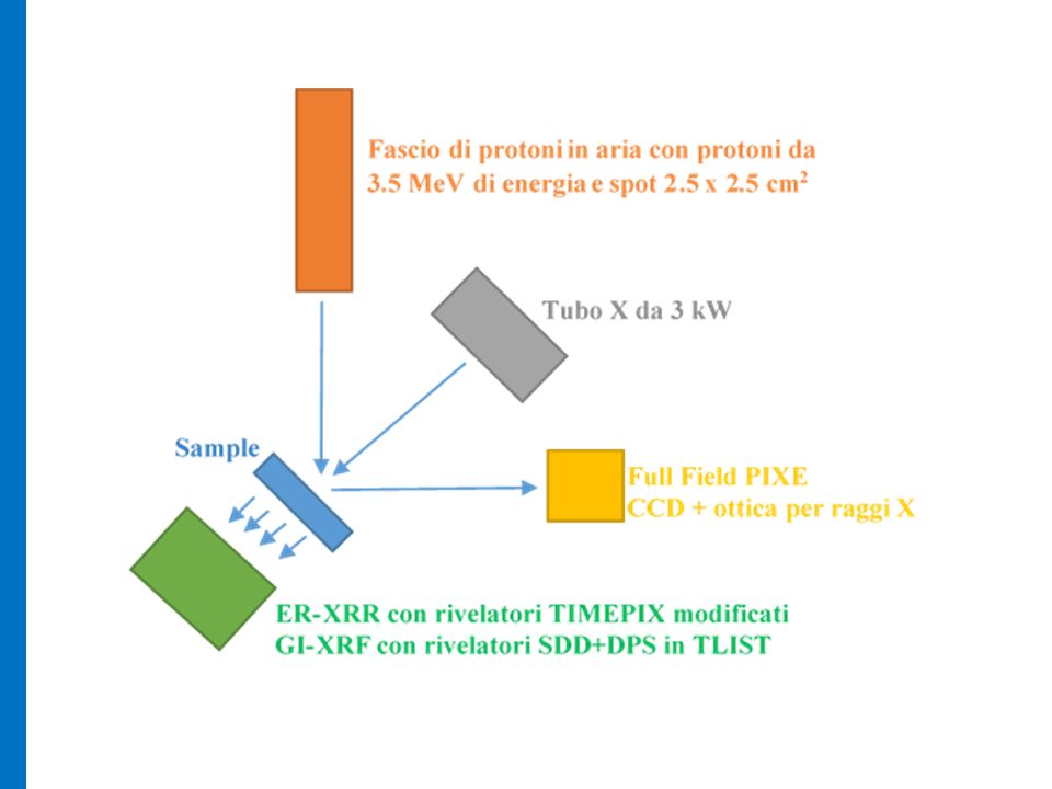
1
CHNET_Imaging (LNS, LABEC, Ferrara) Una stazione multi-tecnica per l’imaging elementale in due dimensioni e per la stratigrafia s FASCI IMPIEGATI 1) Protoni esterni da 3.5 MeV con spot circa 2x2 cm2 su linea 80°; 2) Fasci X di alta intensità da tubo X da 3 kW (già operante alla line 20 °) TECHNICHE DA INTEGRARE 1.Full Field PIXE. Alternativa alla tradizionale scansione con micro-fasci di protoni effettuata con CCD+Ottica per raggi X 2.Radiografia risolta in energia con rivelatori TIMEPIX modificati 3.Misure XRF in geometria radente con fasci laminari.

2
Materials that can benefit of imaging techniques Ancient metals Pottery, glass and enemels Illuminated parchments Paintings

3
Tecniche di Imaging ai LNS X-RAY Microscopy Risoluzione 1 um Micro FF-XRF Risoluzione 20 um

5
Macro e micro FULL FIELD X-RAY Fluorescence

6
Full Field Imaging with polycapillary optics The experimental set-up defines magnification, spatial- resolution and the field of view Advantages with respect to pinholes: very high efficiency Limits with respect pinholes: high cost and no macro-XRF 2D detector Broad X-ray or proton beam Sample XRF image polycapillary

7
Energy Dispersive X-Ray Fluorescence The charge generated on a pixel of the CCD by a photon of a given energy is proportional to its energy. Single-photon measurements allow to minimize the probability of multiple-hit events and to use the CCD as a conventional energy dispersive X-ray detector A single-photon image contains a limited number of illuminated pixels. A multi-image acquisition is necessary to obtain the statistics for the analysis.

8
Processing algorithm: a) identifies these groups in each of the SPC frames. b) searches the presence of a single maximum (single-hit) or of relative maxima in the group (multiple-hits); c) checks if the data, starting from the maxima, follow a monotonic trend and if there is an overlap among the identified distribution. Data Processing The single-photon counting (SPC) frame often present multi-pixel groups that should be corrected.
 searches the presence of a single maximum (single-hit) or of relative maxima in the group (multiple-hits); c) checks if the data, starting from the maxima, follow a monotonic trend and if there is an overlap among the identified distribution. Data Processing The single-photon counting (SPC) frame often present multi-pixel groups that should be corrected..")
9
FOM of data processing Pure targets Ti at: 38 kV and 1.7 mA Bin= 2 (512x512) Readout speed= 1 MHz Titanium target in the Macro-FF-XRF set-up Net Count = 188389 (about 160 cps) Rejected = 6123 (3% of Net count) Pile-up = 520 (0.27% of Net count) due to a spatial overlap Live Time = 1000 sec. (50 frames of 20 seconds each) Total time = 1200 sec. (including readout and processing time)
 Total time = 1200 sec. (including readout and processing time).")
10
Energy Response of the CCD detector The pure targets were irradiated at 35 kV & 1 mA. The acquisition was performed in a single photon mode

11
Energy Resolution of the CCD detector

12
Spatial resolution of the FF-XPC

13
TimePIX detector for fast energy resolved X-ray radiography In order to improve energy resolution and threshold of the system: 1.100 um thick Silicon instead of 300 um 2.Peltier cooling of the detector, under vacuum set-up 3.Removal of the Al coatings on the surface Distinctive aspects of the detector: 1.Very compact geometry; 2.256x256 pixels of 52 um lateral size 3.Up to 850 frame/second Strong limits for XRS applications: 1.Low energy resolution (0,5-1 keV) 2.Low efficiency in the XRF domain (>5 keV)
 2.Low efficiency in the XRF domain (>5 keV)")
14
X-ray Spectroscopy with TimePIX Preliminary X-ray spectra with a standard version of TimePIXE operating with a multi-frame acquisition in photon counting mode

15
Pinholes or polycapillary will be used in the energy resolved set-up for changing magnifications and field of view Spatial and energy resolved approach by a TimePIX detector 10 K frames at 0.05 ms exposure time TIMEPix detector with a polycapillary angular filter

16
Cristallo a mosaicoTubo a raggi X Doppio collimatore Goniometri isocentrici Fascio quasi monocromatico Campione Rivelatore Setup sperimentale per la monocromatizzazione su cristallo

17
Mass attenuation coefficients μ/ρ (cm 2 /g) Energy (KeV) 17 9 keV 10.3 keV Zn K-edge = 9.65 KeV K-edge differential radiography Ferrara: imaging con raggi X monocromatici
 Energy (KeV) 17 9 keV 10.3 keV Zn K-edge = 9.65 KeV K-edge differential radiography Ferrara: imaging con raggi X monocromatici")
18
Anonimo, XX sec. Marina campana Olio su tavola Immagine Low Energy 9keV Radiografie monocromatiche Immagine High Energy 10.3keV Distribuzione dell' elemento: Zinco

19
Laminar beams for grazing incidence XRF and stratigraphic investigations Diode beam alignment SDD detector + Fast DSP (tlist) 3KW X-ray source + optics Polycapillary for quasi parallel beam Pinholes for production of the laminar beam
 3KW X-ray source + optics Polycapillary for quasi parallel beam Pinholes for production of the laminar beam")
20
CHNET_Imaging (LNS) Cognome e NomeQualifica% Romano Francesco PaoloRicercatore CNR80 Pappalardo LigheaRicercatore CNR100 Rizzo FrancescaProf. Associato30 Caliri ClaudiaPhD100 Hellen SantosPost-Doc100 Roberto CatalanoAssegnista Sezione50 Andrea OrlandoBorsista INAF50 FTE totali LNS: 5,1 * In aggiunta altri due laureandi (magistrale)
.")
21
CHNET_Imaging (Firenze e Ferrara) Cognome e NomeQualifica% Massimo ChiariRicercatore INFN10 Lorenzo GiuntiniProf. Ass.70 Mirko MassiDocente Scuola Sec.40 FTE totali LABEC: 1,2 FTE totali FERRARA: 1,3 Cognome e NomeQualifica% Anna ImpallariaDottorando100 Ferruccio PetrucciProf. Ass.30 FTE totali CHNET_IMAGING: 7.7

22
CHNET_Imaging (Piano Finanziario 2015) UnitàMiss.Cons.Invent.Appar.Trasp.Totali LNS3,032,08,01,544,5 LABEC/Firenze2,08,09,519,5 Ferrara2,02,57,512,0 TOTALI710,541,515,501,576,00
 UnitàMiss.Cons.Invent.Appar.Trasp.Totali LNS3,032,08,01,544,5 LABEC/Firenze2,08,09,519,5 Ferrara2,02,57,512,0 TOTALI710,541,515,501,576,00")
23
CHNET_Imaging (LNS): main items Inventario 1. Rivelatore TIMEPIX con modifiche 10.5 k€ (LNS) 2. Gruppo da vuoto 4.5 k€ x 2 (LNS, LABEC) 3. Digital Pulse Processor in TLIST mode 7.5 k (LNS) 4. Shutter Elettro-meccanico per raggi X con 20 ms open/close 5 k€ (LNS) 5. Goniometro motorizzato programmabile 4.5 k€ (LNS) 6. Sistema posizionamento campioni 9.5 k€ (LABEC) Apparati Sistema acquisizione e controllo con CPU NationaI Instruments cRIO 8 k€ Meccanica per radiografie Missioni Misure presso Ferrara, LNS, LANBEC Trasporto Contributo trasporto FF-PIXE da LNS presso LABEC e Ferrara 1.5 k€
 3. Digital Pulse Processor in TLIST mode 7.5 k (LNS) 4. Shutter Elettro-meccanico per raggi X con 20 ms open/close 5 k€ (LNS) 5. Goniometro motorizzato programmabile 4.5 k€ (LNS) 6. Sistema posizionamento campioni 9.5 k€ (LABEC) Apparati Sistema acquisizione e controllo con CPU NationaI Instruments cRIO 8 k€ Meccanica per radiografie Missioni Misure presso Ferrara, LNS, LANBEC Trasporto Contributo trasporto FF-PIXE da LNS presso LABEC e Ferrara 1.5 k€.")
24
CHNET_Imaging: Milestones DescrizioneData completamento Misure FF-PIXE con fasci Protoni presso LNS e LABEC 30-06-2016 Set-up per radiografia risolta in energia con rivelatore TIMEPIX; Test comparativi con Sez. Ferrara su tecniche alternative di radiografia 30-06-2015 Sviluppo e test del sistema GI-XRF30-12-2016 Realizzazione del set-up integrato FF-PIXE, ER-XRR, GI-XRF e misure simultanee integrando le tecniche 30-12-201

Presentazioni simili


 SEZIONI INFN COINVOLTE: +FI.>")


 0.8 mm pitch M = 2, FoV = 25×25 mm 2 50×50 mm 2 LaBr 3 (Ce) 4 mm thick, 3 mm thick window M = 2, FoV = 25×25 mm 2 50×50 mm 2 CsI(Na)>")


>")


