Scaricare la presentazione
La presentazione è in caricamento. Aspetta per favore

1
Nuovi OCT: Prospettive Future
XIV CONGRESSO SOCIETÀ OFTALMOLOGICA CALABRESE Presidente del Congresso G. SCORCIA Organizzatori A. SCRIVANO - S.L. FORMOSO Corso teorico pratico di Semeiotica Strumentale Responsabile Scientifico: Amedeo Lucente Nuovi OCT: Prospettive Future The author declares no competing financial interests

2
Imaging Principi di fisica degli OCT SS-OCT AO-OCT & Imaging

3
Imaging

4
What is Imaging Il termine imaging è stato inventato qualche anno fa negli Stati Uniti, per definire al meglio l’evoluzione dei processi di produzione e riproduzione dell’immagine Identifica l’integrazione tra tutti gli elementi, prodotti, tecnologie e servizi che portano alla realizzazione di una comunicazione visiva Integra insieme fotografia, informatica, grafica, sviluppo, stampa

5
History of Imaging: a flood of innovation
1851 Hermann von Helmholtz direct ophthalmoscope 1871 Adolf von Bäyer Nobel Prize in chemistry 1905, synthesized fluorescein dye 1887 L. Howe “Photographs of the interior of the eye” Trans. Amer. Ophth. Soc. 1887 1915 Francis A. Welch and William Noah Allyn world's first hand-held direct illuminating ophthalmoscope

6
1925/1932 Carl Zeiss by J.W. Nordenson
modern ophthalmoscopy and photography Stroboscopic flash by Harold Edgerton “the man who stopped time” Confocal microscopy by Marvin Minsk father of artificial intelligence Fluorescein angiography (FA) by Harold Novotny and David Alvis The digital camera Kodak Laboratories by Steven Sasson Scanning Laser Ophthalmoscopy SLO by Robert H. Webb Digital photography integrated into a fundus camera by Topcon Optical Coherence Tomography OCT by D. Huang, J. G. Fujimoto et al First angiographer using cSLO (HRA Classic) by Heidelberg Engineering
 by Harold Novotny and David Alvis The digital camera Kodak Laboratories by Steven Sasson Scanning Laser Ophthalmoscopy SLO by Robert H. Webb Digital photography integrated into a fundus camera by Topcon Optical Coherence Tomography OCT by D. Huang, J. G. Fujimoto et al First angiographer using cSLO (HRA Classic) by Heidelberg Engineering.")
8
Device Imaging Single line scan Scans/ second Resolution (microns)
OCT1 1995 100 A-scans x 500 points 100 20 OCT2 2000 100 A-scans x 500 points 100 20 OCT3 Stratus OCT 2002 512 A-scans x1024 points 500 10 Cirrus HD-OCT 4096 A-scans x 1024 points 27,000 5

9
Principi di Fisica

10
Luce Retina

11
Assorbimento luce/tessuti
Finestra ottica d’utilizzo 600nm/1500nm

12
Andamento dell'attenuazione dovuta a 50 mm di acqua, in funzione della lunghezza d'onda; 50 mm di acqua corrispondono circa al percorso di andata e ritorno attraverso l'occhio umano

13
Risoluzione Assiale e Trasversale
Risoluzione Assiale = A1 / A2 Risoluzione Trasversale = T1 / T2 ?

14
Comparison of OCT resolution and imaging depths to those of alternative techniques; the “pendulum” length represents imaging depth, and the “sphere” size represents resolution

15
Field of view in OCT: viewing at the micrometer scale is possible cells such as macrophages can be identified in the plaque Occhio umano 0,1mm = 100 µm Microscopio Ottico 0,2 µm 1 µm = 0,001mm Microscopio Elettronico 0,1nm 1nm = 0,001 µm

16
Comparison of resolution and imaging depth for ultrasound, OCT and confocal microscopy
Standard clinical ultrasound can image deep structures, but has limited resolution. Higher frequencies yield finer resolution, but ultrasonic attenuation is increased limiting image penetration. The axial image resolution in OCT ranges from 1 to 15 µm and is determined by the coherence length of the light source. In most biological tissues, the imaging depth is limited to 2 to 3 mm by attenuation from optical scattering.Confocal microscopy has submicron resolution, but optical scattering limits the imaging depth to a few hundred microns in most tissues.

17
Resolution: limits of OCT
OCT can achieve high axial resolutions independent of numerical aperture. Using low coherence interferometry, the axial resolution is inversely proportional to the bandwidth of the light source. The transverse resolution is given by the focus spot size. The depth of field is determined by the confocal parameter of the focused beam

18
Performance of OCT Roll-off di performance riduzione della potenza del segnale OCT con l’aumentare della profondità ( ̴ 20dB/profondità di 2 mm) Sensibilità rapporto segnale/rumore SNR; intensità riflessa o retrodiffusa fino a ̴ 95dB Velocità d’acquisizione o d’imaging A-scan rate Range dinamico rapporto di potenza del segnale, tra la più forte e la più debole riflessione A-scan che può essere misurata ( ̴ 40-50dB)
")
19
Illumination and observation of the retina by (a) the fundus camera and (b) the SLO
 the fundus camera and (b) the SLO")
20
Confocal Scannig Laser Ophthalmoscopy

21
Schematic Overwiev of AO-SLO

22
Adaptive Optics Scanning Laser Ophthalmoscopy

23
Swept Source OCT

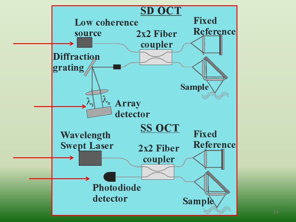
25
Swept Source v/s Sepctral Domine OCT

26
Advantage of SS-OCT - Velocità 5-10 volte maggiore
- Assenza di Roll-off di performance - Miglior rapporto segnale/rumore SNR - Larga area di scansione in retina - Alta qualità in megapixel - Maggiore Depth Resolution

27
Super Resolution OCT Eye (2011) 25, 321–330
 25, 321–330")
28
Adaptive Optics OCT

29
AO & Gemini Observatory
184 milioni di dollari dollari/die per ognuno dei Telescopi. Télescope Gemini North sur le Mauna Kea (Hawaii) Télescope Gemini South sur le Cerro Pachon (Chili)
 Télescope Gemini South. sur le Cerro Pachon (Chili)")
31
AOimage™ AOdetect™-mosaic AOdetect™-artery i2k Retina Stitching SLO
Imagine Eyes rtx1 Imagine Eyes rtx1 (zoom in) Resolution 3-4 μm Resolution μm 50 μm AOdetect™-mosaic AOdetect™-artery AOimage™ i2k Retina Stitching
 Resolution 3-4 μm. Resolution μm. 50 μm. AOdetect™-mosaic. AOdetect™-artery. AOimage™ i2k Retina Stitching.")
32
Adaptive Optics Retinal Camera rtx1

33
AO-OCT

34
Benefit of Adaptive Optics
a) Increased lateral resolution b) Reduced speckle size (granular artifact) c) Increased sensitivity to weak reflections Correction of ocular imperfections across a large pupil results in unprecedented lateral resolution (2–3 mm), sufficient for resolving individual cells en face
 Increased lateral resolution. b) Reduced speckle size (granular artifact) c) Increased sensitivity to weak reflections. Correction of ocular imperfections across a large pupil results in unprecedented. lateral resolution (2–3 mm), sufficient for resolving individual cells en face.")
35
3D resolution (3 × 3 × 3 μm³) Comparison of (top) cell size in a histological cross section of the human retina with (bottom) the resolving capability of the major types of retinal imaging modalities with and without AO. The vertical and horizontal dimensions of the solid black symbols denote, respectively, the lateral and axial resolution of the instruments. Examples shown include the commercial confocal scanning laser ophthalmoscope (cSLO), confocal scanning laser ophthalmoscope with adaptive optics (AO–cSLO), flood illumination with adaptive optics, commercial OCT, ultrahigh-resolution OCT (UHR–OCT), and ultrahigh-resolution OCT with adaptive optics (UHR–AO–OCT). Miller DT1, Kocaoglu OP, Wang Q, Lee S. Eye (Lond) Mar;25(3): doi: /eye
 cell size in a histological cross section of the human retina with (bottom) the resolving capability of the major types of retinal imaging modalities with and without AO. The vertical and horizontal dimensions of the solid black symbols denote, respectively, the lateral and axial resolution of the instruments. Examples shown include the commercial confocal scanning laser ophthalmoscope (cSLO), confocal scanning laser ophthalmoscope with adaptive optics (AO–cSLO), flood illumination with adaptive optics, commercial OCT, ultrahigh-resolution OCT (UHR–OCT), and ultrahigh-resolution OCT with adaptive optics (UHR–AO–OCT). Miller DT1, Kocaoglu OP, Wang Q, Lee S. Eye (Lond) Mar;25(3): doi: /eye")
36
AO-OCT Robert J. Zawadzki IEEE JOURNAL OF SELECTED TOPICS IN QUANTUM ELECTRONICS, VOL. 20, NO. 2, MARCH/APRIL 2014

37
OCT future SS-OCT(1050nm) AS-OCT(1310nm) SLO AO Adaptive Optics
OCM Optical Coherence Microscopy Wide Field Agreement Updateable Algorithms

38
Optical Coherence Microscopy (OCM) combines the coherent detection methods of OCT with confocal microscopy. OCM provides enhanced penetration depth compared to standard confocal microscopy, while dramatically improving the resolution over cross-sectional OCT imaging by James G. Fujimoto

39
MISAM Multifocal Interferometric Synthetic Aperture Microscopy
Yang Xu et al. published 27 Jun 2014 OPTICS EXPRESS MISAM Multifocal Interferometric Synthetic Aperture Microscopy Trade-off between the effective DOF, Depth Of Field, and Transverse Resolution in OCT Multifocal OCT and ISAM, Interferometric Synthetic Aperture Microscopy, at wavelength of 1 µm and whth refractive index of 1.4

40
Il medico deve curare a volte, alleviare spesso, confortare sempre
Il medico deve curare a volte, alleviare spesso, confortare sempre. Edward Livingston Trudeau

41
Thanks for Your attention

Presentazioni simili


>")


>")


 IL PUBBLICO DI CASTELLI.>")

