Scaricare la presentazione
La presentazione è in caricamento. Aspetta per favore

1
Why STI Diagnosis is Important
STIs rapidly spread throughout communities. STIs can be associated with acute illness. STIs can be associated with chronic illness. STIs can be associated with remote illness. Ulcerative STI associated transmission of other illnesses, especially HIV.

2
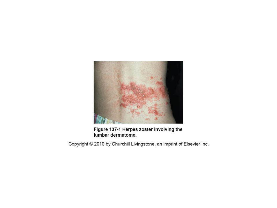
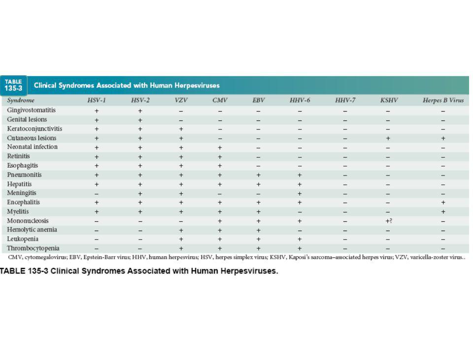
GENITAL ULCER DISEASE (GUD)
I. GENERAL CONSIDERATIONS Genital ulcer disease is one of the major STD syndromes. SETTING UP RAPID, SIMPLE, EASY TO INTERPRET TESTS IS THE CHALLENGE IN GUD DIAGNOSIS

3
DIFFERENTIAL DIAGNOSIS (I)
A. STD-related etiologies and organisms: 1. Genital herpes: Herpes Simplex Virus Type 1 and Type 2 2. Primary syphilis: Treponema pallidum 3. Chancroid: Haemophilus ducreyi 4. Lymphogranuloma venereum (LGV): Chlamydia trachomatis 5. Granuloma inguinale (Donovanosis): Calymmatobacterium granulomatis
: Chlamydia trachomatis. 5. Granuloma inguinale (Donovanosis): Calymmatobacterium. granulomatis.")
4
DIFFERENTIAL DIAGNOSIS (II)
B. Non STD-related etiologies: 1. Non-STD infectious causes of GUD: scabies, common skin infections (e.g. Staph). 2. Non-infectious causes of GUD: aphthous ulcers, Behcet’s syndrome, fixed drug eruption, Reiter’s syndrome, trauma/abrasions. C. No etiology No etiology is found in 20% to 50% of GUD cases, most likely related to the sensitivity of the laboratory tests (affected by self-medication, duration of lesion, technology of the test).
. 2. Non-infectious causes of GUD: aphthous ulcers, Behcet’s syndrome, fixed drug eruption, Reiter’s syndrome, trauma/abrasions. C. No etiology. No etiology is found in 20% to 50% of GUD cases, most likely related to the sensitivity of the laboratory tests (affected by self-medication, duration of lesion, technology of the test).")
5
EPIDEMIOLOGY (1) 1. It is important to know the epidemiological risk factors of disease, including demographic and behavioral characteristics of the patient, and travel abroad or in regions with high rates of syphilis or chancroid.

6
EPIDEMIOLOGY (2) 2. Globally, the most frequent cause of STD-related GUD is genital herpes, followed by syphilis, then chancroid. Lymphogranuloma venereum (LGV) is rare in the U.S. and Europe, and granuloma inguinale (GI, or donovonosis) is almost never encountered in the U.S. and Europe 3. In developing countries, leading causes of GUD are infections with H. ducreyi, followed by infections with T. pallidum and HSV infections. 4. More than one disease is sometimes present in a patient with genital ulcers.
 is rare in the U.S. and Europe, and granuloma inguinale (GI, or donovonosis) is almost never encountered in the U.S. and Europe. 3. In developing countries, leading causes of GUD are infections with H. ducreyi, followed by infections with T. pallidum and HSV infections. 4. More than one disease is sometimes present in a patient with genital ulcers.")
7
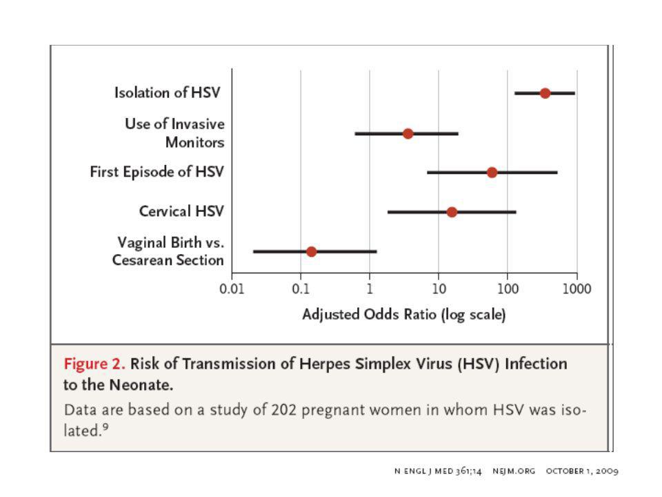
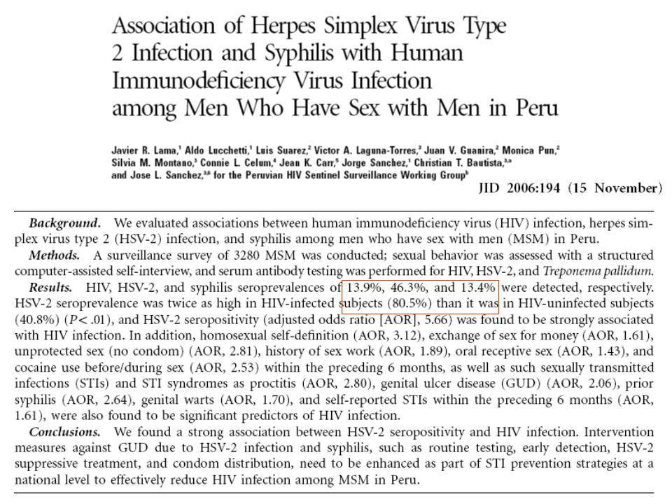
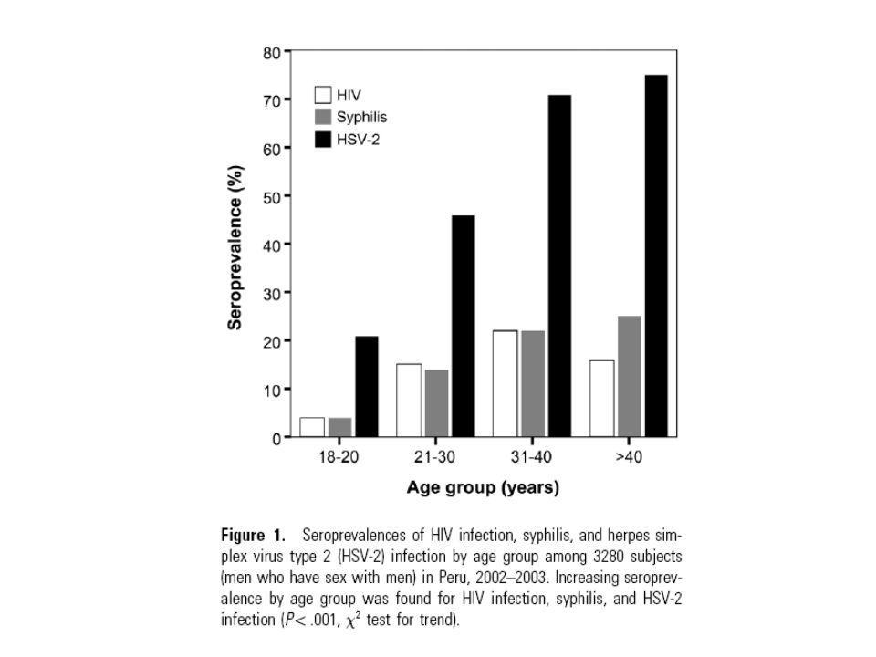
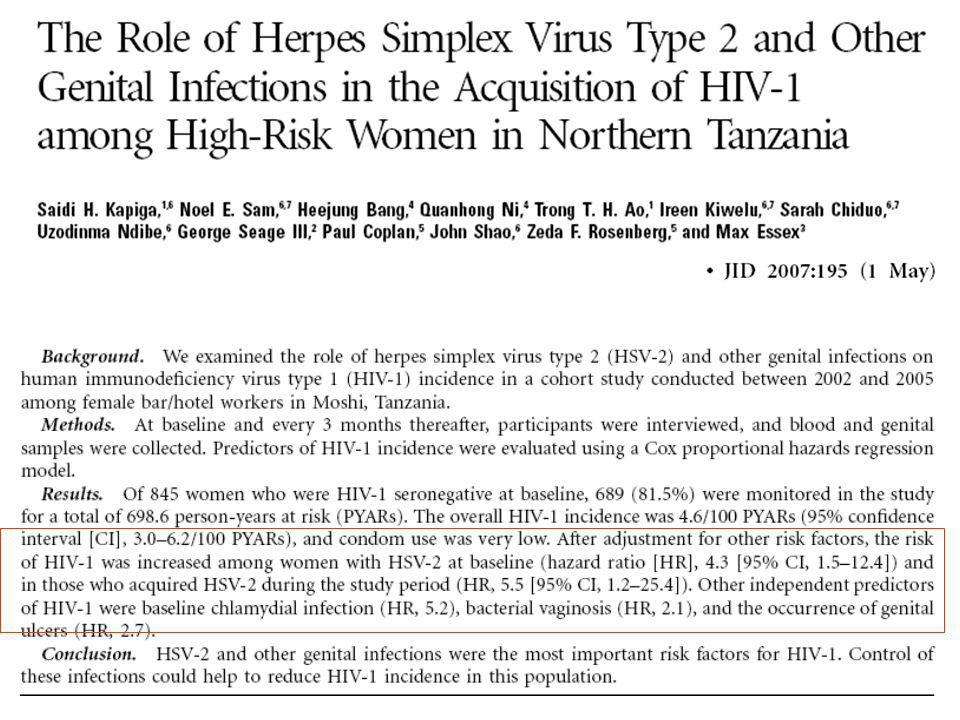
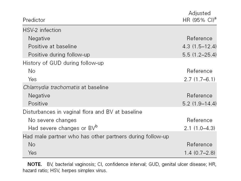
EPIDEMIOLOGY (3) 5. Multiple studies have demonstrated that the presence of GUD increases the risk of HIV infectiousness and susceptibility, resulting in an estimated 2-5-fold increase in HIV transmission rate with GUD. Significant associations with chancroid and syphilis were reported, but the main association was with HSV-2 Active coinfection with HIV-1 and HSV-2 seems to accelerate the progression to AIDS. Also, the transmission of HIV-1 may be facilitated because an increased HIV-1 viral load in blood, plasma, and semen was found in the presence of genital ulcers due to HSV-2

11
BIBLIOGRAFY Dickerson MC, Johnston J, Delea TE, White A, Andrews E. The causal role for genital ulcer disease as a risk factor for transmission of human immunodeficiency virus. An application of the Bradford Hill criteria. Sex Transm Dis Sep-Oct;23(5): Moodley P, Sturm PD, Vanmali T, Wilkinson D, Connolly C, Sturm AW. Association between HIV-1 infection, the etiology of genital ulcer disease, and response to syndromic management. Sex Transm Dis Mar;30(3):241-5. J McClelland RS, Lavreys L, Katingima C, Overbaugh J, Chohan V, Mandaliya K, Ndinya-Achola J, Baeten JM. Contribution of HIV-1 infection to acquisition of sexually transmitted disease: a 10-year prospective study. Infect Dis Feb 1;191(3):333-8.
: Moodley P, Sturm PD, Vanmali T, Wilkinson D, Connolly C, Sturm AW. Association between HIV-1 infection, the etiology of genital ulcer disease, and response to syndromic management. Sex Transm Dis Mar;30(3): J McClelland RS, Lavreys L, Katingima C, Overbaugh J, Chohan V, Mandaliya K, Ndinya-Achola J, Baeten JM. Contribution of HIV-1 infection to acquisition of sexually transmitted disease: a 10-year prospective study. Infect Dis Feb 1;191(3):")
12
DIAGNOSTIC APPROACH (1)
A. Patient history: 1. Lesion history: prodrome, initial presentation (especially presence of vesicles), duration of lesion, pain, other systemic symptoms, use of systemic or topical remedies, any history of similar symptoms in the past or partners with similar symptoms. 2. Medical history: HIV status, skin conditions, drug allergies, medications. 3. Sexual history: gender of partners, number of partners (new, anonymous, serodiscordant), venue for meeting partners, commercial sex exposure, partners with symptoms or signs, partners with known HSV or recent syphilis diagnosis. 4. Travel history
, duration of lesion, pain, other systemic symptoms, use of. systemic or topical remedies, any history of similar symptoms in the. past or partners with similar symptoms. 2. Medical history: HIV status, skin conditions, drug allergies, medications. 3. Sexual history: gender of partners, number of partners (new, anonymous, serodiscordant), venue for meeting partners, commercial. sex exposure, partners with symptoms or signs, partners with known. HSV or recent syphilis diagnosis. 4. Travel history.")
13
DIAGNOSTIC APPROACH (2)
B. Physical exam: 1. Lesion: examine for appearance, distribution, number, size, induration and tenderness. 2. Genital exam: examine genital and perianal area for other lesions. 3. Lymph node(s): note number and location of enlarged nodes, size,tenderness. 4. General exam: thorough examination of oral cavity and skin of torso, palms and soles, and neurologic exam, including cranial nerves.
: note number and location of enlarged nodes, size,tenderness. 4. General exam: thorough examination of oral cavity and skin of torso, palms and soles, and neurologic exam, including cranial nerves.")
14
LESIONS Genital ulcers may present themselves in various forms
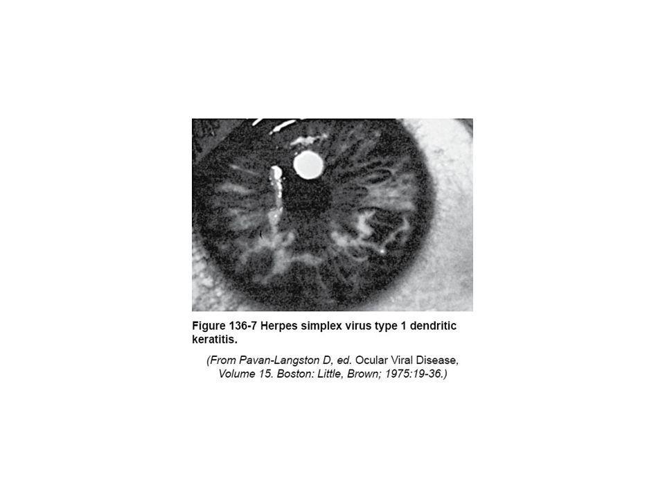
The classic lesion of primary syphilis is a single, painless, indurated ulcer with a clean base and is caused by active infection with T. pallidum The herpetic lesion is characterized by multiple painful lesions which may be recurrent There is, however, significant variability in morphologic presentation, making the clinical interpretation unreliable when used without confirmatory laboratory tests

15
DIAGNOSTIC APPROACH (3)
C. Laboratory testing: General approach. After a sexual history and physical exam of the patient presenting with GUD, the provider needs to consider the complete differential diagnosis and conduct laboratory testing based on clinic testing capability and probability of disease. a) ALL patients with GUD should receive a non-treponemal syphilis serology test (RPR or VDRL). b) If HSV is suspected, a culture or antigen test for HSV should be performed. Consider HSV type-specific serology if history is suggestive of a recurrent lesion or no lesion amenable to culture.
 ALL patients with GUD should receive a non-treponemal syphilis serology test (RPR or VDRL). b) If HSV is suspected, a culture or antigen test for HSV should be performed. Consider HSV type-specific serology if history is suggestive of a recurrent lesion or no lesion amenable to culture.")
16
..laboratory testing c) All patients with sexual risk factors presenting with GUD should be offered counseling and testing for HIV, and screened for other STDs (e.g. chlamydia and gonorrhea). d) Routine testing of all patients with GUD for chancroid is not indicated. Consider if the patient gives a history of travel to an area where chancroid is prevalent, or if the lesion does not respond to treatment, in a patient with negative syphilis serologies.
 All patients with sexual risk factors presenting with GUD should be offered counseling and testing for HIV, and screened for other STDs (e.g. chlamydia and gonorrhea). d) Routine testing of all patients with GUD for chancroid is not indicated. Consider if the patient gives a history of travel to an area where chancroid is prevalent, or if the lesion does not respond to treatment, in a patient with negative syphilis serologies.")
17
Different laboratory tests can be used to discriminate between the causative agents of GUD, and each test differs with respect to sensitivity, specificity, and turnaround time

18
Diagnosi di laboratorio
-Diagnosi diretta -Diagnosi indiretta -importante per HSV -fondamentale per T.pallidum

19
INFORMAZIONI GENERALI
DIAGNOSI DIRETTA INFORMAZIONI GENERALI

20
HSV Culture provides direct evidence for infection and is the "gold standard" for HSV detection, but it may take up to 1 week to get a definitive negative result. Positive results can be obtained within 2 days, and the differentiation into HSV-1 or HSV-2 is possible by using monoclonal antibodies. Direct detection of HSV antigen by immunoassay also enables a fast diagnosis.

21
T. PALLIDUM In vitro culturing of T. pallidum is not possible at all. T. pallidum can be detected by dark-field microscopy examination, but this is a specialized test that is not routinely performed. H. DUCREY H. ducreyi is a fastidious microorganism that is detected by a rather problematic culture technique. Nevertheless, culture is the gold standard for H. ducreyi detection.

22
Polymerase chain reaction
In the past decade, amplification techniques such as PCR have been developed to detect many different infectious agents, including HSV-1, HSV-2, T. pallidum, and H. ducreyi. PCR can be performed for each agent separately or, more efficiently, by a multiplex assay. The advantages of PCR are the direct detection of the pathogen itself, the high sensitivity, and the potentially short turnaround time. A disadvantage is that it should be performed with great care to prevent carryover contamination.

23
Advantages of PCR in GUD diagnosis
PCR represents a great improvement for laboratory diagnosis in the sense that there are fewer patients with genital ulcers for whom no definite cause is found. The turnaround time is shortened from up to 1 week for herpes virus culture to 1 to 3 days for PCR. The PCR is significantly more sensitive than culture of HSV T. pallidum PCR performed early in infection is more informative than usually used serological test (see later). A positive PCR result is indicative of an active H. ducreyi infection. So, when the costs of diagnosis are comparable and the PCR can be performed on a regular basis (i.e., three times per week), PCR detection will become the gold standard for GUD diagnosis.
. A positive PCR result is indicative of an active H. ducreyi infection. So, when the costs of diagnosis are comparable and the PCR can be performed on a regular basis (i.e., three times per week), PCR detection will become the gold standard for GUD diagnosis.")
24
Herpes genitale

25
Pathogenesis of genital HSV infection
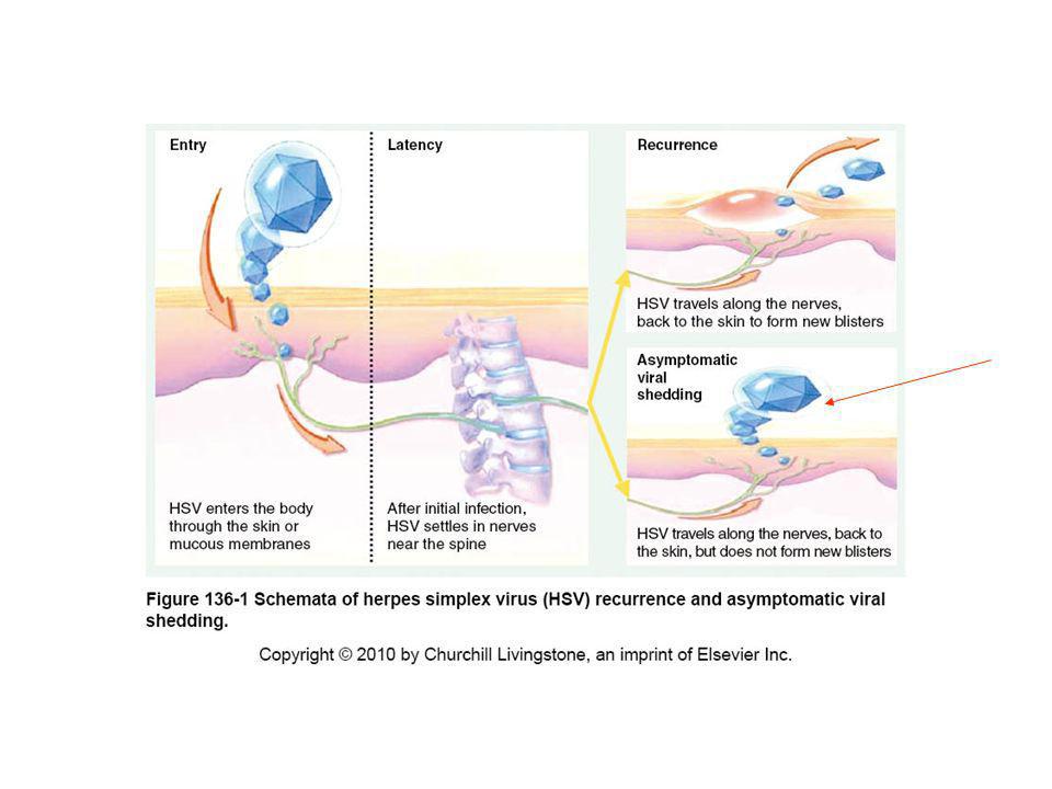
Primary infection with HSV (Sexually transmitted) Viral shedding HSV replication in genital mucosa or skin HSV replication in genital mucosa Anterograde transport of virus to mucosal and cutaneous genital tissues Uptake of HSV by sensory nerves with retrograde transport to sensory ganglia Productive viral replication in sensory neurons Host Immune Response: Circulating and Mucosal Antibody production Cytokine production Cell-mediated immunity Dorsal root ganglia Establishment of non-replicating, latent infection in sensory neurons Limits viral replication at all sites
 Viral shedding. HSV replication in genital mucosa or skin. HSV replication in genital mucosa. Anterograde transport of virus to mucosal and cutaneous genital tissues. Uptake of HSV by sensory nerves with retrograde transport to sensory ganglia. Productive viral replication in sensory neurons. Host Immune Response: Circulating and Mucosal. Antibody production Cytokine production Cell-mediated immunity. Dorsal root ganglia. Establishment of non-replicating, latent infection in sensory neurons. Limits viral replication at all sites.")
32
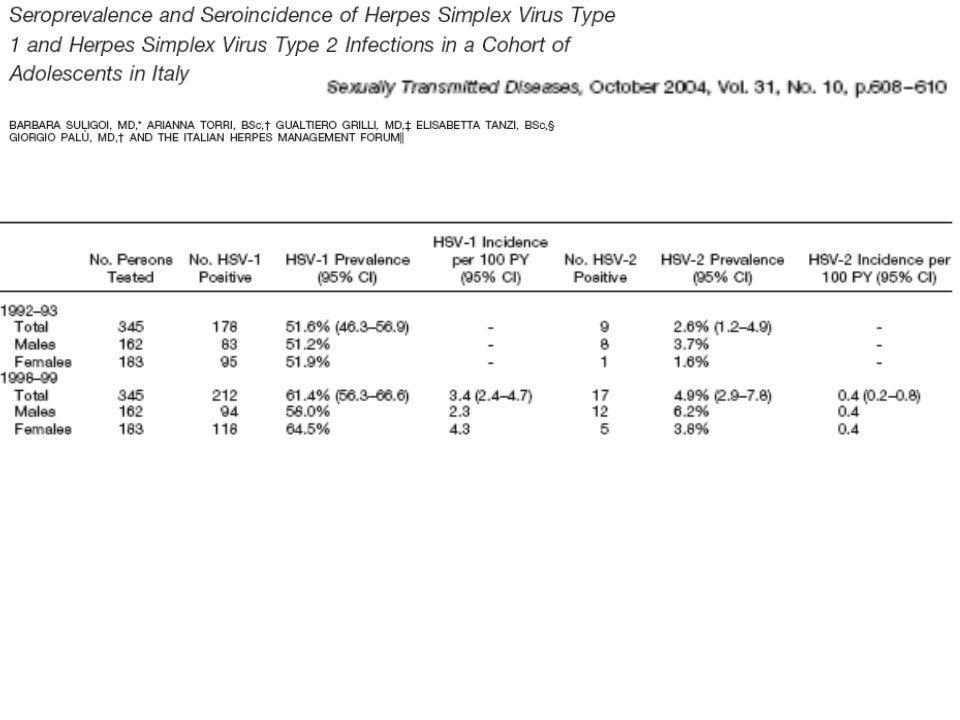
Dimensioni del problema
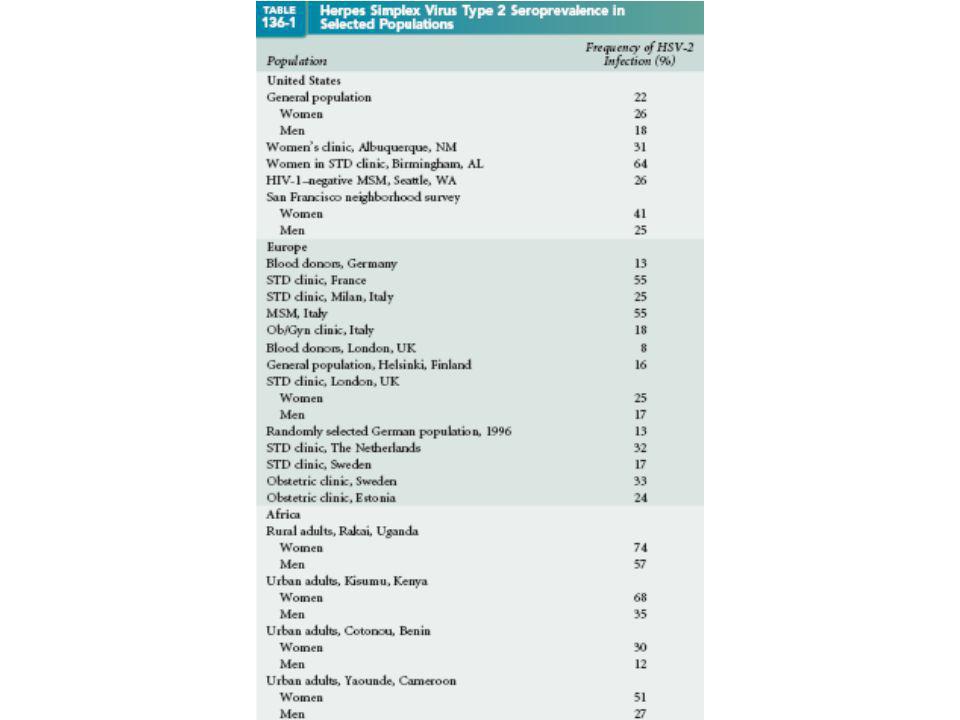
Ogni anno circa americani, in maggioranza adolescenti e giovani, sviluppano l'infezione primaria da virus Herpes simplex genitale (HSV) Un numero di individui compreso tra 25 e 31 milioni è inoltre cronicamente infetto Entrambi i ceppi di HSV, il tipo 1, HSV-1 e il tipo 2, HSV-2, possono causare malattia a livello genitale, ma è l'HSV-2 che provoca la maggioranza delle infezioni genitali recidivanti.
 Un numero di individui compreso tra 25 e 31 milioni è inoltre cronicamente infetto. Entrambi i ceppi di HSV, il tipo 1, HSV-1 e il tipo 2, HSV-2, possono causare malattia a livello genitale, ma è l HSV-2 che provoca la maggioranza delle infezioni genitali recidivanti.")
36
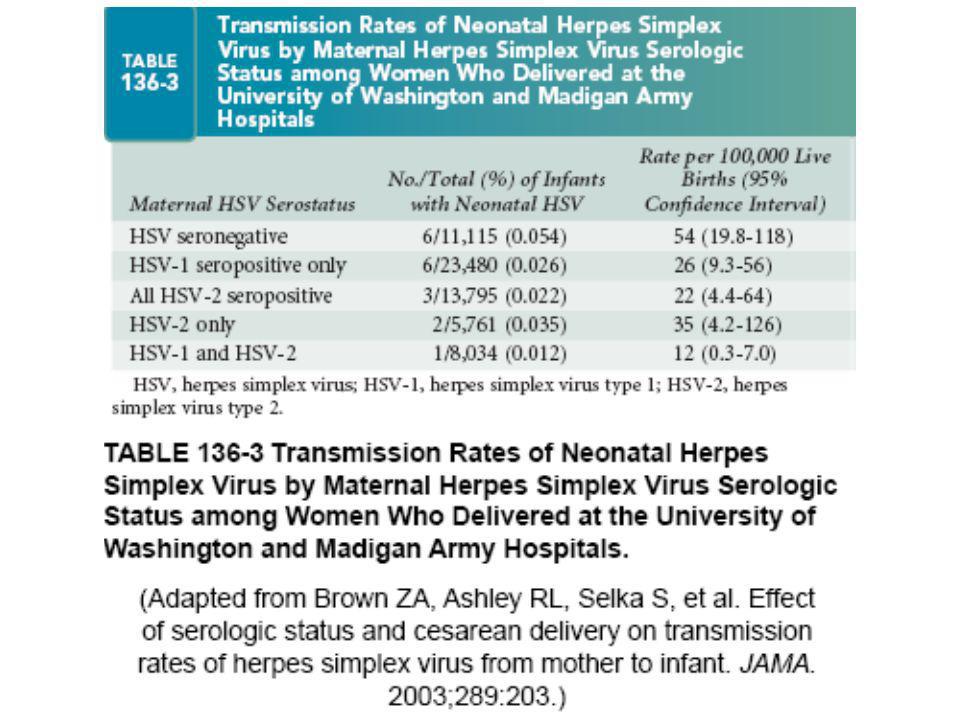
A woman had a normal course of pregnancy
A woman had a normal course of pregnancy. In particular, there were no clinical and laboratory hints to rubella, varicella, syphilis and the acquired immunodeficiency syndrome. The women did not give a history of genital lesions, perineal changes and dysuria during or prior to pregnancy. She was admitted to the hospital since she developed fever, rupture of fetal membranes and premature labor at the 28th week of gestation. At the time of delivery, there were no active herpes lesions. The maternal group B streptococcus (GBS) status was negative. An antibiotic therapy and treatment for fetal lung maturation using betamethasone were started. Because of increasing infection parameters and therapy-resistant labor, the baby was born after weeks of gestation by Caesarian section.
 status was negative. An antibiotic therapy and treatment for fetal lung maturation using betamethasone were started. Because of increasing infection parameters and therapy-resistant labor, the baby was born after weeks of gestation by Caesarian section.")
37
Since the boy developed progressive respiratory insufficiency and had a high oxygen demand of up to 100%, he had to be intubated and supplied with oxygen at the age of 5 h. The laboratory parameters did not indicate infection. However, the placental data revealed histopathological evidence of acute Chorioamnionitis. The chest X-ray on the first day of life revealed signs of respiratory distress syndrome degree III. Because of the negative maternal GBS status, a neonatal GBS pneumonia could be excluded. ………… continuous positive airway pres-sure …………..oxygen for several months …..radiograph of chest indicated broncho-pulmonary dysplasia. At the age of 6 weeks, the ophthalmologic examination, showed a chorioretinal scar in the right eye. Laboratory studies to detect CMV in urine and blood using PCR were negative as were Toxoplasma gondii-specific Ig G and IgM antibodies. From the age of 4 months, the infant developed vesicles on the nose suggestive of recurrent herpes simplex infection. Using PCR, HSV-2 DNA was detected in the secretion of vesicles, but the HSV-2 strain was not isolated in cell culture.

38
The HSV type-specific serostatus using HerpeSelect® 1 and 2 ELISA IgG and recomLine HSV-1 & HSV-2 IgG was determined in the mother and the infant. HSV-2 IgG was positive and HSV-1 IgG was negative in both suggesting previous HSV-2 infection. This means that in the immunoblot both the HSV lysate band and the gG-2 band reacted with positive whereas the gG-1 band was assigned as negative. As the present case demonstrates, prenatal HSV infections may not to be associated with severe congenital abnormalities and can be overlooked easily. They cannot be diagnosed if suitable methods are not used or are not available for routine diagnostics. In the case presented, the chorioretinal scar detected in the 6-week-old infant was taken as an opportunity to exclude congenital T. gondii and CMV infections in the neonate. Although the results were negative, the neonatologists omitted further microbiological diagnostics. The retrospective type-specific serologic diagnosis revealed a previous HSV-2 infection of the mother resulting most likely in a prenatal HSV-2 infection of the infant.

39
Using these methods, the mother was identified to be at risk of infecting her fetus.
Most likely, the mother had a primary HSV-2 infection shortly before the delivery and infected her fetus. The histopathologi-cal evidence of chorioamnionitis indicated ascending infection. Even though the woman could not remember symptomatic genital herpes during pregnancy, she was admitted into the hospital because of a febrile illness accompanied by a premature labor. Case reports of intrauterine HSV infection suggest an active maternal HSV lesion usually referred to as primary lesion or a significant maternal febrile illness in pregnancy as the cause of intrauterine infection. Additionally, primary infection goes unnoticed in more than half of the patients

40
In the present case, it can be speculated whether the premature birth and the respiratory distress syndrome were also caused by HSV-2. A recent seroprevalence study of HSV-2 in Germany showed that between 2.7% and 4.7% of all children aged up to 15 years possess HSV-2-specific antibodies probably acquired by an intrauterine or neonatal infection which may be clinically unapparent or not recognized clinically.

42
E IN ITALIA? - Suligoi et al. Sex. Trasm. Inf., 2000 -Cusini M et al. Sex. Trasm. Dis., 2000

43
Sieroprevalenza per HSV-1 e HSV-2 in tre città italiane Studio di sieroprevalenza HSV, soggetti della popolazione generali di Genova, Roma e Lecce HSV-1 HSV-2 71% positivi 9% positivi negativi negativi

44
Sieroprevalenza dell’HSV-2 in adulti: Italia
(987 soggetti di età> 20 anni della popolazione generale) Studio IHF Sieroprevalenza HSV-1 e HSV-2, 15,0% 5,5% 9,9%
 Studio IHF Sieroprevalenza HSV-1 e HSV-2, ,0% 5,5% 9,9%")
45
HSV-2 seroprevalence by age and gender in a MST clinic, Milan
Study on 919 patients, 661 males and 258 females % HSV-2 + Age in years

50
Diagnosing Genital Herpes - How Do You Do It?
Steps to a Diagnosis There are two general approaches to diagnosing genital herpes: Physical exam and history Laboratory tests

51
WHAT LABORATORY TESTS? A visual examination is only one part of a complete diagnostic work-up. Making a diagnosis of genital herpes by visual inspection and by asking questions is not easy and even experts can be wrong. For one thing, genital herpes does not look the same in every patient; it can "mimic" the appearance of other sexually transmitted diseases (STDs) and other STD's can look like herpes. A mild case of herpes can easily be mistaken for something that has nothing to do with STDs such as a simple rash. Thus confirming a diagnosis with a laboratory test is critical.
 and other STD s can look like herpes. A mild case of herpes can easily be mistaken for something that has nothing to do with STDs such as a simple rash. Thus confirming a diagnosis with a laboratory test is critical.")
52
Manifestations of disease among individuals infected with HSV-2
20% Recognized symptomatic 20% Asymptomatic 60% Unrecognized symptomatic

53
EPISODIO PRIMARIO O RICORRENZA?
LA DIAGNOSI DI INFEZIONE GENITALE DA HSV SI PUO’ OTTENERE SOLO QUANDO LA VALUTAZIONE CLINICA SIA ACCOMPAGNATA DALLA COLTURA VIRALE E DA TEST DI SIEROLOGIA TIPO-SPECIFICA EPISODIO PRIMARIO O RICORRENZA? TIPO DI VIRUS? CORRETTO MANAGEMENT

54
DIAGNOSI DIRETTA: Messa in evidenza di HSV -1 e -2 o di parti di questo (antigeni o acidi nucleici)
")
55
Test di Laboratorio per la Diagnosi Diretta
SENSIBILITA’/SPECIFICITA’ Alta (>90%) Alta Bassa Alta Molto elev Molto elev TEST Isolamento virale (esame colturale) Rilevazione di Antigeni (immunofluorescenza su vetrino, EIA) Rilevazione di Acidi Nucleici
 Alta. Bassa Alta. Molto elev Molto elev. TEST. Isolamento virale. (esame colturale) Rilevazione di Antigeni (immunofluorescenza su vetrino, EIA) Rilevazione di Acidi Nucleici.")
56
Laboratory Tests Testing for the virus directly from the skin is useful if genital signs or symptoms are present at the time you are examined. When lesions or sores are present the physician or health care provider can rub the sore with a swab and submit the sample for detection of herpes simplex virus. There are four main ways laboratories detect HSV from swabs:

57
1. Viral Culture HSV-1 & HSV-2 grow in what is called tissue culture cells or "cell culture." When changes in the cell culture are seen under a microscope, the laboratory does further tests to show that the changes are due to HSV and that the virus is HSV-1 or HSV-2. This picture was taken through a microscope to show cells that have rounded up due to HSV infection. Isolation of HSV from a sore by growth in cell culture is definitive proof that HSV caused the sore.

58
However… Viral culture can take 1-10 days to become positive and even a good sample taken from a lesion with herpes may be negative Some laboratories use a version of viral culture called "shell vial" culture to make the test faster (only 1-2 days) but this test is not as sensitive as standard viral culture. This means the shell vial test has a higher chance than standard culture of being negative even though herpes is present.
 but this test is not as sensitive as standard viral culture. This means the shell vial test has a higher chance than standard culture of being negative even though herpes is present.")
59
ELVIS-ID TEST An interesting version of viral culture is called the ELVISTM (enzyme-linked virus-inducible system) -Id test. This test uses cells containing a HSV-specific gene promoter sequence linked to the reporter gene b-galactosidase. When HSV from a patient's swab infects the ELVIS cells, this enzyme is switched "on" and causes the cells to turn blue. This photo shows ELVIS "blue cells" surrounded by uninfected clear cells.

60
DISADVANTAGES BUT: ELVIS tests are usually completed in 1-2 days
It is more expensive than a standard culture test It may miss some cases of herpes either because there is so much virus in the sample that the cells are destroyed before the test can be completed or because there is very little herpes in the sample

62
2. HSV Fluorescent Antibody Test ("HSV FA")
Fluorescent antibody techniques (FA) are faster than culture and nearly as sensitive for lesions that have newly formed. These tests are less accurate for older, healing lesions. Occasionally, FA can be more sensitive than culture if the virus has not survived transport.
 are faster than culture and nearly as sensitive for lesions that have newly formed. These tests are less accurate for older, healing lesions. Occasionally, FA can be more sensitive than culture if the virus has not survived transport.")
63
3. Diagnosi citologica Cellule ottenute da scraping delle lesioni viene strisciato su vetrino Fissazione in etanolo freddo Colorazione secondo Papanicolaou, Giemsa (Tzanck) o Wright Ricerca di cellule giganti multinucleate con inclusioni intranucleari: sensibilità 60-70%
 o Wright. Ricerca di cellule giganti multinucleate con inclusioni intranucleari: sensibilità 60-70%")
65
4.Rilevazione di Acidi Nucleici: PCR (I)
Rappresenta il metodo diagnostico di scelta nelle infezioni erpetiche del SNC Questo procedimento in condizioni ottimali dimostra una sensibilità del % ed una specificità del % Permette la tipizzazione virale con la scelta di primer opportuni Richiede piccole quantità di materiale clinico Test rapido (poche ore)
")
66
4.Rilevazione di Acidi Nucleici: PCR (II)
SVANTAGGI: I controlli per le cross-contaminazioni sono fondamentali Scarsa disponibilità di saggi commerciali Alti costi Richiede laboratori attrezzati Personale tecnico con esperienza di biologia molecolare

67
Should I ask for a PCR or a culture?
At this time, PCR is less available and more expensive than culture or FA. But PCR is more likely to give a correct answer. The chance of missing herpes in a lesion by culture or FA is much higher than with PCR. If PCR is not affordable or available, a type specific serology blood test is good alternative to detect HSV infection.

68
The New Blood Tests ("Type-Specific Serology")- An Overview
The most important breakthrough in herpes diagnosis in the last few years is the development of serology tests that accurately tell you if you are a carrier of HSV-2. Serology tests detect "antibodies" in the blood. Blood tests obviously do not require swabbing a lesion, so they can be done long after symptoms have faded. The key to accuracy for herpes blood tests is to make sure that the test is a 'type-specific' assay that can tell the difference between HSV-1 and HSV-2 antibodies. Many commercially available tests currently can not make this distinction.

69
Type-specific antibodies
The new blood tests are based on antibodies to two proteins that are part of the HSV-1 and HSV-2 virus envelope. One protein is called glycoprotein gG-1 and it is found only on the outside of the HSV-1 virus or in cells infected with HSV-1 as the virus is produced. The other protein is called glycoprotein gG-2 and is found on HSV-2 virus or in cells infected with HSV-2. These proteins have similar names but they are enough different that antibodies that are produced to one HSV type (say, HSV-1) can not be mistaken for the other type (HSV-2) in the new serology tests. We call antibodies that can react to only one, not both, HSV types "type specific."
 can not be mistaken for the other type (HSV-2) in the new serology tests. We call antibodies that can react to only one, not both, HSV types type specific.")
70
Isn't gG a kind of antibody?
The short name for "glycoprotein G" is a common source of confusion. The main antibody to develop in response to infection is called Immunoglobulin G or "IgG". When scientists were working out the structure of herpes virus, they named the viral glycoproteins alphabetically in order of their discovery. Thus, gG was the seventh glycoprotein to be discovered. As shown in the diagram, other glycoproteins such as gB and gD are also on the outside of the virus.

71
Metodi sierologici tipo-specifici per l’infezione da HSV
Nome Abbr. Riferim. Bibliog. Immunodot enzyme assay gG-IEA Lee et al. 1985; Lee et al. 1986 Western blot WBA Ashley et al. 1988/1991 Immunoblot gG assay gG-blot Sanchez-Martinez et al. 1991a/1991b Indirect gG-2 ELISA gG-2 EIA Ho et al. 1993 Monoclonal antibody blocking RIA MAb-RIA Slomka et al. 1995 RIBATM Strip Immunoblot Assay RIBATM-SIA Alexander et al. 1996; Ashley et al Gull gG-EIA Gull EIA Ashley et al. 1997 MRL gG-EIA MRL-EIA Prince et al., 2000 HSV.ELISA IgG MRL-ELISA Palù et al., 2001

72
Tests to Use ELISA. Few companies make and sell this test kit in the United States and many countries around the world. Some of these tests have been approved by the FDA for diagnosing HSV infection in adults, including pregnant women. The test takes less than a day once the specimen arrives in the laboratory. Immunoblot. This test usually contains the same recombinant gG-1 and gG-2 that is used in the ELISA kits. Some of them are also FDA approved They detect antibodies to both HSV-1 and HSV-2 on a paper strip and are more expensive than the ELISAs. Immunodot. This test is a near-patient test that can be performed in offices and clinics that have laboratory facilities. It is a membrane-based immunoassay for qualitative determination of igG specific for HSV-2. The test employs a semi-purified antibody-binding protein, conjugated to colloidal gold particles and semi purified gG2. Only a couple drops of blood from a finger-stick is needed. This test requires about 10 minutes to perform and read.

73
ELISA These are the packages from Focus Technologies’ tests. Note that the company has a label on the packaging to indicate that the tests are gG Based. This should help the health care provider and his/her laboratory to know that they have purchased one of the new type specific tests.

74
IMMUNOBLOT These are actual immunoblot test strips photographed in the divided container used to perform the test. Each strip is a test for a different patient. The arrows point to the gG-2 (HSV-2) and gG-1 bands (HSV-1) on two of the strips. Most of the tests in the run have HSV-1 antibodies.
 and gG-1 bands (HSV-1) on two of the strips. Most of the tests in the run have HSV-1 antibodies.")
75
Immunoblot IgG assay (HerpeSelect™ 1 and 2)
Controllo interno (siero umano) Antigene comune (HSV) gG1 gG2 1 & 2 1 2 Eqv Neg Neg Neg Invalid Pos Pos Pos Il test e’ considerato valido se: 1)Il controllo interno e’ positivo 2)Se la banda specifica per gG1 e/o gG2 + la banda relativa all’antigene comune per HSV-1 e 2 sono visibili
 Antigene comune (HSV) gG1. gG2. 1 & Eqv. Neg. Neg. Neg. Invalid. Pos. Pos. Pos. Il test e’ considerato valido se: 1)Il controllo interno e’ positivo. 2)Se la banda specifica per gG1 e/o gG2 + la banda relativa all’antigene comune per HSV-1 e 2 sono visibili.")
76
HerpeSelect™ 1 and 2 Immunoblot IgG
Single strip containing serum control common antigen recombinant gG1 recombinant gG2 Test Highlights Single kit tests for both serotypes 20 uL serum Approx. 2 3/4 hrs. for testing Minimal equipment (platform rocker) Best for lower volume testing
 Best for lower volume testing.")
77
IMMUNODOT The biokit HSV-2 test is fast and easy to perform. This photo shows a positive patient result (right side of window) and control dot (left side of window). Normally positives are less red than the control dot, but can be as dark as the control dot. If HSV-2 antibodies are not present, only one red dot appears. Only one red dot represents a "negative" result. If a patient has only HSV-1 antibodies, the test will read "negative" with only one dot being red because this test does not contain gG-1. If neither dot turns red the test is invalid and must be repeated.
 and control dot (left side of window). Normally positives are less red than the control dot, but can be as dark as the control dot. If HSV-2 antibodies are not present, only one red dot appears. Only one red dot represents a negative result. If a patient has only HSV-1 antibodies, the test will read negative with only one dot being red because this test does not contain gG-1. If neither dot turns red the test is invalid and must be repeated.")
78
Western Blot E’ il golden standard per la diagnosi sierologica tipo specifica dell’infezione da HSV:

79
These are Western blot tests from one patient who presented with recurrent HSV-2 infection (Year 0). At 1.1, 1.9, 3.2, and 8.2 years later, serum was drawn and tested again. The left-hand blot in each pair has HSV-1 proteins and the right-hand blot has HSV-2 proteins. Someone trained in Western blot will recognize the gG-2 band and also see that the gG-1 band is missing.

80
Interpreting the gG Serology (1)
Test results from one of the glycoprotein G specific tests (ELISA or Immunoblot) are as follows: HSV-2 Antibodies Present: This means the patient has HSV-2 infection, the virus may be latent in the sacral nerves HSV-2 Antibodies NOT Present: This means with 95-98% accuracy the patient does not have genital herpes unless he acquired it very recently. New studies have shown that by 6 weeks after HSV-2 infection, 60-75% of patients will be positive by ELISA (the HerpeSelect® ) test but for a few patients (about one in 5), it can take as long as 6 months to develop antibodies. If the patient might have acquired herpes very recently, he may need to repeat the antibody test in 6-8 weeks.
 are as follows: HSV-2 Antibodies Present: This means the patient has HSV-2 infection, the virus may be latent in the sacral nerves. HSV-2 Antibodies NOT Present: This means with 95-98% accuracy the patient does not have genital herpes unless he acquired it very recently. New studies have shown that by 6 weeks after HSV-2 infection, 60-75% of patients will be positive by ELISA (the HerpeSelect® ) test but for a few patients (about one in 5), it can take as long as 6 months to develop antibodies. If the patient might have acquired herpes very recently, he may need to repeat the antibody test in 6-8 weeks.")
81
Interpreting the gG Serology (2)
HSV-1 Antibodies Present: This means the patient has HSV-1 infection. The antibody assay cannot tell where HSV-1 is latent. In most people (>90%) it is in the nerves of the mouth and eyes. Some people do have genital HSV-1 and could have HSV-1 antibodies from genital HSV-1 infection. Neither HSV-1 or HSV-2 Antibodies Present: The patient is not infected with either HSV-1 or HSV-2 but he is susceptible to getting infection. There is a small chance that he may have been recently infected, but has not made antibodies yet.
 it is in the nerves of the mouth and eyes. Some people do have genital HSV-1 and could have HSV-1 antibodies from genital HSV-1 infection. Neither HSV-1 or HSV-2 Antibodies Present: The patient is not infected with either HSV-1 or HSV-2 but he is susceptible to getting infection. There is a small chance that he may have been recently infected, but has not made antibodies yet.")
82
Potential population in whom HSV type-specific serology could be useful
To identify subclinical HSV-2 carriers Candidates for behavioural intervention Candidates for antiviral therapy Potentially reduce transmission To identify pregnant women at risk Those who are uninfected but have serologically discordant partners Those already infected

83
Potential population in whom HSV type-specific serology could be useful
To identify asymptomatic individuals in other groups Semen donors Patients entering immunosuppressive therapy Patients with HIV infection To identify candidates for vaccines

84
Suggested ordering guidelines for HSV serological tests
Situation Serologic Comments test(s) Symptomatic, None A virologic test is always better virus positive Symptomatic, Serum pair Misleading in HSV-1+ with new negative (non-typing) HSV-2 infection Serum pair (typing) Very helpful Asymptomatic, Non-typing Helps only if seronegative worried, > 8 weeks post exposure Typing Positive test cannot correlate infection to exposure event High risk, history Non-typing Helpful only if negative negative
 Symptomatic, None A virologic test is always better virus positive. Symptomatic, Serum pair Misleading in HSV-1+ with new negative (non-typing) HSV-2 infection. Serum pair (typing) Very helpful. Asymptomatic, Non-typing Helps only if seronegative worried, > 8 weeks post exposure Typing Positive test cannot correlate infection to exposure event. High risk, history Non-typing Helpful only if negative negative.")
85
Ricadute cliniche della diagnosi di laboratorio
Epidemiologia Prognosi HSV HSV-2 Valutazione del rischio di trasmissione - herpes neonatale partner associazione con HIV e/o con altri patogeni STD Valutazione Genotipica Fenotipica

90
SIFILIDE

91
SIFILIDE: EZIOLOGIA E STORIA
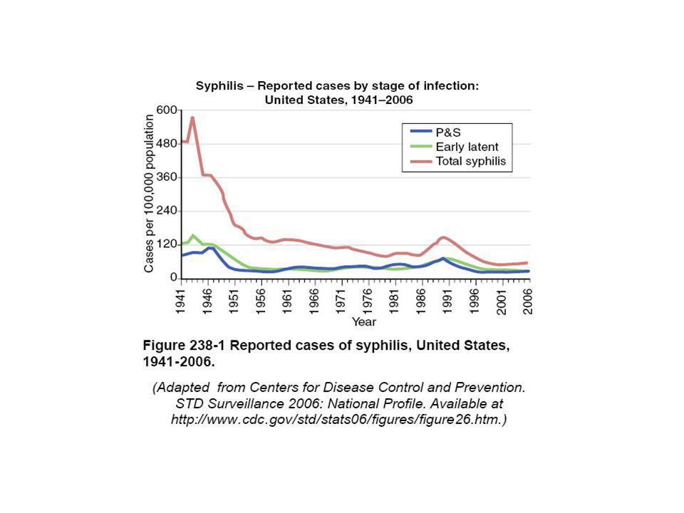
La sifilide, una malattia complessa sessualmente trasmissibile causata dal batterio Treponema pallidum, fu descritta per la prima volta nel XVI secolo e si ritiene che sia stata importata dalle Americhe dopo i primi viaggi degli spagnoli. Nei paesi industrializzati, l’incidenza della sifilide iniziò a calare verso la fine del 1800, per poi avere un altro picco dopo la Prima Guerra mondiale. Dopo la Seconda Guerra, grazie anche alla disponibilità di metodi diagnostici efficaci e al trattamento con antibiotici, la malattia ebbe una nuova riduzione, anche se negli ultimi anni la sua incidenza è andata aumentando sia nei paesi in via di sviluppo che in alcuni paesi europei. Dopo l’AIDS, la sifilide, che ha una incidenza annua di 12 milioni di nuovi malati nel mondo, è la malattia sessualmente trasmissibile con il più alto tasso di mortalità.

92
AGENTE EZIOLOGICO IL Treponema Pallidum, appartiene alla famiglia delle treponematacee, batteri di forma elicoidale, mobili, a divisione trasversale. La loro lunghezza variada 8 a 14 um e la sua larghezza da 0.15 a 0.20 um. La coltura in vitro dei treponemi non è stata ancora realizzata. Per tale motivo il loro metabolismo è poco conosciuto.

93
Nel mondo si registrano 12 milioni di nuovi casi di sifilide all’anno(OMS,1999)
")
94
Aspetti epidemiologici Nel mondo, secondo l’Oms, la sifilide colpisce circa 12 milioni di persone, con grande presenza sia in Africa che in Asia e in America Latina. Il numero di nuovi casi per anno (dal 1995 al 1999) espresso in milioni di persone è il seguente: 1995 1999 Nord America 0.107 Europa occidentale 0.136 Nord Africa e Medio Oriente 0.364 Europa Orientale e Asia Centrale 0.105 Africa Sub Sahariana 3.828 Asia Sud e Sud-est 4.038 Asia orientale e Pacifico 0.244 Australia e Nuova Zelanda 0.008 America latina e Caraibi 2.928

96
Cusini M, Ghislanzoni M, Bernardi C, Carminati G, Zerboni R, Alessi E, Suligoi B. Syphilis outbreak in Milan, Italy. Sex Transm Infect Apr;80(2):154.
:154..")
98
SIFILIDE: ASPETTI GENERALI
La sifilide è una malattia genitale che causa ulcere ed escoriazioni e facilita la trasmissione dell’Aids (rischio di trasmissione del virus HIV è da 2 a 5 volte più elevato) Grazie a un semplice test diagnostico e a una elevata efficacia della cura antibiotica, è oggi una malattia potenzialmente controllabile dai sistemi di sanità pubblica. Se non è trattata adeguatamente però può causare danni al sistema nervoso, ai vasi arteriosi, disordine mentale ed eventualmente, morte. La sifilide si trasmette di persona in persona direttamente attraverso le ferite e le ulcere che si formano nelle zone genitali, rettali e sulla bocca a seguito di contatto sessuale. La malattia può facilmente essere trasmessa
 Grazie a un semplice test diagnostico e a una elevata efficacia della cura antibiotica, è oggi una malattia potenzialmente controllabile dai sistemi di sanità pubblica. Se non è trattata adeguatamente però può causare danni al sistema nervoso, ai vasi arteriosi, disordine mentale ed eventualmente, morte. La sifilide si trasmette di persona in persona direttamente attraverso le ferite e le ulcere che si formano nelle zone genitali, rettali e sulla bocca a seguito di contatto sessuale. La malattia può facilmente essere trasmessa.")
99
STADI DELLA MALATTIA (1)
La SIFILIDE si sviluppa in diversi stadi, ciascuno caratterizzato da sintomi e decorso diverso. Sifilide primaria Tra l’infezione e l’insorgenza dei primi sintomi possono passare da 10 a 90 giorni (mediamente venti giorni). Questo stadio è caratterizzato dalla comparsa di una singola ferita, o da più pustole. Normalmente la ferita è consistente, tonda, piccola e indolore e compare nel punto in cui avviene l’infezione batterica. Questa ferita dura 3-6 settimane e guarisce da sola. Se la malattia non è trattata in questa fase, evolve verso uno stadio secondario. Sifilide secondaria Inizia quando si ha l’insorgenza di una eruzione cutanea in più punti, senza prurito. Questa eruzione può comparire durante la fase di scomparsa della ferita, o anche dopo settimane. L’eruzione è solitamente rossastra o bruna, con macchie sia sui palmi delle mani e dei piedi o in altre parti del corpo. A volte le macchie sono diverse e ricordano eruzioni tipiche di altre malattie. Anche senza alcun trattamento, l’eruzione sparisce da sola. Tra i sintomi tipici di questo stadio possono esserci anche febbre, linfonodi ingrossati, mal di gola, perdita di capelli a chiazze, mal di testa, perdita di peso, dolori muscolari, stanchezza.
. Questo stadio è caratterizzato dalla comparsa di una singola ferita, o da più pustole. Normalmente la ferita è consistente, tonda, piccola e indolore e compare nel punto in cui avviene l’infezione batterica. Questa ferita dura 3-6 settimane e guarisce da sola. Se la malattia non è trattata in questa fase, evolve verso uno stadio secondario. Sifilide secondaria Inizia quando si ha l’insorgenza di una eruzione cutanea in più punti, senza prurito. Questa eruzione può comparire durante la fase di scomparsa della ferita, o anche dopo settimane. L’eruzione è solitamente rossastra o bruna, con macchie sia sui palmi delle mani e dei piedi o in altre parti del corpo. A volte le macchie sono diverse e ricordano eruzioni tipiche di altre malattie. Anche senza alcun trattamento, l’eruzione sparisce da sola. Tra i sintomi tipici di questo stadio possono esserci anche febbre, linfonodi ingrossati, mal di gola, perdita di capelli a chiazze, mal di testa, perdita di peso, dolori muscolari, stanchezza.")
100
STADI DELLA MALATTIA (2)
Sifilide avanzata (stato latente e terziaria) Alla scomparsa dei sintomi del secondo stadio, la persona è ancora malata anche se non mostra più i sintomi evidenti. In questa fase, possono iniziare i danni agli organi interni, al cervello, ai nervi, agli occhi, al cuore e ai vasi sanguigni, al fegato, alle ossa e alle articolazioni. I danni interni possono manifestarsi anche anni dopo la comparsa dei primi sintomi. A questo punto la sifilide entra nel terzo stadio, anche se danni neurologici possono manifestarsi già dal secondo stadio (sifilide neurale). In questa fase l’individuo perde la capacità di controllare i movimenti muscolari, può avere delle paralisi, confusione mentale, cecità graduale e sviluppo di demenza. Il danno può essere tanto serio da portare alla morte. Sifilide congenita A seconda dello stato d’infezione della madre, la malattia può essere trasmessa al feto causando morte in utero (40 per cento dei casi) o la nascita di un bimbo già infetto, con sifilide congenita (70 per cento dei casi). Se la madre ha avuto la malattia nei quattro anni precedenti la gravidanza, il rischio di trasmissione al feto è molto elevato. I sintomi possono anche essere assenti al momento della nascita e comparire successivamente, causando se non trattati adeguatamente anche serie complicazioni allo sviluppo del bambino.
 Alla scomparsa dei sintomi del secondo stadio, la persona è ancora malata anche se non mostra più i sintomi evidenti. In questa fase, possono iniziare i danni agli organi interni, al cervello, ai nervi, agli occhi, al cuore e ai vasi sanguigni, al fegato, alle ossa e alle articolazioni. I danni interni possono manifestarsi anche anni dopo la comparsa dei primi sintomi. A questo punto la sifilide entra nel terzo stadio, anche se danni neurologici possono manifestarsi già dal secondo stadio (sifilide neurale). In questa fase l’individuo perde la capacità di controllare i movimenti muscolari, può avere delle paralisi, confusione mentale, cecità graduale e sviluppo di demenza. Il danno può essere tanto serio da portare alla morte. Sifilide congenita A seconda dello stato d’infezione della madre, la malattia può essere trasmessa al feto causando morte in utero (40 per cento dei casi) o la nascita di un bimbo già infetto, con sifilide congenita (70 per cento dei casi). Se la madre ha avuto la malattia nei quattro anni precedenti la gravidanza, il rischio di trasmissione al feto è molto elevato. I sintomi possono anche essere assenti al momento della nascita e comparire successivamente, causando se non trattati adeguatamente anche serie complicazioni allo sviluppo del bambino.")
101
GRAVIDANZA E SIFILIDE CONGENITA
La sifilide durante la gravidanza può determinare: Aborto Nascita di un feto morto Parto prematuro Morte neonatale Parto a termine con neonati con infezione clinicamente silente (da un terzo e metà dei casi): spesso i neonati non presentano i segni e i sintomi della malattia, che possono comparire dopo mesi o anni oppure rimanere silente per tutta la vita Parto a termine con neonati con infezione clinicamente manifesta
: spesso i neonati non presentano i segni e i sintomi della malattia, che possono comparire dopo mesi o anni oppure rimanere silente per tutta la vita. Parto a termine con neonati con infezione clinicamente manifesta.")
102
EPIDEMIOLOGIA L’aumento dell’incidenza di sifilide negli adulti si ripercuote sull’infezione congenita. Nel 1999 negli USA sono stati registrati 556 casi.

103
DIAGNOSI DI LABORATORIO
La diagnosi di sifilide può essere effettuata utilizzando un’analisi al microscopio di materiali prelevati da una escoriazione o da una ferita del paziente. La diagnosi puo’ essere effettuata anche con test serologico Esistono due tipi di test serologici: NON Treponema-specifici, (Venereal Disease Research Laboratory [VDRL] e Rapid Plasma Reagin [RPR]) Treponema-specifici (p.e. fluorescent treponemal antibody absorbed [FTA-ABS] e T. pallidum particle agglutination [TP-PA]). Il livello di anticorpi rimane nel sangue per mesi e anni anche dopo il trattamento completo della malattia. Dati gli effetti della sifilide contratta prima o durante la gravidanza, lo screening per la presenza di anticorpi anti-Treponema dovrebbe essere effettuato assieme agli altri test serologici nelle prime settimane di gestazione.
 Treponema-specifici (p.e. fluorescent treponemal antibody absorbed [FTA-ABS] e T. pallidum particle agglutination [TP-PA]). Il livello di anticorpi rimane nel sangue per mesi e anni anche dopo il trattamento completo della malattia. Dati gli effetti della sifilide contratta prima o durante la gravidanza, lo screening per la presenza di anticorpi anti-Treponema dovrebbe essere effettuato assieme agli altri test serologici nelle prime settimane di gestazione.")
104
DIAGNOSI DI LABORATORIO
1. Diretta Microscopia in campo oscuro Immunofluorescenza specifica su vetrino (direct fluorescent antibody Treponema pallidum [DFA-TP]) PCR 2.Sierologica Esami non treponemici Esami treponemici
 PCR. 2.Sierologica. Esami non treponemici. Esami treponemici.")
105
DIAGNOSI DIRETTA

106
DARK-FIELD MICROSCOPY
Dark-field microscopy is the most specific technique for diagnosing syphilis when an active chancre is present. However, its accuracy is limited by the experience of the operator performing the test, the number of live treponemes in the lesion, and the presence of nonpathologic treponemes in oral or anal lesions. In preparation for dark-field microscopy, the lesion is cleansed and then abraded gently with a gauze pad. Once a serous exudate appears, it is collected on a glass slide and examined under a microscope equipped with a dark-field condenser. T. pallidum is identified by its characteristic corkscrew appearance. Given the inherent difficulties of dark-field microscopy, negative examinations on three different days are necessary before a lesion may be considered negative for T. pallidum.

108
PCR The polymerase chain reaction (PCR) has been used to detect Treponema pallidum infection. T. pallidum has been found by PCR in the blood (Pietravalle et al, 1999 and Marfin et al, 2001) lymph nodes (Kouznetsov and Prinz, 2002), rashes (Sutton et al, 2001) stomach (Inagaki et al, 1996), aortal wall (O’Regan et al, 2002), or cerebrospinal fluid (Noordhoek et al, 1991) A multiplex PCR assay which simultaneously detects T. pallidum, HSV-1 and H. ducrey has been used to provide an etiological diagnosis for patients with GUD as well as for validating syndromic management algorithms
 lymph nodes (Kouznetsov and Prinz, 2002), rashes (Sutton et al, 2001) stomach (Inagaki et al, 1996), aortal wall (O’Regan et al, 2002), or cerebrospinal fluid (Noordhoek et al, 1991) A multiplex PCR assay which simultaneously detects T. pallidum, HSV-1 and H. ducrey has been used to provide an etiological diagnosis for patients with GUD as well as for validating syndromic management algorithms.")
109
DIAGNOSI INDIRETTA

110
NONTREPONEMAL TESTS Syphilitic infection leads to the production of nonspecific antibodies that react to cardiolipin. This reaction is the basis of traditional nontreponemal tests such as the VDRL (Venereal Disease Research Laboratory) test and rapid plasma reagin test. With nontreponemal tests, false-positive reactions can occur because of pregnancy, autoimmune disorders, and infections. In addition, these tests may show a "prozone" phenomenon in which large amounts of antibody block the antibody-antigen reaction, causing a false-negative test in the undiluted sample. Qualitative nontreponemal tests are widely used for syphilis screening. However, their usefulness is limited by decreased sensitivity in early primary syphilis and during late syphilis, when up to one third of untreated patients may be nonreactive. After adequate treatment of syphilis, nontreponemal tests eventually become nonreactive. However, even with sufficient treatment, patients sometimes have a persistent low-level positive nontreponemal test (referred to as a serofast reaction). Titers are not interchangeable between different test types. Hence, the same nontreponemal test should be used for follow-up evaluations.
 test and rapid plasma reagin test. With nontreponemal tests, false-positive reactions can occur because of pregnancy, autoimmune disorders, and infections. In addition, these tests may show a prozone phenomenon in which large amounts of antibody block the antibody-antigen reaction, causing a false-negative test in the undiluted sample. Qualitative nontreponemal tests are widely used for syphilis screening. However, their usefulness is limited by decreased sensitivity in early primary syphilis and during late syphilis, when up to one third of untreated patients may be nonreactive. After adequate treatment of syphilis, nontreponemal tests eventually become nonreactive. However, even with sufficient treatment, patients sometimes have a persistent low-level positive nontreponemal test (referred to as a serofast reaction). Titers are not interchangeable between different test types. Hence, the same nontreponemal test should be used for follow-up evaluations.")
111
BASIS OF NON-TREPONEMAL TESTS
Capture system Liposomes in suspension visible flocculation with lipoidal antibodies Liposomes in suspension + unattached charcoal particles producing dark coloured flocculation due to trapping of charcoal particles in lattice formed by Ag.Ab complex VDRL antigen coated onto wells of microtitre plates and attached antibody detected by enzyme immunoassay VDRL antigen coated ono well of microtitre plates; attached antibody detected by anti-IgG plus anti-IgM-coated red blood cells Test VDRL RPR EIA (Reagin) SPEA (solid phase erytrocyte adherence)
 SPEA (solid phase erytrocyte adherence)")
112
NON TREPONEMAL TESTS Non Treponemal tests can be qualitative or semiquantitative RPR and VDRL titres are raised in patients with acute infection, reinfection or reactivation About 72.84% of patients with primary or secondary syphilis show a 4-fold decrease in their RPR or VDRL titer 6 months after completing appropriate treatment The rate of seroconversion depends on the pretreatment titre and stage of disease Non treponemal tests are useful not only in identifying active infection but also in monitoring the effectiveness of treatment

113
VDRL VDRL Antigen is a non treponemal preparation specially developed for the rapid detection and semi-quantification by coagulation on a slide of plasma reagins, a group of antibodies detected against tissue components produced by almost every patient infected with Treponema pallidum. The assay is performed by testing the antigen, an association of 0,2% lecithin, 0,03% cardiolipin and 0,9% cholesterol, against unknown samples. The presence or absence of a visible flocculation or agglutination indicates the presence or absence of circulating antibodies in the samples tested. The test permits a rapid screening of a large number of samples so that reactors can be give immediate treatment. In the particular case of blood banks the test allows the quick identification of all serological reactive blood samples.

114
TREPONEMAL-SPECIFIC TESTS
Treponemal-specific tests detect antibodies to antigenic components of T. pallidum. These tests are used primarily to confirm the diagnosis of syphilis in patients with a reactive nontreponemal test. Treponemal-specific tests include the EIA for anti-treponemal IgG, the T. pallidum hemagglutination (TPHA) test, the microhemagglutination test with T. pallidum antigen, the fluorescent treponemal antibody-absorption test (FTA-abs), and the enzyme-linked immunosorbent assay. Treponemal tests have sensitivities and specificities equal to or higher than those for nontreponemal tests. However, treponemal-specific tests are more difficult and expensive to perform, which limits their usefulness as screening tests. False-positive results can occur, especially when the FTA-abs test is used in patients with systemic lupus erythematosus or Lyme disease. Unlike nontreponemal tests, which show a decline in titers or become nonreactive with effective treatment, treponemal-specific tests usually remain reactive for life. Therefore, treponemal-specific test titers are not useful for assessing treatment efficacy.
 test, the microhemagglutination test with T. pallidum antigen, the fluorescent treponemal antibody-absorption test (FTA-abs), and the enzyme-linked immunosorbent assay. Treponemal tests have sensitivities and specificities equal to or higher than those for nontreponemal tests. However, treponemal-specific tests are more difficult and expensive to perform, which limits their usefulness as screening tests. False-positive results can occur, especially when the FTA-abs test is used in patients with systemic lupus erythematosus or Lyme disease. Unlike nontreponemal tests, which show a decline in titers or become nonreactive with effective treatment, treponemal-specific tests usually remain reactive for life. Therefore, treponemal-specific test titers are not useful for assessing treatment efficacy.")
115
BASIS FOR TREPONEMAL TESTS
Antigen Capture system Test Intact treponemes Treponemes fixed onto microscope slides FTA-ABS Purified and sonicated treponemes Attached to red blood cells TPHA Attached to gelatin particles TPPA Attached to microtiter plates EIA Protein separated by PAGE and transferred to filters by WB Immunoblot Recombinant antigens Recombinant EIA Attached to latex particles Latex agglutination

116
TPHA AND FTA-ABS Both the Treponema pallidum haemagglutination assay and the fluorescent Treponema antibody tests are highly specific for Treponema antigens: TPHA - sheep red blood cells coated with T. pallidum are agglutinated by patient's antibody FTA-ABS - T. pallidum is fixed to a microscope slide, antibodies in patient's serum attach and are detected by the addition of fluorescent anti-human immunoglobulin. The test is positive in 90% of patients with primary infection and positive in all patients with secondary or tertiary infection. It can be adapted to detect either IgG or IgM antibody. This test becomes positive in early disease (at about 3-4 weeks after infection - at the same time as the reagin tests). False positives may result from non-sexually transmitted treponemal diseases of the tropics - such as Yaws and Pinta. Differentiation requires a careful history and examination.
. False positives may result from non-sexually transmitted treponemal diseases of the tropics - such as Yaws and Pinta. Differentiation requires a careful history and examination.")
117
ICE Syphilis EIA The ICE Syphilis EIA uses three recombinant T. pallidum antigens (TpN15, TpN17, and TpN47) coated onto the wells of microtiter plate strips The wells are also coated with anti-human immunoglobulin G (IgG) and IgM. The antitreponemal component of the captured antibodies is detected by peroxidase-conjugated recombinant antigen (TpN15, TpN17, and TpN47) EIA is ideally suited for the screening of large numbers of specimens because it can be readily automated, the results are read objectively, and reports may be generated electronically, removing any risk of transcriptional errors
 coated onto the wells of microtiter plate strips. The wells are also coated with anti-human immunoglobulin G (IgG) and IgM. The antitreponemal component of the captured antibodies is detected by peroxidase-conjugated recombinant antigen (TpN15, TpN17, and TpN47) EIA is ideally suited for the screening of large numbers of specimens because it can be readily automated, the results are read objectively, and reports may be generated electronically, removing any risk of transcriptional errors.")
118
New treponemal tests More than 20 companies manifacutre rapid, simple treponemal tests that can use whole blood, serum or plasma “ Most use immunochromatographic strips coated with antigens of T. pallidum Antigen-antibody reactions appear as a coloured line or spot on the membrane. Some use a format similar to RPR where latex particles are coated with treponemal antigens Most of these rapid tets are appropriate for use in primary health-care setting as they require minimal training and give a read-ou in 8-20 min Evaluation are needed

119
Conclusioni (1) In practice serolical tests for syphilis are used for:
Screenin asympomatic individuals with no history suggestive of syphilis, such as pregnant women; Screening genitourinary medicine clinic attenders at recent risk of acquiring a STD infection Screening blood and organ/tissue donors; Detecting or excluding current or past syphilis in patients with HIV infection Testing patients whose history or clinical signs are consitent with syphilis Confirmatory testing of specimens reactive in screening tests for syphilis Assesment of the stage of infection and monitoring the therapeutic response

120
Conclusioni (2) The testing strategy employed varies
Either a non treponemal test alone, a treponemal test alone, or both in combination may be used depending on several factors, including whether the ain is to detect all stages of shyphilis or only infectious syphilis

121
Conclusioni (3) Stage Diagnosis (sensitivity)
Primary syphilis Dark-field microscopy of skin lesion (80%) Nontreponemal tests (78% to 86%) Treponemal-specific tests (76% to 84%) Secondary syphilis Dark-field microscopy of skin lesion (80%) Nontreponemal tests (100%) Treponemal-specific tests (100%) Latent syphilis Nontreponemal tests (95% to 100%) Treponemal-specific tests (97% to 100%) Tertiary (late) syphilis Nontreponemal tests (71% to 73%) Treponemal-specific tests (94% to 96%) Neurosyphilis Cerebrospinal fluid examination
 Nontreponemal tests (78% to 86%) Treponemal-specific tests (76% to 84%) Secondary syphilis Dark-field microscopy of skin lesion (80%) Nontreponemal tests (100%) Treponemal-specific tests (100%) Latent syphilis Nontreponemal tests (95% to 100%) Treponemal-specific tests (97% to 100%) Tertiary (late) syphilis Nontreponemal tests (71% to 73%) Treponemal-specific tests (94% to 96%) Neurosyphilis Cerebrospinal fluid examination.")
122
SCREENING VDRL e TPHA sono i test di screening per eccellenza
I test non treponemici utilizzati in combinazione con quelli treponemici hanno un valore predittivo alto e i risultati positivi sono probabilmente indicativi di un’ infezione reale

123
DIAGNOSI DELLA SIFILIDE CONGENITA
Per stabilire se un neonato da madre portatrice dell’infezione non adeguatamente trattata, sia effettivamente infetto le indicazioni diagnostiche si basano: sulla ricerca delle IgM specifiche (fluorescent treponemal antibody absorption test used with fractionated serum, FTA-ABS 19S IgM test). sulla evidenziazione diretta del microrganismo mediante PCR o test di immunofluorescenza su siero e liquido cefalorachidiano del neonato. I neonati di madri sieroreattive devono essere trattati con penicillina, indipendentemente dal fatto che la madre fosse stata o meno trattata in gravidanza. La sieronegatività al parto può non essere un indice certo di non-infezione materna. Quindi, in caso di madre a Rischio, bisogna ritestare la donna e il figlio, nei mesi successivi al parto.
. sulla evidenziazione diretta del microrganismo mediante PCR o test di immunofluorescenza su siero e liquido cefalorachidiano del neonato. I neonati di madri sieroreattive devono essere trattati con penicillina, indipendentemente dal fatto che la madre fosse stata o meno trattata in gravidanza. La sieronegatività al parto può non essere un indice. certo di non-infezione materna. Quindi, in caso di madre a. Rischio, bisogna ritestare la donna e il figlio, nei mesi successivi al parto.")
124
PREVENZIONE DELLA SIFILIDE CONGENITA
La prevenzione della sifilide congenita si attua mediante il controllo sierologico in gravidanza e il trattamento delle donne infette e dei loro partners. La sifilide congenita si può evitare quindi praticando i test sierologici, in quanto un adeguato trattamento farmacologico effettuato alla madre prima della fine del 4° mese di gravidanza evita l’ infezione al feto.

127
Diagnosis IN SUMMURY Dark-field microscopy is important in evaluating moist cutaneous lesions, such as the chancre of primary syphilis or the condyloma lata of secondary syphilis. When dark-field microscopy is not available, direct immunofluorescence staining of fixed smears (direct fluorescent antibody Treponema pallidum [DFA-TP]) is an option. Both procedures detect the causative organism at a rate of approximately 85-92%. In suspected acquired syphilis, perform nontreponemal serology screening using Venereal Disease Research Laboratory (VDRL), rapid plasma reagin (RPR), or the recently developed ICE Syphilis recombinant antigen test. Then, test sera yielding a positive or equivocal reaction by the fluorescent treponemal antibody-absorption (FTA-ABS), quantitative VDRL/RPR, and microhemagglutination assay Treponema pallidum (MHA-TP) tests. For evaluation of infants with suspected congenital syphilis, the 19S immunoglobulin M FTA-ABS serology test or the Captia Syphilis-M test currently is recommended. Every pregnant woman should undergo a nontreponemal test at her first prenatal visit, and women at high risk of exposure should have a repeat test in the third trimester and again at delivery.
 is an option. Both procedures detect the causative organism at a rate of approximately 85-92%. In suspected acquired syphilis, perform nontreponemal serology screening using Venereal Disease Research Laboratory (VDRL), rapid plasma reagin (RPR), or the recently developed ICE Syphilis recombinant antigen test. Then, test sera yielding a positive or equivocal reaction by the fluorescent treponemal antibody-absorption (FTA-ABS), quantitative VDRL/RPR, and microhemagglutination assay Treponema pallidum (MHA-TP) tests. For evaluation of infants with suspected congenital syphilis, the 19S immunoglobulin M FTA-ABS serology test or the Captia Syphilis-M test currently is recommended. Every pregnant woman should undergo a nontreponemal test at her first prenatal visit, and women at high risk of exposure should have a repeat test in the third trimester and again at delivery.")
128
CONCLUSIONI Il controllo della sifilide dipende dalla combinazione di competenze: Cliniche Epidemiologiche Laboratoristiche

130
CANCROIDE O ULCERA MOLLE

131
CHANCROID INTRODUCTION
Chancroid is a sexually transmitted disease caused by infection with the bacterium Haemophilus ducreyi. This organism causes one or more ulcers on the genitalia and are associated with inguinal lymphadenitis. The affected lymph nodes may progress towards abscess formation. Co-existent infection with other organisms e.g. Herpes simplex, Treponema pallidum and Chlamydia trachomatis is common. A diagnosis of chancroid is based on the typical clinical findings and exclusion of other conditions. Strictly speaking, a definitive diagnosis of chancroid should only be made where H. ducreyi is recovered from genital ulcers.

132
2. EPIDEMIOLOGY Chancroid is endemic in tropical and subtropical countries, but it is sporadic in temperate countries. The disease was prevalent in war-times. Recently, there have been outbreaks in the United States, Canada and some European countries. It is thought that prostitutes constitute an important reservoir of infection. The disease is also more common in the uncircumcised men, and in unhygienic and low socioeconomic conditions. Asymptomatic carriers exist.

133
3. AETIOLOGY The causative organism, Haemophilus ducreyi, was first described by Ducrey in 1889. It is a Gram negative coccoid-bacillary rod predominantly located in the extra cellular spaces. H. ducreyi is a fastidious organism, requiring stringent conditions for a successful growth in vitro. Chancroid is almost exclusively a sexually transmitted disease. However, there have been reports that susceptible medical personnel acquired extragenital lesions by accidental inoculations

134
5. DIFFERENTIAL DIAGNOSIS
The differential diagnosis includes syphilitic chancre, herpes genitalia, granuloma inguinale, secondary pyogenic infection of traumatic lesion, excoriated scabies, neoplasm and allergic conditions. Syphilis and genital herpes must be differentiated from chancroid. One of these co-exists in about 10% of patients with chancroid. Typical chancroidal ulcers are undermined, invariably tender and are deeper than herpetic ulcers. In chancre, the ulcer is indurated and painless. LGV gives multilocular buboes but ulcers are inconspicuous.

135
6. INVESTIGATIONS A diagnosis of Chancroid can be made when the clinical features are typical and other venereal causes of genital ulcerations have been excluded. Investigations like viral culture for Herpes simplex; dark ground examination of a smear from the ulcer for T. pallidum and serological tests for syphilis. Gram-stained smear from the ulcer can also be performed in suitable cases looking for the Gram negative coccobacillary rods which form long trails within mucous strands giving a 'shoal of fish' appearance. Culture plates containing enriched GC media are useful for cultivation of this fastidious organism, and can be available upon arrangement with the laboratory.

136
Laboratory Studies (1) Gram stain
The sensitivity of Gram stain to diagnose chancroid has been reported to range from 10-90%. The classic description of H ducreyi on Gram stain is that of a "school of fish" with small, pleomorphic, gram-negative rods. Most experts agree that Gram stain has limited utility in diagnosing chancroid. Culture Definitive diagnosis requires culturing H ducreyi on special culture medium that is not always readily available. Even with the proper medium, the sensitivity is not higher than 80%. Serology: Serologic testing has been hindered by the inability to distinguish acute from past exposure. Newer diagnostic techniques are evolving rapidly. DNA probes and polymerase chain reaction (PCR) appear promising, with high sensitivity and specificity.
 appear promising, with high sensitivity and specificity.")
137
Laboratory study (2) On primary isolation media, growth may be visible at 24 hours but identifiable colonies may not be seen until after 48 to 72 hours of inoculation. Culture plates should not be discarded as negative until after at least five days of inoculation. A Gram-stained smear should be performed on colonies suspected of being H. ducreyi. Gram-negative bacilli compatible with H. ducreyi should be biochemically tested. In contrast to other haemophilus species, H. ducreyi requires hemin (X factor) for growth and thus is positive in the porphyrin test. H ducreyi does not require NAD (V factor) for growth. The use of specific monoclonal antibodies to detect bacterial antigens is sensitive, specific and less time consuming but are not widely available. Polymerase chain reaction (PCR) is now also employed to achieve a higher diagnostic accuracy. Biopsy of the ulcer is rarely performed.
 for growth and thus is positive in the porphyrin test. H ducreyi does not require NAD (V factor) for growth. The use of specific monoclonal antibodies to detect bacterial antigens is sensitive, specific and less time consuming but are not widely available. Polymerase chain reaction (PCR) is now also employed to achieve a higher diagnostic accuracy. Biopsy of the ulcer is rarely performed.")
138
LINFOGRANULOMA VENEREO

139
Background Lymphogranuloma venereum (LGV) is a sexually transmitted disease that primarily infects the lymphatics. The disease originally was described in 1833 by Wallace. It was defined as a clinical and pathological entity in 1913 by Durand, Nicolas, and Favre. LGV synonyms include lymphopathia venerea, tropical bubo, climatic bubo, strumous bubo, poradenitis inguinales, Durand-Nicolas-Favre disease, and lymphogranuloma inguinale.

140
Pathophysiology LGV is caused by Chlamydia trachomatis. It gains entrance through skin breaks and abrasions, or it crosses the epithelial cells of mucous membranes. The organism travels via the lymphatics to multiply within mononuclear phagocytes in regional lymph nodes. Transmission is predominantly sexual. However, transmission by fomites, nonsexual personal contact, and laboratory accidents has been documented. The creation of aerosols of this organism has been associated with infection and pulmonary symptoms.

141
Frequency In the US: Sporadic cases occur in North America, Europe, Australia, and most of Asia. The majority of cases in the US involve recent travel to an endemic area where the patient was sexually active; therefore, obtaining a travel history is important. Historically, the average number of LGV cases in the US has been fewer than 600 per year. Internationally: LGV is endemic in East and West Africa, India, Southeast Asia, South America, and the Caribbean.

142
Causes The causal organism is C trachomatis, serovars L1, L2, and L3.
Serovar L2 is the most common cause. Risk factors Unprotected sex Anal intercourse Residing in or visiting tropical/developing countries Prostitution

143
DIAGNOSI

144
Laboratory findings Initial laboratory analysis may reveal mild leukocytosis. These nonspecific results do not aid the clinician in the diagnosis of LGV. Previously, the Frei test was the only method available to identify a chlamydial infection. Currently, the Frei intradermal test is only of historical interest. The test was based on a positive hypersensitivity to an intradermal standardized antigen, lymphogranuloma venereum, which indicated past or present chlamydial infection. The Frei test would become positive 2-8 weeks after infection. Unfortunately, the Frei antigen is common to all chlamydial species and is not specific to LGV. Commercial manufacturing of Frei antigen was discontinued in 1974.

145
Laboratory findings Complement fixation (CF) is more sensitive than the Frei skin test, but it has some cross-reactivity with other chlamydial species. CF sensitivity is 80% for LGV. A test titer of 1:16 is strongly suggestive of LGV and a titer of > 1:64 indicates active LGV. A 4-fold rise or fall in titer further supports the diagnosis. The microimmunofluorescence test for the L-type serovar of C trachomatis is the most sensitive and specific test. Availability of this test is the limiting factor. Dermatopathology is not pathognomonic for LGV, and cytology using Giemsa stain or iodine stain fails to provide a high percentage of diagnoses. Definitive diagnosis may be made by aspiration of the bubo and growth of the aspirated material in cell culture. C trachomatis can be cultured in as many as 30% of cases.
 is more sensitive than the Frei skin test, but it has some cross-reactivity with other chlamydial species. CF sensitivity is 80% for LGV. A test titer of 1:16 is strongly suggestive of LGV and a titer of > 1:64 indicates active LGV. A 4-fold rise or fall in titer further supports the diagnosis. The microimmunofluorescence test for the L-type serovar of C trachomatis is the most sensitive and specific test. Availability of this test is the limiting factor. Dermatopathology is not pathognomonic for LGV, and cytology using Giemsa stain or iodine stain fails to provide a high percentage of diagnoses. Definitive diagnosis may be made by aspiration of the bubo and growth of the aspirated material in cell culture. C trachomatis can be cultured in as many as 30% of cases.")
146
Granuloma inguinale (Donovanosi)
")
147
Granuloma inguinale Background: Granuloma inguinale (GI) is primarily a sexually transmitted disease in which characteristic intracellular inclusions called Donovan bodies may be seen. It usually manifests as genital lesions, which are indolent, progressive, ulcerative, and granulomatous. Pathophysiology: GI is caused by Calymmatobacterium granulomatis, a gram-negative pleomorphic bacillus. The mode of transmission is primarily through sexual contact, although GI may be obtained through a fecal route or by passage through an infected birth canal. It is considered to be only mildly contagious, and repeated exposure may be necessary for clinical infection to occur.
 is primarily a sexually transmitted disease in which characteristic intracellular inclusions called Donovan bodies may be seen. It usually manifests as genital lesions, which are indolent, progressive, ulcerative, and granulomatous. Pathophysiology: GI is caused by Calymmatobacterium granulomatis, a gram-negative pleomorphic bacillus. The mode of transmission is primarily through sexual contact, although GI may be obtained through a fecal route or by passage through an infected birth canal. It is considered to be only mildly contagious, and repeated exposure may be necessary for clinical infection to occur.")
148
Frequency In the US: Fewer than 100 cases are reported annually, many of which are thought to be due to foreign travel. Internationally: GI is endemic in Western New Guinea, the Caribbean, Southern India, South Africa, Southeast Asia, Australia, and Brazil.

149
DIAGNOSIS a) Calymmatobacterium granulomatis can not be cultured.
b) The diagnosis is made by the identification of intracellular “Donovan bodies” on a biopsy or smear of lesion exudate using a Wright’s stain. Lesions are slowly destructive and granulomatous.
 The diagnosis is made by the identification of intracellular Donovan bodies on a biopsy or smear of lesion exudate using a Wright’s stain. Lesions are slowly destructive and granulomatous.")
150
CONCLUSIONI

151
ESAMI DI PRIMO LIVELLO: UN RIASSUNTO
L’OMS ha avviato, in collaborazione con la Banca Mondiale e l’Undp, un programma speciale, la “Sexually Transmitted Diseases Diagnostics Initiative (SDI)” con lo scopo di valutare i test disponibili e mettere a punto linee guida per una diagnostica rapida ed efficace da effettuarsi anche nei paesi poveri che hanno limitate risorse sanitarie. L’iniziativa ha prodotto la pubblicazione di un Manuale Operativo di test rapidi di laboratorio da effettuarsi nei diversi casi (Laboratory-based Evaluation of Rapid Syphilis Diagnostics Manual of Operations). The tests to be included in this evaluation should have the following operational characteristics: 1. Rapid -- test result is available in less than 15 min. 2. Simple-- test can be performed in a single or 2 steps, requiring minimal training and no equipment 3. Easy to interpret --card or strip format with visual readout
 con lo scopo di valutare i test disponibili e mettere a punto linee guida per una diagnostica rapida ed efficace da effettuarsi anche nei paesi poveri che hanno limitate risorse sanitarie. L’iniziativa ha prodotto la pubblicazione di un Manuale Operativo di test rapidi di laboratorio da effettuarsi nei diversi casi (Laboratory-based Evaluation of Rapid Syphilis Diagnostics Manual of Operations). The tests to be included in this evaluation should have the following operational characteristics: 1. Rapid -- test result is available in less than 15 min. 2. Simple-- test can be performed in a single or 2 steps, requiring minimal training and no equipment. 3. Easy to interpret --card or strip format with visual readout.")
152
ESAMI DI PRIMO LIVELLO: UN RIASSUNTO
HSV alcuni tests sierologici tipo- specifici (biokit), MA con prudenza SIFILIDE non treponemal tests new treponemal tests LGV Test di Frei
, MA con prudenza. SIFILIDE non treponemal tests. new treponemal tests. LGV Test di Frei.")
153
ESAMI DI SECONDO LIVELLO: UN RIASSUNTO
ESSENZIALI IN OGNI CASO PER RAGGOCLIERE IL MAGGIOR NUMERO POSSIBILE DI INFORMAZIONI E PROCEDERE AL CORRETTO MANAGEMENT DEL PAZIENTE CON ULCERE GENITALI

Presentazioni simili


 064825120 - fax.>")


>")

