Scaricare la presentazione
La presentazione è in caricamento. Aspetta per favore

1
Institute of HeartMath, Boulder Creek, CA, USA
The Psychophysiology of Appreciation: Implications for Organizational Contexts based on research by Carlo A. Pruneti, Ph.D. Dept. of Clinical and Esperimental Medicine, University of Parma, Italy Rollin McCraty, Ph.D. Institute of HeartMath, Boulder Creek, CA, USA Thank you _____, and thanks to all of you for being here. Although I am serving as a conduit for Rollin McCraty this morning, I am not completely detached from the topic area, given my long standing interest in embodied knowledge and its influence on human behaviors, and my involvement with Leslie Sekerka’s research into Appreciative Inquiry, which you will hear about shortly. I believe that what I will be sharing with you over the next few minutes is very exciting for both theory and practice in organizational studies.

2
Dual Systems and Bi-directional Flow
Before getting into the meat of the presentation, however, we need a little review of high school anatomy. As you might recall, the human nervous system consist of two interrelated information conduits between the brain and most major organs – the sympathetic and parasympathetic systems. Pictures from The Autonomic Nervous System, Hudler (1998)
")
3
Parasympathetic versus Sympathetic
Parasympathetic "Rest & Digest" Sympathetic "Fight or Flight" Decrease Heart Rate Increase Heart Rate Decrease Force of Contraction Increase Force of Contraction Decrease Blood Pressure Increase Blood Pressure Miosis (Pupil Constriction) Mydriasis (Pupil Dilation) Spasm of Accommodation Paralysis of Accommodation Bronchoconstriction Bronchodilation Increase Gut Activity Decrease Gut Activity Increase Secretions Decrease Secretions Vasoconstriction Vasodilatation No innervations to Sweat glands Increase Sweating And it is because of the interaction of these two systems that we can navigate and respond to our environment. In the face of danger, for example, and even as we are still consciously making sense of what assails us, information flowing through both the sympathetic and parasympathetic systems gets us ready to flee or fight, anticipating our conscious reactions and enabling us to take action quickly. Once the danger is past, the same systems guide our bodies and minds to seek rest and renewal.
 Mydriasis (Pupil Dilation) Spasm of Accommodation. Paralysis of Accommodation. Bronchoconstriction. Bronchodilation. Increase Gut Activity. Decrease Gut Activity. Increase Secretions. Decrease Secretions. Vasoconstriction. Vasodilatation. No innervations to Sweat glands. Increase Sweating. And it is because of the interaction of these two systems that we can navigate and respond to our environment. In the face of danger, for example, and even as we are still consciously making sense of what assails us, information flowing through both the sympathetic and parasympathetic systems gets us ready to flee or fight, anticipating our conscious reactions and enabling us to take action quickly. Once the danger is past, the same systems guide our bodies and minds to seek rest and renewal.")
4
Ascending Heart Signals
Amygdala: Emotional Memory Thalamus: Synchronizes cortical activity In the brain-as-control-center research era, it was believed that information flowed only in one direction – brain out. Over two decades of research has discounted such thinking, however, showing that other body systems (e.g., heart, gut, skin), are capable of generating, storing, recognizing, and responding to electrical activation patterns that are not unlike many brain processes. The systems, in other words, are knowledge processors Research has further shown that the S & P systems involve bi-directional flows of information, and that … Medulla: Blood pressure and ANS regulation Inhibits cortical function Facilitates cortical function Heart Rhythms >>> Atrial Peptide Oxytocin Pulse (Biophysical) ECG (Electromagnetic) Dopamine Epinephrine Norepinephrine © Copyright Institute of HeartMath
, are capable of generating, storing, recognizing, and responding to electrical activation patterns that are not unlike many brain processes. The systems, in other words, are knowledge processors. Research has further shown that the S & P systems involve bi-directional flows of information, and that … Medulla: Blood pressure and ANS regulation. Inhibits cortical function. Facilitates cortical function. Heart Rhythms >>> Atrial Peptide. Oxytocin. Pulse (Biophysical) ECG (Electromagnetic) Dopamine. Epinephrine. Norepinephrine. © Copyright 2001 Institute of HeartMath.")
5
Synchronized electrical activity (i. e
Synchronized electrical activity (i.e., knowledge sharing) between the brain and other body systems underlies our ability to perceive, feel, focus, learn, reason and perform at our best. Stress is the disruption in the harmonious synchronization of nervous system activity. It is the synchronized flow on information between brain cavity systems and other body systems through the dual-path nervous system that allows us to perceive, feel, focus, learn, reason, and respond to our environment. Research has further shown that the dual-path nature of communication between brain and body systems allows for synchronization or asynchronization of information sharing, and … that our health and performance are affected by whether or not information sharing is synchronized. Stress emerges when harmonious synchronization of nervous system activity is disrupted.
 between the brain and other body systems underlies our ability to perceive, feel, focus, learn, reason and perform at our best. Stress is the disruption in the harmonious synchronization of nervous system activity. It is the synchronized flow on information between brain cavity systems and other body systems through the dual-path nervous system that allows us to perceive, feel, focus, learn, reason, and respond to our environment. Research has further shown that the dual-path nature of communication between brain and body systems allows for synchronization or asynchronization of information sharing, and … that our health and performance are affected by whether or not information sharing is synchronized. Stress emerges when harmonious synchronization of nervous system activity is disrupted.")
6
In Summary: Emotions such as anger, frustration, or anxiety, lead to erratic and disordered heart rhythms, indicating less synchronization in the reciprocal action between the parasympathetic and sympathetic branches of the autonomic nervous system (ANS). Positive emotions, such as appreciation, or care, are associated with a highly ordered or coherent patterns in the heart rhythm, reflecting greater synchronization between the two branches of the ANS, and a shift in autonomic balance toward increased parasympathetic activity (McCraty, Atkinson, & Tiller, 1995; McCraty, Atkinson, Tiller, Rein, & Watkins, 1995; Tiller, McCraty, & Atkinson, 1996). The bottom line is that emotions such as anger and anxiety can cause erratic and disordered information flows between the brain cavity and other body systems, which in turn can have adverse effects on our ability concentrate, learn, reason, and make sense of our environment. Conversely, positive emotions such as appreciation and contentment are associated with highly ordered or coherent patterns of information flow through the P & S systems, and can enhance our ability to function. This is exciting stuff – think about all the fun that cognitivists can have with this stuff.
. Positive emotions, such as appreciation, or care, are associated with a highly ordered or coherent patterns in the heart rhythm, reflecting greater synchronization between the two branches of the ANS, and a shift in autonomic balance toward increased parasympathetic activity (McCraty, Atkinson, & Tiller, 1995; McCraty, Atkinson, Tiller, Rein, & Watkins, 1995; Tiller, McCraty, & Atkinson, 1996). The bottom line is that emotions such as anger and anxiety can cause erratic and disordered information flows between the brain cavity and other body systems, which in turn can have adverse effects on our ability concentrate, learn, reason, and make sense of our environment. Conversely, positive emotions such as appreciation and contentment are associated with highly ordered or coherent patterns of information flow through the P & S systems, and can enhance our ability to function. This is exciting stuff – think about all the fun that cognitivists can have with this stuff.")
7
Heart Rate Variability is:
The HeartMath Institute, under RM’s leadership, has focused on the heart knowledge system, and in particular on finding ways to influence the heart system directly, and use it to influence brain system cognitive and emotional outcomes. They are trying, in other words, to supplement traditional brain-focused techniques (i.e, meditation) for infusing welfare into organizations. A key measure they have developed is heart rate variability – the subtle variations in the heart rate caused by lack of harmony between P & S system activity. A measure of neurocardiac function that reflects heart-brain interactions and autonomic nervous system dynamics. McCraty & Singer, 2002
 for infusing welfare into organizations. A key measure they have developed is heart rate variability – the subtle variations in the heart rate caused by lack of harmony between P & S system activity. A measure of neurocardiac function that reflects heart-brain interactions and autonomic nervous system dynamics. McCraty & Singer,")
8
Heart Rate Variability
1 2 -0.5 0.5 1.5 m Volts Heart Rate Variability 2.5 seconds of heartbeat data .859 sec. .793 sec. .726 sec. 70 BPM 76 BPM 83 BPM © Copyright 1997 Institute of HeartMath As the graph shows, the elapsed time between heart beats in a person is indicative of a second set of information that influences actual heart rate, and which is linked to harmony or cacophony between the P & S systems.

9
Overview of Blood Circulation
Red –oxygenated blood Blue – deoxygenated blood Pulmonary system –where blood flows through the arteries and veins of the lungs to pick up oxygen Arterial system - distributes blood - Muscular - high pressure - aorta acts is an auxiliary pump Capillaries - exchange of nutrients and gases Venous system - blood reservoir, low pressure Heart - 4 chambers, two pumps; pulmonary and systemic

10
The Heartbeat Valves Valves
All four chambers relaxed-blood passively entering all chambers - diastole. Contraction of the left atrium and right atrium at the same time (atrial contraction or “atrial kick”) causes blood from the atria to enter the ventricles (Adding to the blood from passive filling. Maximum amount of blood ventricles will have) – atrial systole. Pressure in the ventricles greater than in the atria, which closes the valves so no blood can back flow into the atriums. Ventricles contract which opens the outflow valves so the blood can go to either the pulmonary or systemic system – ventricular systole. Pressure in ventricles decreases, valves close to protect against back flow. Valves
 causes blood from the atria to enter the ventricles (Adding to the blood from passive filling. Maximum amount of blood ventricles will have) – atrial systole. Pressure in the ventricles greater than in the atria, which closes the valves so no blood can back flow into the atriums. Ventricles contract which opens the outflow valves so the blood can go to either the pulmonary or systemic system – ventricular systole. Pressure in ventricles decreases, valves close to protect against back flow. Valves.")
11
Electrical Pathways SA node - pacemaker of the heart.
- “fires” an action potential 60 – 80 times a minute spontaneously. - firing rate = heart rate (HR) and changes with needs of the body. - regulated by the autonomic nervous system. - initiates atrial contraction via the posterior internodal tract, middle internodal tract, and Bachmann’s Bundle. - “activates” AV node AV node - delays SA signal before passing to the ventricles. - initiates ventriclular contraction via conduction pathways. - abnormal pathways result in abnormal-looking electrocardiogram
 and changes with needs of the body. - regulated by the autonomic nervous system. - initiates atrial contraction via the posterior internodal tract, middle internodal tract, and Bachmann’s Bundle. - activates AV node. AV node. - delays SA signal before passing to the ventricles. - initiates ventriclular contraction via conduction pathways. - abnormal pathways result in abnormal-looking electrocardiogram.")
12
Cardiac Action Potential
Action potentials (AP) in the heart - an electrocardiogram (ECG) is the AVERAGE of all the action potentials - when group of cells depolarizes: chain reaction, rest of heart depolarizes, unless cells have not yet recovered from last depolarization (refractory period: limit to peak heart rate) - normal AP starts at AP node 0) Na+ channels open; K+ channels close Na+ channels close; Ca++ channels open Ca++ close; some K+ channels open K+ channels open And many, many other channels. Cardiac Action Potential
 in the heart. - an electrocardiogram (ECG) is the AVERAGE of all the action potentials. - when group of cells depolarizes: chain reaction, rest of heart depolarizes, unless cells have not yet recovered from last depolarization (refractory period: limit to peak heart rate) - normal AP starts at AP node. 0) Na+ channels open; K+ channels close. Na+ channels close; Ca++ channels open. Ca++ close; some K+ channels open. K+ channels open. And many, many other channels. Cardiac Action Potential.")
13
Components of the ECG ECG = electrocardiogram
- electrodes placed on the body record electrical signals from the heart. Depolarization – positively increases in voltage from resting voltage. Repolarization – decrease in voltage back to the resting voltage. Components of the ECG

14
ECG Measurements

15
Autonomic Nervous System Effects on the Heart
Autonomic nervous system is the “automatic” part of the central nervous system. -regulates all body functions including heart rate and blood pressure. Two arms of the autonomic nervous system. -Sympathetic=“fight or flight” responses -Parasympathetic=relaxation and recovery -Parasympathetic travels on vagus nerve, so parasympathetic also called vagal. Parasympathetic Nervous System (PNS), inhibits cardiac action potentials Sympathetic Nervous System (SNS), stimulates cardiac action potentials
, inhibits cardiac action potentials. Sympathetic Nervous System (SNS), stimulates cardiac action potentials.")
16
Single Channel Normal ECG
QRS complex ECG recordings can have one view (single channel) or many. t wave p wave
 or many. t wave. p wave.")
17
Keywords Parasympathetic Nervous System Vagal APC or SVE Bigeminy VPCs
Atrium Ventricle SA node AV node ECG Components P wave QRS complex T wave Sympathetic Nervous System Parasympathetic Nervous System Vagal APC or SVE Bigeminy VPCs VT VF Keywords

18
Background (HRV) Decreased heart rate variability
Abnormal heart rate variability Identify patients with autonomic abnormalities who are at increased risk of arrhythmic events. Arrhythmic Events = VT or VF HRV = Heart Rate Variability Background (HRV)
")
19
Simplified Model of Cardiovascular Autonomic Control
Renin angiotensin system Heart Rate Cardiac output Blood pressure Parasympathetic Nervous system Sympathetic The feedback loops are responsible for determining the actual heart rate.

20
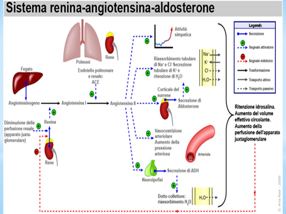
Regolazione del Sistema Renina - Angiotensina
Il complesso sistema rennina-angiotensina presiede alla regolazione della pressione arteriosa, cioè della forza esercitata dal sangue sulle pareti delle arterie, da cui dipende l'adeguata perfusione di sangue a tutti i distretti corporei; tale pressione è influenzata, tra l'altro, dalla quantità di sangue che il cuore spinge quando pompa, dalla sua forza di contrazione e dalle resistenze che si oppongono al libero scorrere del torrente ematico. Ebbene, il sistema renina-angiotensina agisce da un lato incrementando il volume del sangue (attraverso lo stimolo su sintesi e rilascio di aldosterone dalla corteccia surrenale), e dall'altro inducendo vasocostrizione. La vasocostrizione - vale a dire la diminuzione del lume dei vasi sanguigni - indotta dal sistema renina-angiotensina, aumenta significativamente la pressione arteriosa. Ci accorgiamo di questo fenomeno quando innaffiando l'orto con un tubo di gomma ne riduciamo il calibro con le dita per aumentare la distanza raggiunta dal getto d'acqua. Altrettanto intuitivo è il fatto che questo, e con esso la pressione idrica, aumenta e diminuisce mano a mano che apriamo o chiudiamo, rispettivamente, il rubinetto. Lo stesso effetto è indotto dall'aldosterone, ormone sintetizzato dalla corteccia del surrene sotto lo stimolo del sistema renina-angiotensina. L'aldosterone agisce infatti sulla parte distale dei nefroni (unità funzionali del rene), dove determina una diminuzione dell'escrezione di sodio e di acqua, ed un aumento dell'escrezione di potassio e ioni idrogeno. La ritenzione di sodio e acqua da parte del rene aumenta il volume plasmatico e la pressione arteriosa, proprio come nell'esempio dell'acqua e del rubinetto.
, e dall altro inducendo vasocostrizione. La vasocostrizione - vale a dire la diminuzione del lume dei vasi sanguigni - indotta dal sistema renina-angiotensina, aumenta significativamente la pressione arteriosa. Ci accorgiamo di questo fenomeno quando innaffiando l orto con un tubo di gomma ne riduciamo il calibro con le dita per aumentare la distanza raggiunta dal getto d acqua. Altrettanto intuitivo è il fatto che questo, e con esso la pressione idrica, aumenta e diminuisce mano a mano che apriamo o chiudiamo, rispettivamente, il rubinetto. Lo stesso effetto è indotto dall aldosterone, ormone sintetizzato dalla corteccia del surrene sotto lo stimolo del sistema renina-angiotensina. L aldosterone agisce infatti sulla parte distale dei nefroni (unità funzionali del rene), dove determina una diminuzione dell escrezione di sodio e di acqua, ed un aumento dell escrezione di potassio e ioni idrogeno. La ritenzione di sodio e acqua da parte del rene aumenta il volume plasmatico e la pressione arteriosa, proprio come nell esempio dell acqua e del rubinetto.")
22
HR Fluctuations Fluctuations in HR (HRV) are mediated by sympathetic (SNS) and parasympathetic (PNS) inputs to the SA node. Rapid fluctuations in HR usually reflect PNS control only (respiratory sinus arrhythmia). Slower fluctuations in HR reflect combined SNS and PNS + other psychological and emotional influences. Autonomic nervous system - sympathetic nervous system (SNS) and parasympathetic nervous system (PNS) - responsible for bringing about changes in the body in response to external changes through mostly involuntary actions. SNS - “fight or flight” response by releasing epinephrine (also known as adrenaline). PNS - returns the body to normal by releasing acetylcholine. Respiratory Sinus Arrythmia (RSA) - a natural cycle of speeding up and slowing down of heart rate caused by breathing
. Slower fluctuations in HR reflect combined SNS and PNS + other psychological and emotional influences. Autonomic nervous system. - sympathetic nervous system (SNS) and parasympathetic nervous system (PNS) - responsible for bringing about changes in the body in response to external changes through mostly involuntary actions. SNS - fight or flight response by releasing epinephrine (also known as adrenaline). PNS - returns the body to normal by releasing acetylcholine. Respiratory Sinus Arrythmia (RSA) - a natural cycle of speeding up and slowing down of heart rate caused by breathing.")
23
Rapid Fluctuations in HR Are Vagally Mediated
“Rapid” fluctuations in HR are at >10 cycles/min (respiratory frequencies) Vagal effect on HR mediated by acetylcholine binding which has an immediate effect on SA node. If HR patterns are normal, rapid fluctuations in HR are vagally modulated Vagus nerve - one of the 12 pairs of nerves originating in the brain - can directly stimulate the sinoatrial (SA) node. The release of acetycholine onto the SA node results in a change in channel properties, which then decreases the inward current so the action potential cycle last longer (slower HR) Cycles/min – think of a sinusoidal wave; how many complete “cycles” (such as peak to peak) are occurring per minute Rapid Fluctuations in HR Are Vagally Mediated
 Vagal effect on HR mediated by acetylcholine binding which has an immediate effect on SA node. If HR patterns are normal, rapid fluctuations in HR are vagally modulated. Vagus nerve. - one of the 12 pairs of nerves originating in the brain. - can directly stimulate the sinoatrial (SA) node. The release of acetycholine onto the SA node results in a change in channel properties, which then decreases the inward current so the action potential cycle last longer (slower HR) Cycles/min – think of a sinusoidal wave; how many complete cycles (such as peak to peak) are occurring per minute. Rapid Fluctuations in HR Are Vagally Mediated.")
24
Acetylcholine Binding
Acetylcholine – directly binds to the receptor in the SA node which causes an immediate change in heart rate. Acetylcholine Binding The Acetylcholine Neurotransmitter binds to a receptor on a muscle once released from a neuron.

25
Slower Fluctuations in HR Reflect Both SNS and Vagal Influences
“Slower” fluctuations in HR are <10 cycles per min. SNS effect on HR is mediated by norepinephrine release which has a delayed effect on SA node Both SNS and vagal nerve traffic fluctuate at >10 cycles/min, but the time constant for changes in SNS tone to affect HR is too long to affect HR at normal breathing frequencies. Slower Fluctuations in HR Reflect Both SNS and Vagal Influences

26
Sympathetic activation takes too long to affect RSA
NE – norepinephrine RSA - respiratory sinus arrhythmia There are many more steps here compared with acetylcholine receptor binding. NE takes longer to increase heart rate than acetylcholine does to decrease heart rate. *Don’t need to know details, just know this takes longer! NE blinds to the beta-receptor (Alpha subunit of G-protein). After binding, G protein links to second messenger (adenyl cyclase) which converts ATP to cAMP. cAMP activates protein kinase A which breaks ATP to ADP+phosphate which phosphorylates the pacemaker channels and increases HR
. After binding, G protein links to second messenger (adenyl cyclase) which converts ATP to cAMP. cAMP activates protein kinase A which breaks ATP to ADP+phosphate which phosphorylates the pacemaker channels and increases HR.")
27
Assessment of HRV Approach 1 Physiologist’s Paradigm
HR data collected over short period of time (~5-20 min), with or without interventions, under carefully controlled laboratory conditions. Physiologist - biologist that specializes in the study of the processes that occur within living organisms. HR = heart rate Assessment of HRV
, with or without interventions, under carefully controlled laboratory conditions. Physiologist - biologist that specializes in the study of the processes that occur within living organisms. HR = heart rate. Assessment of HRV.")
28
HRV Perspectives Longer-term HRV-quantifies changes in HR over periods of >5min. Intermediate-term HRV-quantifies changes in HR over periods of <5 min. Short-term HRV-quantifies changes in HR from one beat to the next Ratio HRV-quantifies relationship between two HRV indices.

29
Sources of Heart Rate Variability
Extrinsic Motor Activity Mental Stress Physical Stress Intrinsic Periodic Rhythms Respiratory sinus arrhythmia Baroreceptor reflex regulation Thermoregulation Neuroendocrine secretion Circadian rhythms Other, unknown rhythms Sleep apnea – brief breathing interruptions during sleep. Baroreceptor reflex regulation – short-term blood pressure regulation Theremoregulation – body temperature regulation Circadian rhythms – biological processes that take around 24 hours to complete

30
Ways to Quantify HRV Approach 1: How much variability is there?
Time Domain and Geometric Analyses Approach 2: What are the underlying rhythms? What physiologic process do they represent? How much power does each underlying rhythm have? Frequency Domain Analysis Approach 3: How much complexity or self-similarity is there? Non-Linear Analyses

31
Time Domain HRV Longer-term HRV
SDNN-Standard deviation of N-N intervals in msec (Total HRV) SDANN-Standard deviation of mean values of N-Ns for each 5 minute interval in msec (Reflects circadian, neuroendocrine and other rhythms + sustained activity) N-N - normal-to-normal To calculate a standard deviation of a series of numbers: Find the average Calculate the differences between each number and the average Square each difference (so they don’t cancel out) Sum Divide by number of datapoints Take square root
 SDANN-Standard deviation of mean values of N-Ns for each 5 minute interval in msec (Reflects circadian, neuroendocrine and other rhythms + sustained activity) N-N - normal-to-normal. To calculate a standard deviation of a series of numbers: Find the average. Calculate the differences between each number and the average. Square each difference (so they don’t cancel out) Sum. Divide by number of datapoints. Take square root.")
32
Time Domain HRV Intermediate-term HRV
SDNNIDX-Average of standard deviations of N-Ns for each 5 min interval in ms (Combined SNS and PNS HRV) Coefficient of variance (CV)- SDNNIDX/AVNN. Heart rate normalized SDNNIDX.
 Coefficient of variance (CV)- SDNNIDX/AVNN. Heart rate normalized SDNNIDX.")
33
Time Domain HRV Short-term HRV
rMSSD-Root mean square of successive differences of N-N intervals in ms pNN50-Percent of successive N-N differences >50 ms Calculated from differences between successive N-N intervals Reflect PNS influence on HR Root mean square – average change from one beat to the next

34
Geometric HRV HRV Index-Measure of longer-term HRV
Wider triangle – more HRV Narrower triangle – less HRV HRV Index-Measure of longer-term HRV From Farrell et al, J am Coll Cardiol 1991;18:687-97

35
Examples of Normal and Abnormal
Geometric HRV

36
Based on autoregressive techniques or fast Fourier transform (FFT).
Partitions the total variance in heart rate into underlying rhythms that occur at different frequencies. These frequencies can be associated with different intrinsic, autonomically-modulated periodic rhythms. FFT – transforms data from the time domain into the frequency domain (next slide shows an example) Frequency Domain HRV
 Frequency Domain HRV.")
37
What are the Underlying Rhythms?
One rhythm 5 seconds/cycle or 12 times/min In the time domain, the plot can be drawn using one sinusoidal wave with a frequency of 0.2 Hz. Therefore, in the frequency domain, there is a peak at 0.2 Hz. 5 seconds/cycle= 1/5 cycle/second 1/5 cycle/second= 0.2 Hz

38
What are the Underlying Rhythms?
Three Different Rhythms High Frequency = 0.25 Hz (15 cycles/min Low Frequency = 0.1 Hz (6 cycles/min) Very Low Frequency = Hz (1 cycle/min)
 Very Low Frequency = Hz. (1 cycle/min)")
39
Ground Rules for Measuring Frequency Domain HRV
Only normal-to-normal (NN) intervals included At least one normal beat before and one normal beat after each ectopic beat is excluded Cannot reliably compute HRV with >20% ectopic beats With the exception of ULF, HRV in a 24-hour recording is calculated on shorter segments (5 min) and averaged. ULF – Ultra Low frequency Ground Rules for Measuring Frequency Domain HRV
 intervals included. At least one normal beat before and one normal beat after each ectopic beat is excluded. Cannot reliably compute HRV with >20% ectopic beats. With the exception of ULF, HRV in a 24-hour recording is calculated on shorter segments (5 min) and averaged. ULF – Ultra Low frequency. Ground Rules for Measuring Frequency Domain HRV.")
40
Frequency Domain HRV Longer-Term HRV Total Power (TP)
Sum of all frequency domain components. Ultra low frequency power (ULF) At >every 5 min to once in 24 hours. Reflects circadian, neuroendocrine, sustained activity of subject, and other unknown rhythms.
 At >every 5 min to once in 24 hours. Reflects circadian, neuroendocrine, sustained activity of subject, and other unknown rhythms.")
41
Frequency Domain HRV Intermediate-term HRV
Very low frequency power (VLF) At ~20 sec-5 min frequency Reflects activity of renin-angiotensin system, vagal activity, activity of subject. Exaggerated by sleep apnea. Abolished by atropine Low frequency power (LF) At 3-9 cycles/min Baroreceptor influences on HR, mediated by SNS and vagal influences. Abolished by atropine. Renin-angiotensin system – regulates blood pressure via the kidney Atropine – inhibits acetylcholine actions.
 At ~20 sec-5 min frequency. Reflects activity of renin-angiotensin. system, vagal activity, activity of subject. Exaggerated by sleep apnea. Abolished. by atropine. Low frequency power (LF) At 3-9 cycles/min Baroreceptor influences on HR, mediated by SNS and vagal influences. Abolished by atropine. Renin-angiotensin system – regulates blood pressure via the kidney. Atropine – inhibits acetylcholine actions.")
42
Frequency Domain HRV Short-term HRV High frequency power (HF)
At respiratory frequencies (9-24 cycles/minute, respiratory sinus arrhythmia but may also include non-respiratory sinus arrhythmia). Normally abolished by atropine. Vagal influences on HR with normal patterns.
. Normally abolished by atropine. Vagal influences on HR with normal patterns.")
43
Emotions Reflected in Heart Rhythm Patterns
60 70 80 90 HEART RATE 1 50 100 150 200 TIME (SECONDS) FRUSTRATION APPRECIATION © Copyright 1997 Institute of HeartMath Research has further shown that heart rate variability can shift significantly, with negative emotions being associated with erratic shifts that can adversely affect the functioning of other systems, while positive emotions engender smooth flows and synchronized S & P activity – a state that HeartMath labels psychophysiological coherence.
 FRUSTRATION. APPRECIATION. © Copyright 1997 Institute of HeartMath. Research has further shown that heart rate variability can shift significantly, with negative emotions being associated with erratic shifts that can adversely affect the functioning of other systems, while positive emotions engender smooth flows and synchronized S & P activity – a state that HeartMath labels psychophysiological coherence.")
44
Increased Heart Rhythm Coherence Improves Cognitive Performance
Auditory Discrimination Task Mean Reaction Times 37 0.4 * Mean Reaction Times (msec.) Having identified heart rate variability as a key indicator of psychophysiological coherence, researchers at HeartMath have tested its influence on organizationally relevant cognitive performance outcomes (such as the auditory discrimination reaction times shown here), and found significant improvements when heart rate variability is reduced.
 Having identified heart rate variability as a key indicator of psychophysiological coherence, researchers at HeartMath have tested its influence on organizationally relevant cognitive performance outcomes (such as the auditory discrimination reaction times shown here), and found significant improvements when heart rate variability is reduced.")
45
The Power to Change Performance
The Freeze-Frame Tool: Positive emotion refocusing technique The Heart Lock-In Tool: Emotional Restructuring Technique Coherent Communication: Increases Team coherence Boosting Organizational Climate: What it is and specific ways to improve it. The Freeze Framer: Heart rhythm feedback that reflects nervous system dynamics. HeartMath’s research has of necessity also identified individual and group-level interventions that can be used to influence the heart system directly to enhance psychophysiological coherence, and in the process have presented the organizational sciences with powerful tools by which to promote organizational well-being that complement more traditional cognition- and attitude-focused interventions.

46
Embodied and Distributed
Cortex Sub cortical Areas Medulla body, and ways of managing them to promote individual and organizational well-being. Cartesian mind-body duality has been under attack for some time across multiple fronts, and this research is very compelling in promoting a view of knowledge that goes beyond the brain and what is consciously processable. In terms of practice, the research makes clear and accessible some of the mechanisms unleashed by AI and other managerial practices that are rooted in positive psychology. And to the jaundiced manager who rails against positive psychology as touchy-feely mumbo jumbo, it provides the hard evidence that may just replace the cynicism with enthusiasm. SYMPATHETIC PARASYMPATHETIC Embodied and Distributed Knowledge Systems Skin Arteries Lungs Etc. Hormones Blood Pressure Etc.

Presentazioni simili

 Brussels, 26 settembre 2013.>")


>")


