Scaricare la presentazione
La presentazione è in caricamento. Aspetta per favore

1
La Glicolisi Dipartimento di Oncologia Sperimentale e Applicazioni Cliniche “Sezione di Biochimica” Facoltà di Medicina e Chirurgia “Laboratorio di Vitaminologia” U.O. di Analisi Microbiologiche, Virologiche e Parassitologiche A.O.U.P “P. Giaccone” Prof. Gennaro Taibi

2
Il glucosio entra nelle cellule, secondo gradiente di concentrazione
Il glucosio entra nelle cellule, secondo gradiente di concentrazione. L’ingresso è mediato da specifici trasportatori (GLUT). Il glucosio una volta entrato nella cellula viene fosforilato a glucosio-6-P.
. Il glucosio una volta entrato nella cellula viene fosforilato a glucosio-6-P.")
3
Tutte le cellule fanno glicolisi
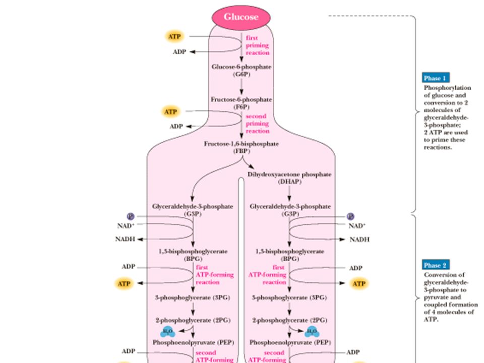
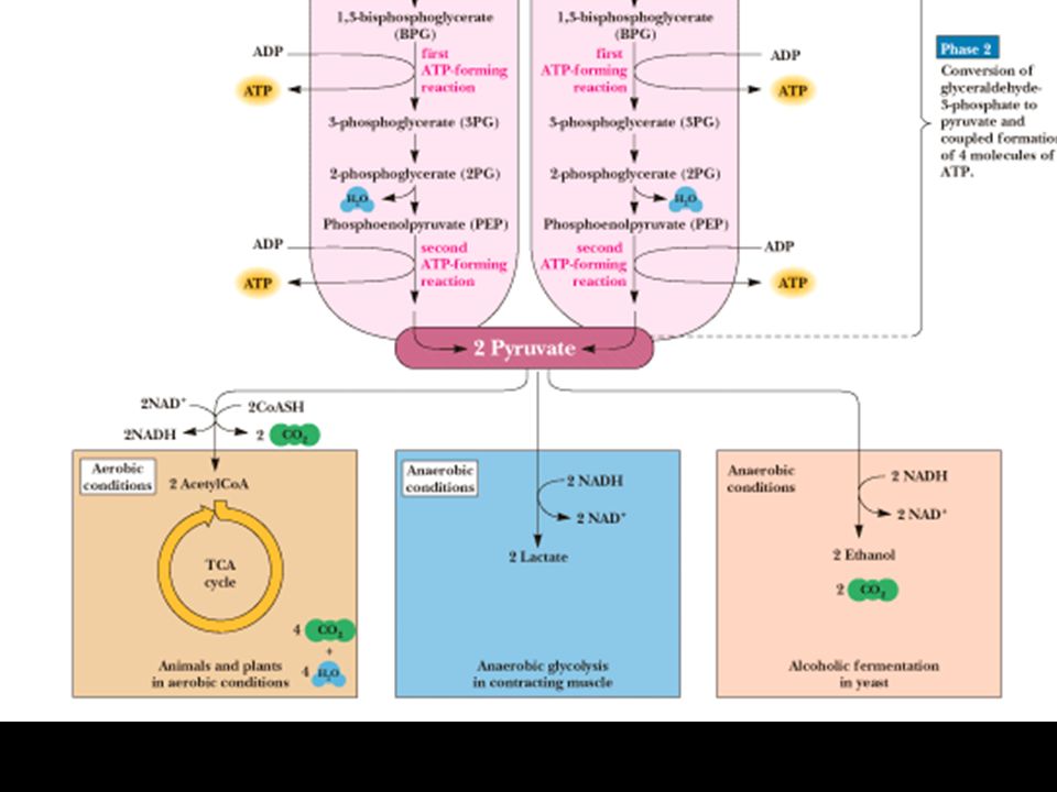
La glicolisi è costituita da 10 reazioni che avvengono con velocità e finalità differenti E’ possibile identificare due fasi: FASE I In questa fase si utilizzano due molecole di ATP ed una molecola di glucoso viene convertita in due molecole di Gliceraldeide-3-P FASE II In questa fase l’energia liberata nel processo viene conservata dalla cellula con produzione di 4 molecole di ATP e 4 di NADH I prodotti finali della glicolisi sono due molecole di piruvato Il piruvato può avere destini differenti

4
Fosforilazione del glucoso
Prima fase Fosforilazione del glucoso Catalizzata dalla esochinasi o dalla sua isoforma glucochinasi E’ una reazone di innesco in cui viene consumata una molecola di ATP L’esochinasi (e la glucochinasi) agiscono per fosforilare il glucoso e trattenerlo all’interno delle cellule La Km per il glucosio è 0.1 mM; la cellula ha 4 mmol di glucoso, quindi l’esochinasi è normalmente attiva La glucochinasi (Kmglucosio = 10 mM) agisce solo quando la [glucoso] nella cellula è alta. L’esochinasi è altamente regolata: inibita allostericamente dal prodotto glucoso-6-P
 agiscono per fosforilare il glucoso e trattenerlo all’interno delle cellule. La Km per il glucosio è 0.1 mM; la cellula ha 4 mmol di glucoso, quindi l’esochinasi è normalmente attiva. La glucochinasi (Kmglucosio = 10 mM) agisce solo quando la [glucoso] nella cellula è alta. L’esochinasi è altamente regolata: inibita allostericamente dal prodotto glucoso-6-P.")
7
FASE I una molecola di glucoso viene convertita in due molecole di Gliceraldeide-3-P

8
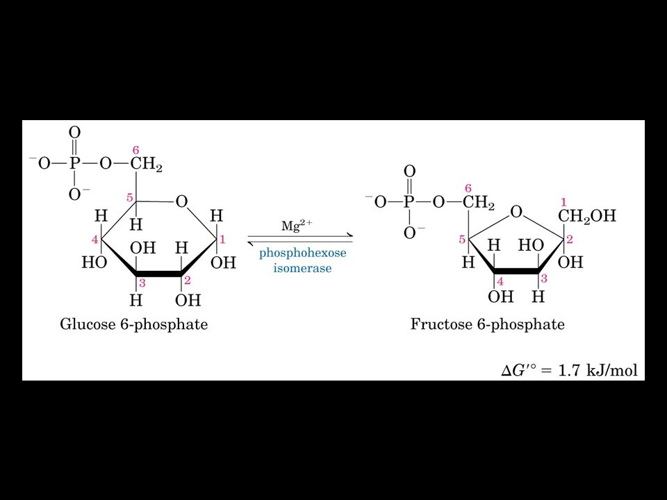
Fosfoglucoisomerasi Isomerizzazione di una molecola di
Glucoso-6-P a Fructoso-6-P 1° Questa reazione favorisce la successiva reazione di fosforilazione al C1 (sarebbe più sfavorevole come emiacetale –OH), 2° l’isomerizazione activa il C-3 per la successiva scissione (aldolasi)
, 2° l’isomerizazione activa il C-3 per la successiva scissione (aldolasi)")
10
Fosfofrutto-1-chinasi
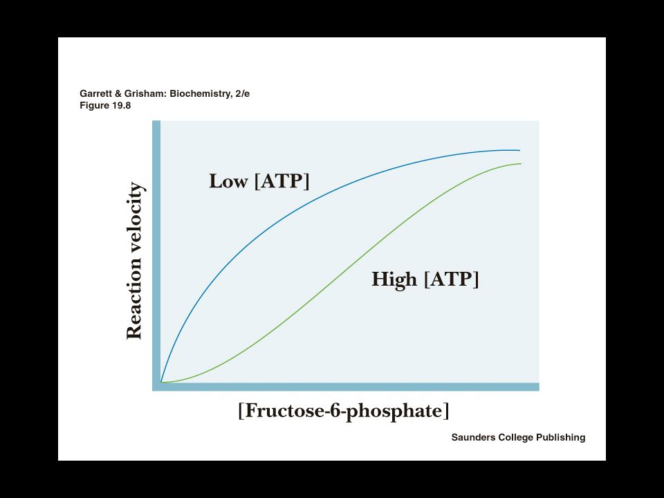
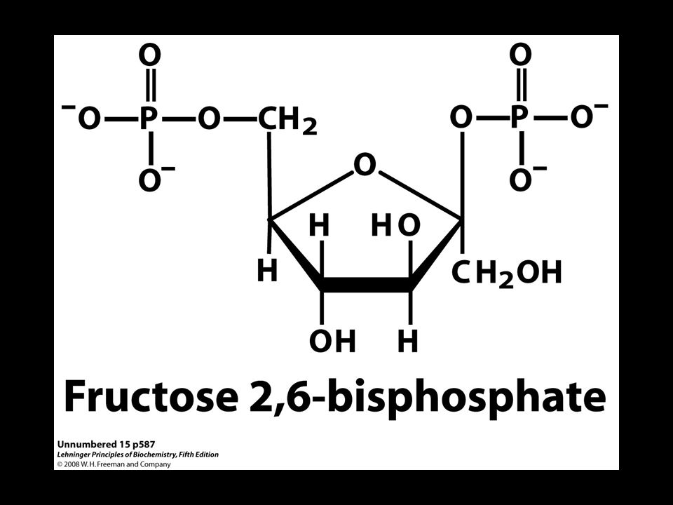
E’ la seconda reazione di innesco della glicolisi ed è la tappa regolativa Ha un DG negativo, PFK è altamente regolata ATP inibisce, AMP reverte l’inibizione Il citrato è un inibitore allosterico Fruttoso-2,6-bisfosfato è un attivatore allosterico L’attività PFK-1 aumenta quando la carica energetica è bassa L’attività PFK-1 decrementa quando la carica energetica è alta

13
FIGURE 15-14c Phosphofructokinase-1 (PFK-1) and its regulation
FIGURE 15-14c Phosphofructokinase-1 (PFK-1) and its regulation. (c) Summary of the regulators affecting PFK-1 activity.
 and its regulation. (c) Summary of the regulators affecting PFK-1 activity.")
14
FIGURE Regulation of fructose 1,6-bisphosphatase (FBPase-1) and phosphofructokinase-1 (PFK-1). The important role of fructose 2,6-bisphosphate in the regulation of this substrate cycle is detailed in subsequent figures.

16
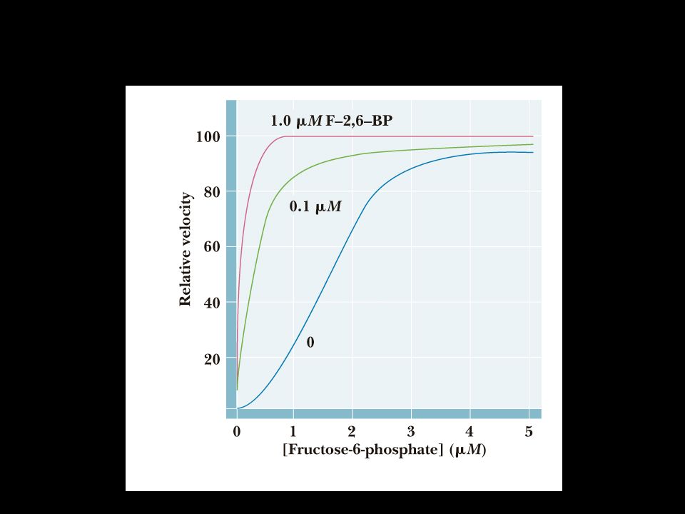
FIGURE 15-16a Role of fructose 2,6-bisphosphate in regulation of glycolysis and gluconeogenesis. Fructose 2,6-bisphosphate (F26BP) has opposite effects on the enzymatic activities of phosphofructokinase-1 (PFK-1, a glycolytic enzyme) and fructose 1,6-bisphosphatase (FBPase-1, a gluconeogenic enzyme). (a) PFK-1 activity in the absence of F26BP (blue curve) is half-maximal when the concentration of fructose 6-phosphate is 2 mM (that is, K0.5 = 2 mM). When 0.13 μM F26BP is present (red curve), the K0.5 for fructose 6-phosphate is only 0.08 mM. Thus F26BP activates PFK-1 by increasing its apparent affinity for fructose 6-phosphate (see Figure 15-14b).
 has opposite effects on the enzymatic activities of phosphofructokinase-1 (PFK-1, a glycolytic enzyme) and fructose 1,6-bisphosphatase (FBPase-1, a gluconeogenic enzyme). (a) PFK-1 activity in the absence of F26BP (blue curve) is half-maximal when the concentration of fructose 6-phosphate is 2 mM (that is, K0.5 = 2 mM). When 0.13 μM F26BP is present (red curve), the K0.5 for fructose 6-phosphate is only 0.08 mM. Thus F26BP activates PFK-1 by increasing its apparent affinity for fructose 6-phosphate (see Figure 15-14b)..")
17
FIGURE 15-16b Role of fructose 2,6-bisphosphate in regulation of glycolysis and gluconeogenesis. Fructose 2,6-bisphosphate (F26BP) has opposite effects on the enzymatic activities of phosphofructokinase-1 (PFK-1, a glycolytic enzyme) and fructose 1,6-bisphosphatase (FBPase-1, a gluconeogenic enzyme). (b) FBPase-1 activity is inhibited by as little as 1 μM F26BP and is strongly inhibited by 25 μM. In the absence of this inhibitor (blue curve) the K0.5 for fructose 1,6-bisphosphate is 5 μM, but in the presence of 25 μM F26BP (red curve) the K0.5 is >70 μM. Fructose 2,6-bisphosphate also makes FBPase-1 more sensitive to inhibition by another allosteric regulator, AMP.
 has opposite effects on the enzymatic activities of phosphofructokinase-1 (PFK-1, a glycolytic enzyme) and fructose 1,6-bisphosphatase (FBPase-1, a gluconeogenic enzyme). (b) FBPase-1 activity is inhibited by as little as 1 μM F26BP and is strongly inhibited by 25 μM. In the absence of this inhibitor (blue curve) the K0.5 for fructose 1,6-bisphosphate is 5 μM, but in the presence of 25 μM F26BP (red curve) the K0.5 is >70 μM. Fructose 2,6-bisphosphate also makes FBPase-1 more sensitive to inhibition by another allosteric regulator, AMP..")
18
FIGURE 15-16c Role of fructose 2,6-bisphosphate in regulation of glycolysis and gluconeogenesis. Fructose 2,6-bisphosphate (F26BP) has opposite effects on the enzymatic activities of phosphofructokinase-1 (PFK-1, a glycolytic enzyme) and fructose 1,6-bisphosphatase (FBPase-1, a gluconeogenic enzyme). (c) Summary of regulation by F26BP.
 has opposite effects on the enzymatic activities of phosphofructokinase-1 (PFK-1, a glycolytic enzyme) and fructose 1,6-bisphosphatase (FBPase-1, a gluconeogenic enzyme). (c) Summary of regulation by F26BP..")
19
FIGURE 15-17a Regulation of fructose 2,6-bisphosphate level
FIGURE 15-17a Regulation of fructose 2,6-bisphosphate level. (a) The cellular concentration of the regulator fructose 2,6-bisphosphate (F26BP) is determined by the rates of its synthesis by phosphofructokinase-2 (PFK-2) and its breakdown by fructose 2,6-bisphosphatase (FBPase-2).
 The cellular concentration of the regulator fructose 2,6-bisphosphate (F26BP) is determined by the rates of its synthesis by phosphofructokinase-2 (PFK-2) and its breakdown by fructose 2,6-bisphosphatase (FBPase-2).")
20
FIGURE 15-17b Regulation of fructose 2,6-bisphosphate level
FIGURE 15-17b Regulation of fructose 2,6-bisphosphate level. (b) Both enzyme activities are part of the same polypeptide chain, and they are reciprocally regulated by insulin and glucagon.
 Both enzyme activities are part of the same polypeptide chain, and they are reciprocally regulated by insulin and glucagon.")
21
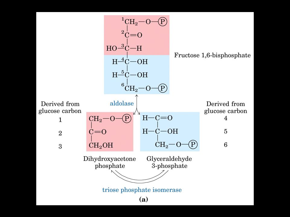
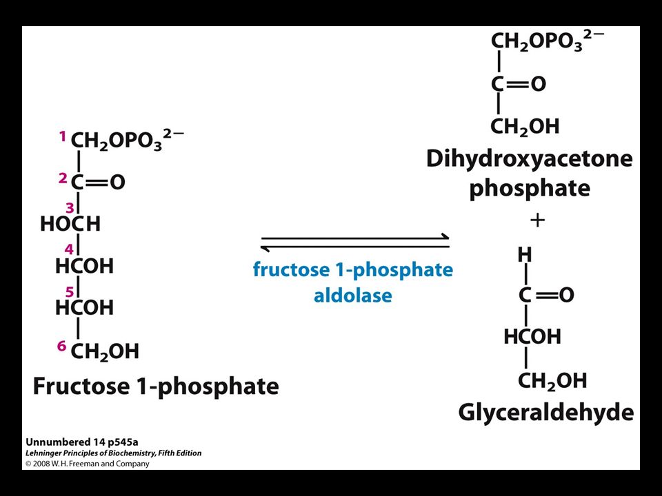
Una molecola a C6 viene scissa in due molecole da C3
Aldolasi Una molecola a C6 viene scissa in due molecole da C3 Il meccanismo di reazione prevede la formazione di una base di Schiff tra il carbonile del substrato e una lisina nel sito attivo dell’enzima.

24
Trioso fosfato isomerasi
Il Diossiacetone-P viene convertito in Gliceraldeide 3-P

25
FASE II due molecole di Gliceraldeide-3-P vengono convertite in due molecole di acido piruvico

26
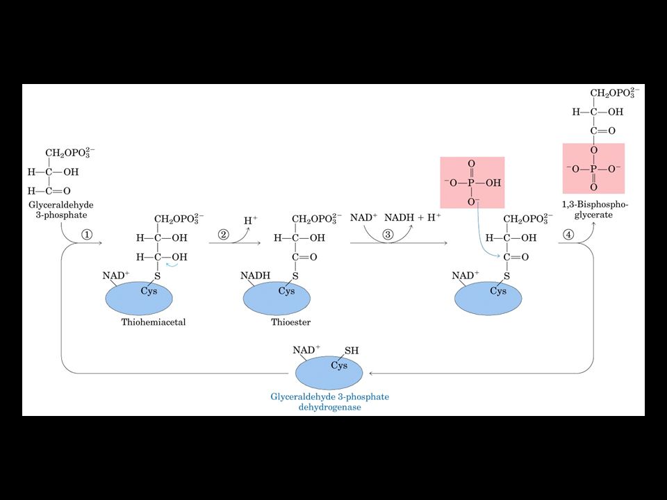
La Gliceraldeide 3-P Deidrogenasi

28
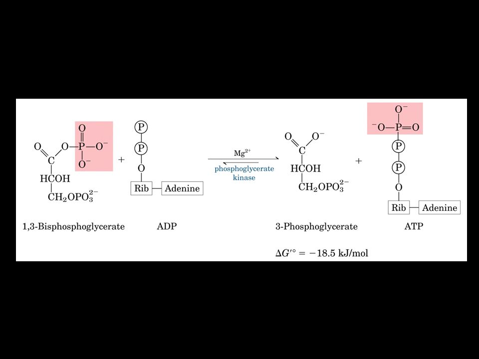
FOSFOGLICERICO CHINASI
Viene sintetizzata una molecola di ATP da un fosfato ad alta energia E’ una fosforilazione a livello del substrato Negli eritrociti questa reazione viene bypassata per la formazione del 2,3-BPG

30
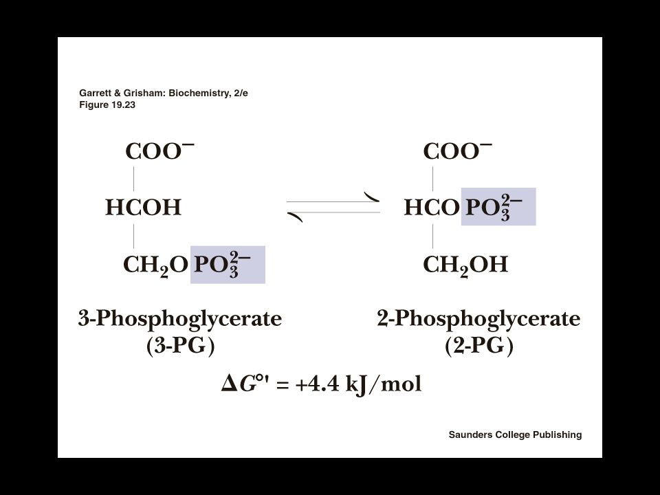
FOSFOGLICERATO MUTASI
Trasferisce il gruppo fosfato dal C-3 al C-2 del 3-fosfoglicerato

32
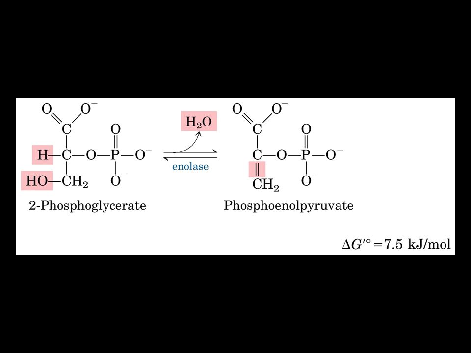
ENOLASI da acido 2-P-glicerico a fosfoenolpiruvico
E’ una reazione di deidratazione che comporta la produzione di un composto ad alta energia: la forma enolica fosforilata dell’acido piruvico

34
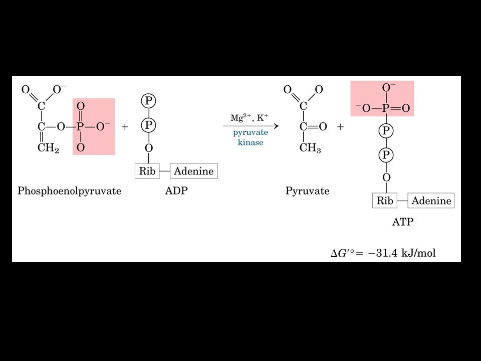
Nella reazione da PEP a Piruvico si forma una molecola di ATP
Piruvato chinasi Nella reazione da PEP a Piruvico si forma una molecola di ATP L’enzima è attivato allostericamente da AMP e F-1,6-P e inibito da ATP e acetil-CoA

37
Regulation of pyruvate kinase
Regulation of pyruvate kinase. The enzyme is allosterically inhibited by ATP, acetyl-CoA, and long-chain fatty acids (all signs of an abundant energy supply), and the accumulation of fructose 1,6-bisphosphate triggers its activation. Accumulation of alanine, which can be synthesized from pyruvate in one step, allosterically inhibits pyruvate kinase, slowing the production of pyruvate by glycolysis. The liver isozyme (L form) is also regulated hormonally. Glucagon activates cAMP-dependent protein kinase , which phosphorylates the pyruvate kinase L isozyme, inactivating it. When the glucagon level drops, a protein phosphatase (PP) dephosphorylates pyruvate kinase, activating it. This mechanism prevents the liver from consuming glucose by glycolysis when blood glucose is low; instead, the liver exports glucose. The muscle isozyme (M form) is not affected by this phosphorylation mechanism. FIGURE Regulation of pyruvate kinase. The enzyme is allosterically inhibited by ATP, acetyl-CoA, and long-chain fatty acids (all signs of an abundant energy supply), and the accumulation of fructose 1,6-bisphosphate triggers its activation. Accumulation of alanine, which can be synthesized from pyruvate in one step, allosterically inhibits pyruvate kinase, slowing the production of pyruvate by glycolysis. The liver isozyme (L form) is also regulated hormonally. Glucagon activates cAMP-dependent protein kinase (PKA; see Figure 15-35), which phosphorylates the pyruvate kinase L isozyme, inactivating it. When the glucagon level drops, a protein phosphatase (PP) dephosphorylates pyruvate kinase, activating it. This mechanism prevents the liver from consuming glucose by glycolysis when blood glucose is low; instead, the liver exports glucose. The muscle isozyme (M form) is not affected by this phosphorylation mechanism.
, and the accumulation of fructose 1,6-bisphosphate triggers its activation. Accumulation of alanine, which can be synthesized from pyruvate in one step, allosterically inhibits pyruvate kinase, slowing the production of pyruvate by glycolysis. The liver isozyme (L form) is also regulated hormonally. Glucagon activates cAMP-dependent protein kinase , which phosphorylates the pyruvate kinase L isozyme, inactivating it. When the glucagon level drops, a protein phosphatase (PP) dephosphorylates pyruvate kinase, activating it. This mechanism prevents the liver from consuming glucose by glycolysis when blood glucose is low; instead, the liver exports glucose. The muscle isozyme (M form) is not affected by this phosphorylation mechanism. FIGURE Regulation of pyruvate kinase. The enzyme is allosterically inhibited by ATP, acetyl-CoA, and long-chain fatty acids (all signs of an abundant energy supply), and the accumulation of fructose 1,6-bisphosphate triggers its activation. Accumulation of alanine, which can be synthesized from pyruvate in one step, allosterically inhibits pyruvate kinase, slowing the production of pyruvate by glycolysis. The liver isozyme (L form) is also regulated hormonally. Glucagon activates cAMP-dependent protein kinase (PKA; see Figure 15-35), which phosphorylates the pyruvate kinase L isozyme, inactivating it. When the glucagon level drops, a protein phosphatase (PP) dephosphorylates pyruvate kinase, activating it. This mechanism prevents the liver from consuming glucose by glycolysis when blood glucose is low; instead, the liver exports glucose. The muscle isozyme (M form) is not affected by this phosphorylation mechanism.")
38
Two alternative fates for pyruvate.
Pyruvate can be converted to glucose and glycogen via gluconeogenesis or oxidized to acetyl-CoA for energy production. The first enzyme in each path is regulated allosterically; acetyl-CoA, produced either by fatty acid oxidation or by the pyruvate dehydrogenase complex, stimulates pyruvate carboxylase and inhibits pyruvate dehydrogenase. FIGURE Two alternative fates for pyruvate. Pyruvate can be converted to glucose and glycogen via gluconeogenesis or oxidized to acetyl-CoA for energy production. The first enzyme in each path is regulated allosterically; acetyl-CoA, produced either by fatty acid oxidation or by the pyruvate dehydrogenase complex, stimulates pyruvate carboxylase and inhibits pyruvate dehydrogenase.

39
REGOLAZIONE DELLA GLICOLISI
Tre siti principali di regolazione: Esochinasi Fosfofrutto 1-chinasi Piruvato chinasi

40
Il regolatore della Fosfofruttochinasi-1 (PFK1)
")
41
La Fosfofruttochinasi-2 (PFK2) sintetizza il Fruttosio 2,6-P
 sintetizza il Fruttosio 2,6-P")
42
La Fosfofruttochinasi-2 è regolata
per fosforilazione/defosforilazione

43
Regolazione della sintesi e della degradazione del Fruttosio 2,6 P

46
Regolazione dell’attività catalitica della piruvico chinasi

47
Effect of type 1 diabetes on carbohydrate and fat metabolism in an adipocyte.
Normally, insulin triggers the insertion of GLUT4 transporters into the plasma membrane by the fusion of GLUT4-containing vesicles with the membrane, allowing glucose uptake from the blood. When blood levels of insulin drop, GLUT4 is resequestered in vesicles by endocytosis. In insulin-dependent diabetes mellitus, these normal processes are inhibited as indicated by X. The lack of insulin prevents glucose uptake via GLUT4; as a consequence, cells are deprived of glucose and blood glucose is elevated. Lacking glucose for energy supply, adipocytes break down triacylglycerols stored in fat droplets and supply the resulting fatty acids to other tissues for mitochondrial ATP production. Two byproducts of fatty acid oxidation in the liver accumulate and are released into the blood, providing fuel for the brain but also decreasing blood pH, causing ketoacidosis. The same sequence of events takes place in muscle, except that myocytes do not store triacylglycerols and instead take up fatty acids that are released into the blood by adipocytes. FIGURE 14-9 Effect of type 1 diabetes on carbohydrate and fat metabolism in an adipocyte. Normally, insulin triggers the insertion of GLUT4 transporters into the plasma membrane by the fusion of GLUT4-containing vesicles with the membrane, allowing glucose uptake from the blood. When blood levels of insulin drop, GLUT4 is resequestered in vesicles by endocytosis. In type 1 (insulin-dependent) diabetes mellitus, these normal processes are inhibited as indicated by X. The lack of insulin prevents glucose uptake via GLUT4; as a consequence, cells are deprived of glucose and blood glucose is elevated. Lacking glucose for energy supply, adipocytes break down triacylglycerols stored in fat droplets and supply the resulting fatty acids to other tissues for mitochondrial ATP production. Two byproducts of fatty acid oxidation in the liver (acetoacetate and β-hydroxybutyrate, see p. 666) accumulate and are released into the blood, providing fuel for the brain but also decreasing blood pH, causing ketoacidosis. The same sequence of events takes place in muscle, except that myocytes do not store triacylglycerols and instead take up fatty acids that are released into the blood by adipocytes.
 diabetes mellitus, these normal processes are inhibited as indicated by X. The lack of insulin prevents glucose uptake via GLUT4; as a consequence, cells are deprived of glucose and blood glucose is elevated. Lacking glucose for energy supply, adipocytes break down triacylglycerols stored in fat droplets and supply the resulting fatty acids to other tissues for mitochondrial ATP production. Two byproducts of fatty acid oxidation in the liver (acetoacetate and β-hydroxybutyrate, see p. 666) accumulate and are released into the blood, providing fuel for the brain but also decreasing blood pH, causing ketoacidosis. The same sequence of events takes place in muscle, except that myocytes do not store triacylglycerols and instead take up fatty acids that are released into the blood by adipocytes.")
48
Destini metabolici dell’acido piruvico
In condizioni aerobiche: trasloca nel mitocondrio e viene avviato al ciclo di Krebs In condizioni anaerobiche: sarà substrato della LDH per formare lattato e riossidare il NADH

49
Destini metabolici del NADH
In condizioni aerobiche: gli elettroni del NADH vengono trasferiti nel mitocondrio attraverso uno dei sistemi shuttle e avviati alla catena di trasporto degli e- In condizioni anaerobiche: il NADH verrà riossidato dalla lattico deidrogenasi (LDH), ristabilendo la quota di NAD+ necessario affinchè la via glicolitica possa continuare
, ristabilendo la quota di NAD+ necessario affinchè la via glicolitica possa continuare.")
50
FERMENTAZIONE LATTICA

51
Shuttle del Glicerolo 3-P

52
Shuttle Malato-Aspartato

53
Alla glicolisi possono essere avviati anche altri substrati
Fruttoso Mannoso Galattoso

54
FIGURE Entry of dietary glycogen, starch, disaccharides, and hexoses into the preparatory stage of glycolysis.

57
Conversione del Galattosio in UDP Glucosio e successivamente in Glucosio 1-P
FIGURE Conversion of galactose to glucose 1-phosphate. The conversion proceeds through a sugar-nucleotide derivative, UDP-galactose, which is formed when galactose 1-phosphate displaces glucose 1-phosphate from UDP-glucose. UDP-galactose is then converted by UDP-glucose 4-epimerase to UDP-glucose, in a reaction that involves oxidation of C-4 (pink) by NAD+, then reduction of C-4 by NADH; the result is inversion of the configuration at C-4. The UDP-glucose is recycled through another round of the same reaction. The net effect of this cycle is the conversion of galactose 1-phosphate to glucose 1-phosphate; there is no net production or consumption of UDP-galactose or UDP-glucose.
 by NAD+, then reduction of C-4 by NADH; the result is inversion of the configuration at C-4. The UDP-glucose is recycled through another round of the same reaction. The net effect of this cycle is the conversion of galactose 1-phosphate to glucose 1-phosphate; there is no net production or consumption of UDP-galactose or UDP-glucose.")
58
I° Tappa: Fosforilazione del Galattosio a Galattosio 1-P
FIGURE (part 1) Conversion of galactose to glucose 1-phosphate. The conversion proceeds through a sugar-nucleotide derivative, UDP-galactose, which is formed when galactose 1-phosphate displaces glucose 1-phosphate from UDP-glucose. UDP-galactose is then converted by UDP-glucose 4-epimerase to UDP-glucose, in a reaction that involves oxidation of C-4 (pink) by NAD+, then reduction of C-4 by NADH; the result is inversion of the configuration at C-4. The UDP-glucose is recycled through another round of the same reaction. The net effect of this cycle is the conversion of galactose 1-phosphate to glucose 1-phosphate; there is no net production or consumption of UDP-galactose or UDP-glucose.
 Conversion of galactose to glucose 1-phosphate. The conversion proceeds through a sugar-nucleotide derivative, UDP-galactose, which is formed when galactose 1-phosphate displaces glucose 1-phosphate from UDP-glucose. UDP-galactose is then converted by UDP-glucose 4-epimerase to UDP-glucose, in a reaction that involves oxidation of C-4 (pink) by NAD+, then reduction of C-4 by NADH; the result is inversion of the configuration at C-4. The UDP-glucose is recycled through another round of the same reaction. The net effect of this cycle is the conversion of galactose 1-phosphate to glucose 1-phosphate; there is no net production or consumption of UDP-galactose or UDP-glucose.")
59
II° Tappa: Uridilazione del Galattosio 1-P per intervento di una uridiltransferasi
FIGURE (part 2) Conversion of galactose to glucose 1-phosphate. The conversion proceeds through a sugar-nucleotide derivative, UDP-galactose, which is formed when galactose 1-phosphate displaces glucose 1-phosphate from UDP-glucose. UDP-galactose is then converted by UDP-glucose 4-epimerase to UDP-glucose, in a reaction that involves oxidation of C-4 (pink) by NAD+, then reduction of C-4 by NADH; the result is inversion of the configuration at C-4. The UDP-glucose is recycled through another round of the same reaction. The net effect of this cycle is the conversion of galactose 1-phosphate to glucose 1-phosphate; there is no net production or consumption of UDP-galactose or UDP-glucose.
 Conversion of galactose to glucose 1-phosphate. The conversion proceeds through a sugar-nucleotide derivative, UDP-galactose, which is formed when galactose 1-phosphate displaces glucose 1-phosphate from UDP-glucose. UDP-galactose is then converted by UDP-glucose 4-epimerase to UDP-glucose, in a reaction that involves oxidation of C-4 (pink) by NAD+, then reduction of C-4 by NADH; the result is inversion of the configuration at C-4. The UDP-glucose is recycled through another round of the same reaction. The net effect of this cycle is the conversion of galactose 1-phosphate to glucose 1-phosphate; there is no net production or consumption of UDP-galactose or UDP-glucose.")
60
III° Tappa: Ossidazione del gruppo alcolico al C-4
FIGURE (part 3) Conversion of galactose to glucose 1-phosphate. The conversion proceeds through a sugar-nucleotide derivative, UDP-galactose, which is formed when galactose 1-phosphate displaces glucose 1-phosphate from UDP-glucose. UDP-galactose is then converted by UDP-glucose 4-epimerase to UDP-glucose, in a reaction that involves oxidation of C-4 (pink) by NAD+, then reduction of C-4 by NADH; the result is inversion of the configuration at C-4. The UDP-glucose is recycled through another round of the same reaction. The net effect of this cycle is the conversion of galactose 1-phosphate to glucose 1-phosphate; there is no net production or consumption of UDP-galactose or UDP-glucose.
 Conversion of galactose to glucose 1-phosphate. The conversion proceeds through a sugar-nucleotide derivative, UDP-galactose, which is formed when galactose 1-phosphate displaces glucose 1-phosphate from UDP-glucose. UDP-galactose is then converted by UDP-glucose 4-epimerase to UDP-glucose, in a reaction that involves oxidation of C-4 (pink) by NAD+, then reduction of C-4 by NADH; the result is inversion of the configuration at C-4. The UDP-glucose is recycled through another round of the same reaction. The net effect of this cycle is the conversion of galactose 1-phosphate to glucose 1-phosphate; there is no net production or consumption of UDP-galactose or UDP-glucose.")
61
E sua successiva riduzione con produzione dell’epimero “UDP-Glucosio”
FIGURE (part 4) Conversion of galactose to glucose 1-phosphate. The conversion proceeds through a sugar-nucleotide derivative, UDP-galactose, which is formed when galactose 1-phosphate displaces glucose 1-phosphate from UDP-glucose. UDP-galactose is then converted by UDP-glucose 4-epimerase to UDP-glucose, in a reaction that involves oxidation of C-4 (pink) by NAD+, then reduction of C-4 by NADH; the result is inversion of the configuration at C-4. The UDP-glucose is recycled through another round of the same reaction. The net effect of this cycle is the conversion of galactose 1-phosphate to glucose 1-phosphate; there is no net production or consumption of UDP-galactose or UDP-glucose.
 Conversion of galactose to glucose 1-phosphate. The conversion proceeds through a sugar-nucleotide derivative, UDP-galactose, which is formed when galactose 1-phosphate displaces glucose 1-phosphate from UDP-glucose. UDP-galactose is then converted by UDP-glucose 4-epimerase to UDP-glucose, in a reaction that involves oxidation of C-4 (pink) by NAD+, then reduction of C-4 by NADH; the result is inversion of the configuration at C-4. The UDP-glucose is recycled through another round of the same reaction. The net effect of this cycle is the conversion of galactose 1-phosphate to glucose 1-phosphate; there is no net production or consumption of UDP-galactose or UDP-glucose.")
Presentazioni simili