Scaricare la presentazione
La presentazione è in caricamento. Aspetta per favore

1
RAMAN, MINERALI ED AMBIENTE
C.Rinaudo Dipartimento di Scienze dell’ambiente e della vita-Università degli Studi del Piemonte Orientale “Amedeo Avogadro”

2
PRINCIPI DELLA SPETTROSCOPIA RAMAN
Nel 1928 il fisico indiano C. V. Raman aveva osservato che quando un materiale viene colpito da un fascio di radiazioni monocromatiche una piccola parte della radiazione diffusa presenta una lunghezza d’onda diversa da quella della radiazione incidente. In particolare la radiazione diffusa è costituita da: diffusione Rayleigh alla stessa lunghezza d'onda della radiazione incidente, molto intensa; Righe Stokes a lunghezze d'onda inferiori rispetto a quella della radiazione incidente; Righe anti-Stokes a lunghezze d'onda superiori rispetto a quella della radiazione incidente.

3
Raman aveva osservato che la frequenza delle bande Stokes e anti-Stokes era indipendente dalla lunghezza d’onda della sorgente eccitatrice, ma dipendeva solo dalle vibrazioni dei legami chimici che costituiscono il composto in esame. Questo fa sì che lo spettro Raman costituisca una “impronta digitale” del campione in esame.

4
Strumentazione µ-Raman

5
Le vibrazioni possono essere di: stiramento simmetrico
stiramento asimmetrico oppure di: deformazione I modi di deformazione possono essere: scissoring (a forbice nel piano); rocking (oscillazione nel piano); wagging (ondeggio fuori del piano); twisting (torsione fuori dal piano).
; rocking (oscillazione nel piano); wagging (ondeggio fuori del piano); twisting (torsione fuori dal piano).")
6
Spettroscopia µ-Raman applicata aminerali importanti da un punto di vista ambientale:
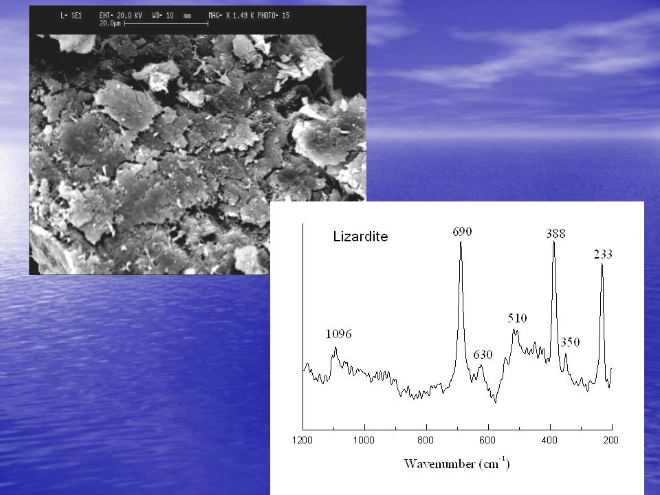
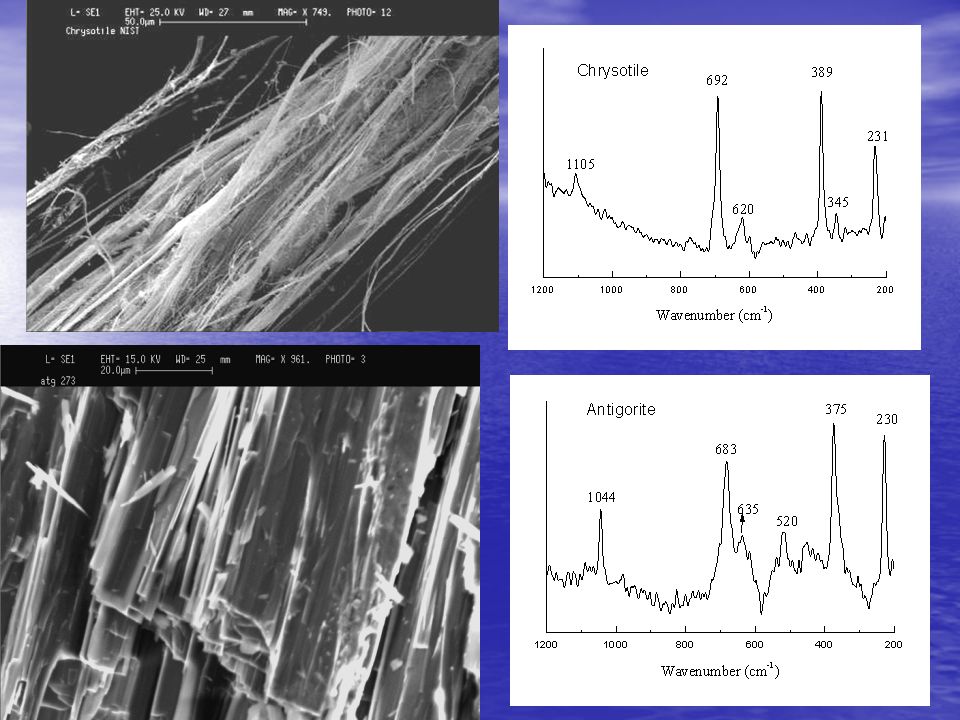
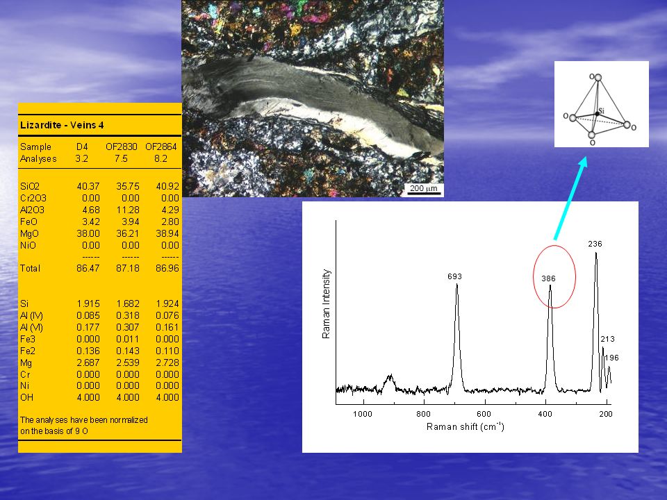
i serpentini La struttura cristallina è formata da strati tetraedrici, da strati ottaedrici e da un interstrato vuoto. Al centro di ogni tetraedro c’è il Si, al centro di ogni ottaedro prevalentemente Mg. Tetrahedra SiO4 OH- groups Mg2+

7
FLUORO-EDENITE FLUORO-EDENITE Characterization by Micro-Raman
Per compensare le differenze di dimensioni dello strato ottaedrico rispetto a quello tetraedrico, nei minerali del serpentino sono adottate tre stategie: vicarianza di Al-Si nello strato tetraedrico e modeste rotazioni dei tetraedri : lizardite; arrotolamento degli strati: crisotilo; rotazione dello strato tetraedrico ogni 8 tetraedri: antigorite. incidence of pleural mesothelioma. spectral region > 1000 cm-1: as Si-Ob-Si spectral region ~ 900 cm-1: s O-SiO- band 760 cm-1: s Si-Ob-Si band 679 cm-1: 1 (Ag) Si-Ob-Si Fig. 1 – prismatic and acicular fluoro-edenite. Characterization by Micro-Raman Fig. 4 – Raman spectra obtained on fluoro-edenite (see Fig. 1); deconvolutions carried out using OPUS software. Micro-Raman spectra of fluoro-edenite (Fig. 4) were recorded at the Department of Environmental and Life Science (University of Eastern Piedmont) using a Jobin Yvon µ-Raman HR-800 LabRaman spectrometer, equipped with a He-Ne (λ = nm) laser, a CCD air-cooled detector, and an Olympus BX 41 microscope. Bands at frequencies < 650 cm-1 are the result of vibrational modes of the octahedral MO6 (M= octahedral cation) and of deformation modes of the ribbons (Si4O11) and the F-O bonds. Its chemical formula is: A(Na0.56K0.15)B(Na0.30Ca1.62Mg0.03Mn0.05)C(Mg4.68Fe2+0.19Fe3+0.10Ti4+0.03) T(Si7.42Al0.58)O22O3(F1.98Cl0.02)2 FLUORO-EDENITE Fluoro-edenite is a new amphibole found in lavas from Mt Etna (Biancavilla-Sicily). Widely used in the past as a building material, it has been implicated in an abnormal incidence of pleural mesothelioma. spectral region > 1000 cm-1: as Si-Ob-Si spectral region ~ 900 cm-1: s O-SiO- band 760 cm-1: s Si-Ob-Si band 679 cm-1: 1 (Ag) Si-Ob-Si Fig. 1 – prismatic and acicular fluoro-edenite. Characterization by Micro-Raman Fig. 4 – Raman spectra obtained on fluoro-edenite (see Fig. 1); deconvolutions carried out using OPUS software. Micro-Raman spectra of fluoro-edenite (Fig. 4) were recorded at the Department of Environmental and Life Science (University of Eastern Piedmont) using a Jobin Yvon µ-Raman HR-800 LabRaman spectrometer, equipped with a He-Ne (λ = nm) laser, a CCD air-cooled detector, and an Olympus BX 41 microscope. Bands at frequencies < 650 cm-1 are the result of vibrational modes of the octahedral MO6 (M= octahedral cation) and of deformation modes of the ribbons (Si4O11) and the F-O bonds. Its chemical formula is: A(Na0.56K0.15)B(Na0.30Ca1.62Mg0.03Mn0.05)C(Mg4.68Fe2+0.19Fe3+0.10Ti4+0.03) T(Si7.42Al0.58)O22O3(F1.98Cl0.02)2 FLUORO-EDENITE
 Si-Ob-Si. Fig. 1 – prismatic and acicular fluoro-edenite. Characterization by Micro-Raman. Fig. 4 – Raman spectra obtained on fluoro-edenite (see Fig. 1); deconvolutions carried out using OPUS software. Micro-Raman spectra of fluoro-edenite (Fig. 4) were recorded at the Department of Environmental and Life Science (University of Eastern Piedmont) using a Jobin Yvon µ-Raman HR-800 LabRaman spectrometer, equipped with a He-Ne (λ = nm) laser, a CCD air-cooled detector, and an Olympus BX 41 microscope. Bands at frequencies < 650 cm-1 are the result of vibrational modes of the octahedral MO6 (M= octahedral cation) and of deformation modes of the ribbons (Si4O11) and the F-O bonds. Its chemical formula is: A(Na0.56K0.15)B(Na0.30Ca1.62Mg0.03Mn0.05)C(Mg4.68Fe2+0.19Fe3+0.10Ti4+0.03) T(Si7.42Al0.58)O22O3(F1.98Cl0.02)2. FLUORO-EDENITE. Fluoro-edenite is a new amphibole found in lavas from Mt Etna (Biancavilla-Sicily). Widely used in the past as a building material, it has been implicated in an abnormal incidence of pleural mesothelioma. spectral region > 1000 cm-1: as Si-Ob-Si. spectral region ~ 900 cm-1: s O-SiO- band 760 cm-1: s Si-Ob-Si. band 679 cm-1: 1 (Ag) Si-Ob-Si. Fig. 1 – prismatic and acicular fluoro-edenite. Characterization by Micro-Raman. Fig. 4 – Raman spectra obtained on fluoro-edenite (see Fig. 1); deconvolutions carried out using OPUS software. Micro-Raman spectra of fluoro-edenite (Fig. 4) were recorded at the Department of Environmental and Life Science (University of Eastern Piedmont) using a Jobin Yvon µ-Raman HR-800 LabRaman spectrometer, equipped with a He-Ne (λ = nm) laser, a CCD air-cooled detector, and an Olympus BX 41 microscope. Bands at frequencies < 650 cm-1 are the result of vibrational modes of the octahedral MO6 (M= octahedral cation) and of deformation modes of the ribbons (Si4O11) and the F-O bonds. Its chemical formula is: A(Na0.56K0.15)B(Na0.30Ca1.62Mg0.03Mn0.05)C(Mg4.68Fe2+0.19Fe3+0.10Ti4+0.03) T(Si7.42Al0.58)O22O3(F1.98Cl0.02)2. FLUORO-EDENITE.")
8
νs (SiOSi) νas (SiOSi)
 νas (SiOSi)")
11
Crisotilo Antigorite Lizardite 1105 m 1096 m - 1044 f 692 mf 683 mf 690 mf 620 m 635 m 630 m 520 m 510 m 389 mf 375 mf mf 345 m 350 m 231 f 230 mf 233 mf

12
Ctl

14
Lizardite bastite Lizardite vena Lizardite mesh SiO2 40.95 41.40 40.00 38.41 38.35 37.28 Cr2O3 1.59 1.82 0.00 0.48 0.45 Al2O3 1.42 1.17 4.91 6.97 7.29 9.50 MgO 39.78 40.36 39.00 37.55 37.77 37.51 Totale 86.43 86.77 87.21 86.64 86.79 87.46 Cationi calcolati sulla base di 7 ossigeni anidri Si 1.942 1.952 1.875 1.817 1.808 1.742 Cr 0.060 0.068 0.000 0.018 0.017 Al IV 0.058 0.048 0.125 0.183 0.192 0.258 Al VI 0.022 0.146 0.206 0.213 0.265 Al 0.080 0.065 0.271 0.389 0.405 0.523 Fe+2 0.107 0.130 0.147 0.114 0.106 Mg 2.812 2.836 2.724 2.648 2.654 2.612 Ni

15
SPETTROSCOPIA µ-RAMAN APPLICATA AGLI ANFIBOLI FIBROSI
Tetraedri SiO4 gruppi OH- Ca2+, Na+ (coord. 8) Fe2+, Mg2+ (coord. 6) Fe2+, Mg2+, Fe3+, Al3+, Ti4+ Antofillite: (Mg,Fe2+)7[Si8O22(OH)2] Actinolite: Ca2(Mg,Fe2+)5[Si8O22(OH)2] Amosite: (Fe2+,Mg)7[Si8O22(OH)2] Tremolite: Ca2Mg5[Si8O22(OH)2] Crocidolite: Na2Fe2+3Fe3+2[Si8O22(OH)2]
 Fe2+, Mg2+ (coord. 6) Fe2+, Mg2+, Fe3+, Al3+, Ti4+ Antofillite: (Mg,Fe2+)7[Si8O22(OH)2] Actinolite: Ca2(Mg,Fe2+)5[Si8O22(OH)2] Amosite: (Fe2+,Mg)7[Si8O22(OH)2] Tremolite: Ca2Mg5[Si8O22(OH)2] Crocidolite: Na2Fe2+3Fe3+2[Si8O22(OH)2]")
16
Antophyllite νs (SiOSi) νas (SiOSi)
 νas (SiOSi)")
17
Actinolite νs(SiOSi) Immagine SEM
 Immagine SEM")
18
Amosite νs (SiOSi) νas (SiOSi)
 νas (SiOSi)")
19
Tremolite νs (SiOSi) νas (SiOSi)
 νas (SiOSi)")
20
Crocidolite νs (SiOSi) νas (SiOSi) SEM image
 νas (SiOSi) SEM image")
21
νs νas Amosite 659 1020 Anthophyllite 674 1044 Actinolite 669 1062
Antophyllite Actinolite Amosite νs νas Amosite 659 1020 Anthophyllite 674 1044 Actinolite 669 1062 Tremolite 676 1062, 1031 Crocidolite 664,577 1082 Chrysotile 692 1105 Tremolite Chrysotile Crocidolite

22
ALTRI MINERALI FIBROSI NON CLASSIFICATI AMIANTI
FLUORO-EDENITE Biancavilla (CT) CARLOSTURANITE Serpentiniti di Sampeyre (Val Varaita) Località Monte Calvario
 CARLOSTURANITE. Serpentiniti di Sampeyre. (Val Varaita) Località Monte Calvario.")
23
Fluoro-edenite A(Na,K)B(Na,Ca,Mg, Mn)2C(Mg,Fe2+,Fe3+Ti4+)5T(Si,Al)O22O3(F,Cl)2 Tremolite
B(Na,Ca,Mg, Mn)2C(Mg,Fe2+,Fe3+Ti4+)5T(Si,Al)O22O3(F,Cl)2 Tremolite")
24
C E D A B

25
Analisi chimiche semi-quantitative ottenute con SEM-WDS
CAMPIONE 3RIF 4 9 10 31 48 WT % P2O5 0.00 0.01 0.03 0.05 0.02 SiO2 52.83 52.47 53.93 51.91 52.92 51.46 TiO2 0.55 0.59 0.04 0.73 0.62 Al2O3 3.81 4.17 3.04 4.07 3.87 4.41 MgO 23.60 22.61 23.05 22.63 22.78 22.60 CaO 10.73 11.07 10.36 10.87 11.03 10.77 MnO 0.46 0.53 0.47 0.54 0.48 0.57 FeO 2.25 2.32 2.49 2.48 2.34 2.59 Na2O 3.07 3.28 3.18 3.03 3.25 K2O 0.82 0.81 0.84 0.86 0.83 F 4.11 4.56 4.63 4.35 4.28 4.55 Cl 0.07 0.09 0.08 0.06

27
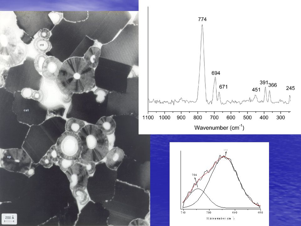
Carlosturanite

30
Carlosturanite 127

31
Applicazione su cementi-amianti
CC: Calcite Ch: crisotilo Cr: Crocidolite

32
Crocidolite

33
Spettro Raman del crisotilo
Spettro Raman del cemento

34
In conclusione la tecnica Raman permette una rapida identificazione di fasi mineralogiche con composizione chimica e struttura cristallina poco differenti e, tramite l’abbinamento ad un microscopio ottico, permette di individuare con precisione le singole fasi mineralogiche in campioni polimineralici; non necessita di preparazione del campione, quindi permette di asssociare l’immagine ottica alla fase minerale.

35
Applicazioni a campioni istologici
cellula fibra

37
Desidererei rngraziare:
La dott.ssa Daniela Gastaldi La dott.ssa Simona Cairo La dott.ssa Elisa Fornero Il dott. Mario Allegrina Il dott. Giovanni Albertazzi Senza il cui prezioso lavoro questi risultati non sarebbero stati raggiunti

38
GRAZIE PER LA VOSTRA ATTENZIONE

39
PUBBLICAZIONI Belluso, E., Fornero, E., Albertazzi, G., Cairo, S., Rinaudo, C. (2006) Micro-Raman as method to distinguish carlosturanite from serpentine minerals. Canadian Mineralogist (in press). Groppo, C., Rinaudo, C., Cairo, S., Gastaldi, D. & Compagnoni, R. (2006): Micro-Raman Spectroscopy for a quick and reliable identification of serpentine minerals from ultramafics, European Journal of Mineralogy, 18, Rinaudo C., S. Cairo, D. Gastaldi, A. Gianfagna, S. Mazziotti Tagliani, G. Tosi , C.Conti (2006) Characterization of fluoro-edenite by µ-Raman and µ-FTIR spectroscopy, Mineralogical Magazine 70(3), Rinaudo, C., Gastaldi, D., Belluso, E. & Capella S. (2005): Application of Raman Spectroscopy on asbestos fibre identification, Neues Jahrbuch für Mineralogie, 128/1, Rinaudo, C., Belluso, E. & Gastaldi, D. (2004): Assessment of the use of Raman spectroscopy for the determination of amphibole asbestos, Mineralogical Magazine, 68, Rinaudo, C., Gastaldi, D. & Belluso, E. (2003): Characterization of chrysotile, antigorite and lizardite by FT-Raman Spectroscopy, Canadian Mineralogist, 41, Rinaudo, C., Gastaldi, D. & Belluso, E. (2003): La spettroscopia Raman: tecnica di identificazione rapida di fibre di asbesto, Siti contaminati, 2,
 Micro-Raman as method to distinguish carlosturanite from serpentine minerals. Canadian Mineralogist (in press). Groppo, C., Rinaudo, C., Cairo, S., Gastaldi, D. & Compagnoni, R. (2006): Micro-Raman Spectroscopy for a quick and reliable identification of serpentine minerals from ultramafics, European Journal of Mineralogy, 18, Rinaudo C., S. Cairo, D. Gastaldi, A. Gianfagna, S. Mazziotti Tagliani, G. Tosi , C.Conti (2006) Characterization of fluoro-edenite by µ-Raman and µ-FTIR spectroscopy, Mineralogical Magazine 70(3), Rinaudo, C., Gastaldi, D., Belluso, E. & Capella S. (2005): Application of Raman Spectroscopy on asbestos fibre identification, Neues Jahrbuch für Mineralogie, 128/1, Rinaudo, C., Belluso, E. & Gastaldi, D. (2004): Assessment of the use of Raman spectroscopy for the determination of amphibole asbestos, Mineralogical Magazine, 68, Rinaudo, C., Gastaldi, D. & Belluso, E. (2003): Characterization of chrysotile, antigorite and lizardite by FT-Raman Spectroscopy, Canadian Mineralogist, 41, Rinaudo, C., Gastaldi, D. & Belluso, E. (2003): La spettroscopia Raman: tecnica di identificazione rapida di fibre di asbesto, Siti contaminati, 2,")
Presentazioni simili


2HPO4 è … 132 g 114 g>")

