Scaricare la presentazione
La presentazione è in caricamento. Aspetta per favore

1
Subunità 30S: rRNA 16S 21 proteine Subunità 30S: rRNA 23S rRNA 5S
Protein synthesis by the ribosome Ribosomes translate the genetic information encoded in messenger RNA (mRNA) to assemble amino acids into proteins. Ribosomes are made up of two subunits, both of which consist of ribosomal RNA (rRNA) and many proteins (r-proteins). In the upper panel of the figure, the backbone traces of the rRNAs (yellow and grey) and the r-proteins (bronze and blue) are shown at the interfaces of the small (30S) and large (50S) ribosomal subunits from the thermophilic bacterium Thermus thermophilus. The small subunit contains the decoding site where the mRNA sequence (magenta) is read in blocks of three nucleotides, called codons. Each codon denotes one of twenty different amino acids, and each amino acid is ferried to the ribosome by its own transfer RNA (tRNA) or set of tRNAs. Every tRNA has an anticodon sequence that makes a specific match with the corresponding mRNA codon. The mRNA passes through two narrow channels on the 30S subunit to be displayed at the interface decoding site, where it interacts with the tRNA anticodon (see figure, lower panel). Protein synthesis is initiated when the start codon on the mRNA is guided into the peptidyl site (P site) on the 30S subunit to interact with the initiator tRNA (green) charged with the amino acid methionine. The interfaces of the small and large subunits then come into contact as shown in the vertical section through the P site (see figure, lower panel). Then, the second mRNA codon, in the adjacent aminoacyl site (A site), accepts the next tRNA with its amino acid. The base-pairing match between the tRNA anticodon and mRNA codon is checked by the decoding site within the A site of the small subunit. If the match is accepted, the aminoacyl end of the tRNA is swung into the catalytic site, the peptidyl-transferase centre, on the large subunit, where a peptide bond is formed between the methionine and the second amino acid. The ribosome then moves one codon along the mRNA, bringing the third codon into the A site; this translocates the tRNA holding the dipeptide into the P site, and the deacylated initiator tRNA is moved into the exit site (E site) from where it is ejected. The cycle is repeated up to several hundred times, and the peptide chain grows from the peptidyl-transferase centre through the tunnel in the large subunit to emerge at the back of the ribosome (red arrow). Eventually the ribosome reaches a stop codon on the mRNA, signalling the completion and release of the peptide chain. All these stages in the initiation, elongation and release of the peptide chain are helped by protein factors, usually with the expenditure of energy in the form of guanosine nucleotide triphosphate (GTP).
 to assemble amino acids into proteins. Ribosomes are made up of two subunits, both of which consist of ribosomal RNA (rRNA) and many proteins (r-proteins). In the upper panel of the figure, the backbone traces of the rRNAs (yellow and grey) and the r-proteins (bronze and blue) are shown at the interfaces of the small (30S) and large (50S) ribosomal subunits from the thermophilic bacterium Thermus thermophilus. The small subunit contains the decoding site where the mRNA sequence (magenta) is read in blocks of three nucleotides, called codons. Each codon denotes one of twenty different amino acids, and each amino acid is ferried to the ribosome by its own transfer RNA (tRNA) or set of tRNAs. Every tRNA has an anticodon sequence that makes a specific match with the corresponding mRNA codon. The mRNA passes through two narrow channels on the 30S subunit to be displayed at the interface decoding site, where it interacts with the tRNA anticodon (see figure, lower panel). Protein synthesis is initiated when the start codon on the mRNA is guided into the peptidyl site (P site) on the 30S subunit to interact with the initiator tRNA (green) charged with the amino acid methionine. The interfaces of the small and large subunits then come into contact as shown in the vertical section through the P site (see figure, lower panel). Then, the second mRNA codon, in the adjacent aminoacyl site (A site), accepts the next tRNA with its amino acid. The base-pairing match between the tRNA anticodon and mRNA codon is checked by the decoding site within the A site of the small subunit. If the match is accepted, the aminoacyl end of the tRNA is swung into the catalytic site, the peptidyl-transferase centre, on the large subunit, where a peptide bond is formed between the methionine and the second amino acid. The ribosome then moves one codon along the mRNA, bringing the third codon into the A site; this translocates the tRNA holding the dipeptide into the P site, and the deacylated initiator tRNA is moved into the exit site (E site) from where it is ejected. The cycle is repeated up to several hundred times, and the peptide chain grows from the peptidyl-transferase centre through the tunnel in the large subunit to emerge at the back of the ribosome (red arrow). Eventually the ribosome reaches a stop codon on the mRNA, signalling the completion and release of the peptide chain. All these stages in the initiation, elongation and release of the peptide chain are helped by protein factors, usually with the expenditure of energy in the form of guanosine nucleotide triphosphate (GTP).")
2
Struttura degli aminoglicosidi
Aminociclitolo

3
Meccanismo d’azione degli aminoglicosidi

4
Trasporto degli aminoglicosidi attraverso la membrana plasmatica dei batteri
ENERGY- DEPENDENT PHASE I cationi bivalenti iperosmolarità pH anaerobiosi ENERGY- DEPENDENT PHASE II Sintesi di proteine di membrana alterate

5
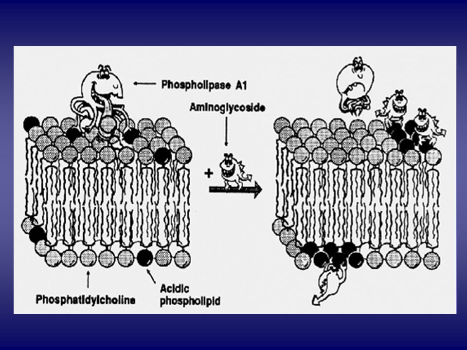
Ipotesi sull’azione battericida degli aminoglicosidi
The Davis model explains the bactericidal activity of aminoglycosides. The Davis model of aminoglycoside action proposes that low concentrations of aminoglycosides induce protein misreading an d that the misread (abnormal) proteins allow higfher concentrations of aminoglycosides to enter the cell and halt protein synthesis. A. Initially, AG are present at low concentration inside the bacterial cell, despite therapeutic (high) extracellular concentration of drug, because the drug molecules have poor uptake across the bacterial membranes. B. Low intracellular concentrations of AG bind to bacterial ribosomes and cause incorporation of incorrect aminoacids (misreading) into nascent polypeptides. C. The abnormal proteins insert into the bacterial membranes, forming pores and causing membrane damage. D. The damaged membranes allow additional AG molecules to flood into the cell, causing complete inhibition of ribosome activity. The effect is irreversible, perhaps because of trapping of drug inside the cell (“caging”). The membrane damage cannot be repaired because new proteins cannot be synthesized, and cell death ensues.
 proteins allow higfher concentrations of aminoglycosides to enter the cell and halt protein synthesis. A. Initially, AG are present at low concentration inside the bacterial cell, despite therapeutic (high) extracellular concentration of drug, because the drug molecules have poor uptake across the bacterial membranes. B. Low intracellular concentrations of AG bind to bacterial ribosomes and cause incorporation of incorrect aminoacids (misreading) into nascent polypeptides. C. The abnormal proteins insert into the bacterial membranes, forming pores and causing membrane damage. D. The damaged membranes allow additional AG molecules to flood into the cell, causing complete inhibition of ribosome activity. The effect is irreversible, perhaps because of trapping of drug inside the cell ( caging ). The membrane damage cannot be repaired because new proteins cannot be synthesized, and cell death ensues.")
6
The decoding site at the interface of the 30S ribosomal subunit
The decoding site at the interface of the 30S ribosomal subunit. Nucleotides A1492 and A1493 are shown in the conformation that they adopt after having flipped out of the 16S rRNA helix 44 to interact with an mRNA codon (magenta) and its cognate tRNA anticodon (green) at the ribosomal aminoacyl site66. The conformational changes induced at these nucleotides and at G530 are indicated by the arrows. The aminoglycoside antibiotics paromomycin (red) and geneticin (blue) bind within helix 44 as shown48, and induce a similar, but not identical, conformational shift in A1492 and A1493 (not shown). Nucleotides G1405 and A1408, methylation of which confers aminoglycoside resistance, are indicated. Escherichia coli rRNA nucleotide numbering is used throughout.
 and its cognate tRNA anticodon (green) at the ribosomal aminoacyl site66. The conformational changes induced at these nucleotides and at G530 are indicated by the arrows. The aminoglycoside antibiotics paromomycin (red) and geneticin (blue) bind within helix 44 as shown48, and induce a similar, but not identical, conformational shift in A1492 and A1493 (not shown). Nucleotides G1405 and A1408, methylation of which confers aminoglycoside resistance, are indicated. Escherichia coli rRNA nucleotide numbering is used throughout.")
7
Streptomycin RESISTENZA LysAsn LysGln SUBUNITÀ 30S DIPENDENZA
Proteina S12 LysGln DIPENDENZA

8
Meccanismi di resistenza agli aminoglicosidi
1- Alterazioni del trasporto di membrana 2 – Alterazione dei siti di legame ribosomiali 3 – Inattivazione enzimatica

9
Inattivazione enzimatica degli aminoglicosidi

10
CELLULE TUBULARI PROSSIMALI DI RATTO
trattato con gentamicina (10 mg/kg/g x 7 gg)
")
11
SEQUENZA DEGLI EVENTI Incorporazione degli AG (mediante pinocitosi) da parte delle cellule epiteliali del tubulo prossimale Trasporto e accumulo degli AG nei lisosomi, e sviluppo di fosfolipidosi lisosomiale Rottura (?) dei lisosomi, necrosi cellulare e fosfolipiduria Necrosi tubulare/rigenerazione tubulare Necrosi tubulare acuta Insufficienza renale
 dei lisosomi, necrosi cellulare e fosfolipiduria. Necrosi tubulare/rigenerazione tubulare. Necrosi tubulare acuta. Insufficienza renale.")
13
APOPTOSI IN CELLULE TUBULARI PROSSIMALI
trattate con gentamicina (10 mg/kg/g x 10 gg)
")
14
NEFROTOSSICITA’ DA AMINOGLICOSIDI
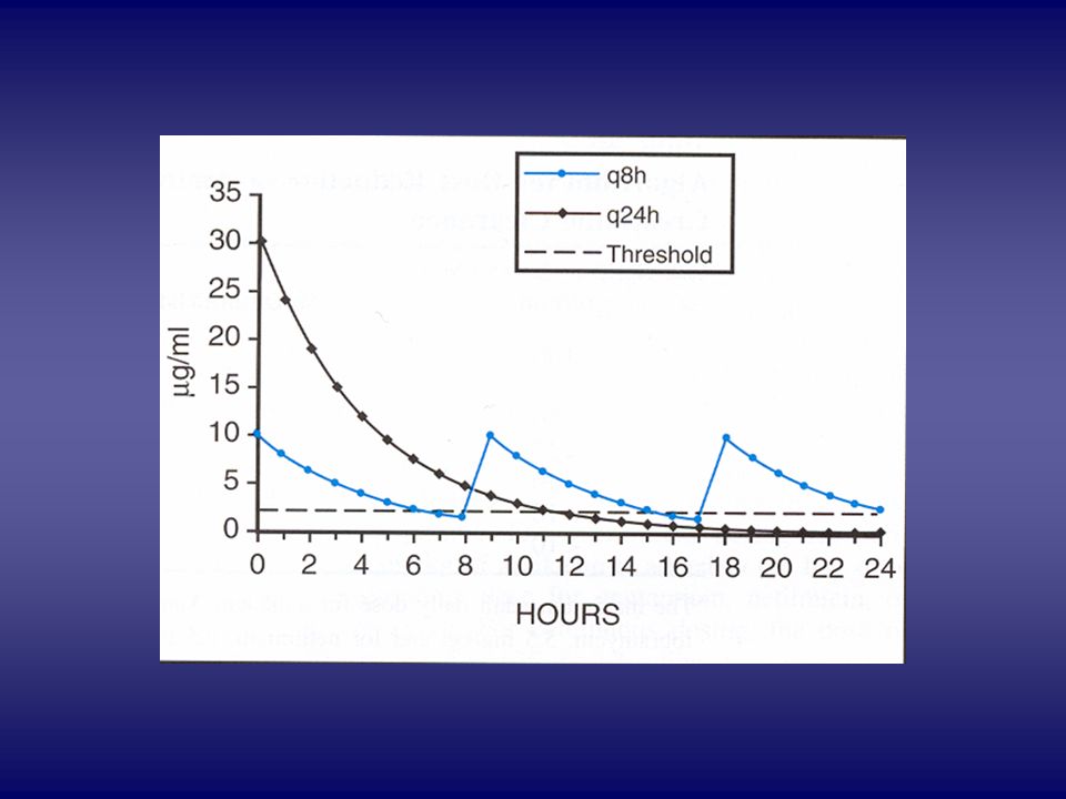
Incidenza 0-50% in dipendenza da: fattori legati al paziente fattori legati al farmaco schema di somministrazione trattamenti concomitanti GENTAMICINA NETILMICINA AMIKACINA

16
3 - Blocco neuromuscolare
Effetti collaterali degli aminoglicosidi 1- Nefrotossicità 2 - Ototossicità 3 - Blocco neuromuscolare 4 - reazioni allergiche

17
Struttura delle tetracicline
I GENERAZIONE II GENERAZIONE

18
Meccanismo d’azione delle tetracicline

19
Meccanismi di resistenza alle tetracicline
Diminuzione dei livelli intracellulari di TC Ingresso Acquisizione di sistemi di estrusione Diminuzione dell’accesso delle TC ai siti di legame ribosomiali Inattivazione delle TC

20
Resistenza alle tetracicline
Geni tet (27) Geni otr (3) Gene tcr3(1) Otr (A) Otr (B) Otr (C) ? Tet (X) Enzima inattivante le TC 17 8 Ribosomal protection proteins Tet (U) ? Proteine di trasporto
 Geni otr (3) Gene tcr3(1) Otr (A) Otr (B) Otr (C) Tet (X) Enzima inattivante le TC Ribosomal protection proteins. Tet (U) Proteine di trasporto.")
21
Struttura delle glicilcicline
The two main tetracycline-resistance mechanisms are ribosomal protection and active drug efflux2>. Tigecycline is not affected by either of these mechanisms, which is thought to be because of steric hindrance due to the large 9-t-butyl-glycylamido side chain (Fig. 1)2-5>. It has been shown to have in vitro and in vivo activity
2-5>. It has been shown to have in vitro and in vivo activity.")
22
Effetti collaterali delle tetracicline
Effetti GI Irritazione GI Superinfezioni colite pseudomembranosa da C. difficile

23
Fotosensibilizzazione
Effetti collaterali delle tetracicline Fotosensibilizzazione Effetti GI Epatotossicità Nefrotossicità sintesi proteica ed effetto catabolico Diabete insipido nefrogenico da demeclociclina Effetto tossico diretto da TC parzialmente degradate

24
Fotosensibilizzazione
Effetti collaterali delle tetracicline Fotosensibilizzazione Effetti GI Epatotossicità Effetto in gestazione Nefrotossicità Effetto sui denti

25
Meccanismo d’azione del cloramfenicolo
Resistenza acetilazione da parte della cloramfenicolo-acetiltransferasi

26
Effetti tossici e collaterali del cloramfenicolo
Tossicità ematologica Sindrome del bambino grigio: dovuta a scarsa capacità di glicurono-coniugazione nel neonato e escrezione renale inadeguata Encefalopatia; cardiomiopatia: dovute a inibizione della sintesi proteica mitocondriale Inibizione degli enzimi farmaco-metabolizzanti epatici

27
Struttura dei macrolidi
ERITROMICINA CLARITROMICINA AZITROMICINA DIRITROMICINA

28
Meccanismo d’azione dei macrolidi

29
Meccanismi di resistenza ai macrolidi
Estrusione attraverso meccanismi attivi di pompa Produzione di metilasi che modifica il bersaglio ribosomiale (geni Erm fenotipo MLSB) Alterazioni cromosomiche modificazioni proteine ribosomiali Inattivazione per idrolisi
 Alterazioni cromosomiche modificazioni proteine ribosomiali. Inattivazione per idrolisi.")
30
Effetti tossici e collaterali dei macrolidi
Reazioni allergiche Febbre, eosinofilia, eruzioni cutanee Epatite colestatica Disturbi gastro-intestinali Aritmie cardiache Allungamento del tratto QT Tachicardia ventricolare Disturbi dell’udito

31
Struttura dei ketolidi
Fig. 3. Structure–activity relationships of ketolides.

32
Interazioni dei ketolidi con rRNA
Fig. 1. Proposed interactions of 23S rRNA with 14-membered macrolides.

33
Ketolides: the future of the macrolides. , Pages 493-500 Angela M
Ketolides: the future of the macrolides?, Pages Angela M. Nilius and Zhenkun Ma

34
Vantaggi dei ketolidi rispetto ai macrolidi
affinità del legame al ribosoma (legame a due diversi domini del rRNA 23S) potenza durata effetto post-antibiotico mancata interazione con i sistemi di efflusso mancata induzione di geni Erm
 potenza. durata effetto post-antibiotico. mancata interazione con i sistemi di efflusso. mancata induzione di geni Erm.")
35
Binding sites of antibiotics on the bacterial ribosome
Binding sites of antibiotics on the bacterial ribosome. The 30S ribosomal subunit is shown on the left and the 50S ribosomal subunit is shown on the right. The antibiotic-binding sites were initially determined by biochemical and genetic techniques; subsequently, many sites were revealed in greater detail by X-ray crystallography. At the overlapping sites, antibiotic binding is usually mutually exclusive (for example, for macrolide, lincosamide and streptogramin B compounds), however, streptogramin A and B compounds bind synergistically at adjacent sites. Subunit models are based on the Thermus thermophilus 70S ribosome structure29. In this figure, for clarity, part of the r-protein L9 has been omitted. Ribosomal RNAs are shown in yellow and grey and r-proteins in bronze and blue.
, however, streptogramin A and B compounds bind synergistically at adjacent sites. Subunit models are based on the Thermus thermophilus 70S ribosome structure29. In this figure, for clarity, part of the r-protein L9 has been omitted. Ribosomal RNAs are shown in yellow and grey and r-proteins in bronze and blue.")
36
Struttura degli oxazolidinoni

37
Antibiotici attivi sulla sintesi proteica
I. ANTIBIOTICI ATTIVI SUI RIBOSOMI 1) ANTIBIOTICI CHE LEGANO LA SUBUNITÀ 30S Aminoglicosidi Tetracicline 2) ANTIBIOTICI CHE LEGANO LA SUBUNITÀ 50S Cloramfenicolo Macrolidi Lincosamidi Streptogramine Pleuromutiline Oxazolidinoni II. ANTIBIOTICI CHE NON SI LEGANO AI RIBOSOMI Acido fusidico Puromicina Mupirocina
 ANTIBIOTICI CHE LEGANO LA SUBUNITÀ 30S. Aminoglicosidi. Tetracicline. 2) ANTIBIOTICI CHE LEGANO LA SUBUNITÀ 50S. Cloramfenicolo. Macrolidi. Lincosamidi. Streptogramine. Pleuromutiline. Oxazolidinoni. II. ANTIBIOTICI CHE NON SI LEGANO AI RIBOSOMI. Acido fusidico. Puromicina. Mupirocina.")
38
Meccanismo d’azione dell’acido fusidico
Ile-tRNA 70S initiation complex Mupirocin SChematic representation of oxazolidinone mechanism of action. (a) Initiation of protein synthesis occurs when the 30S ribosomal subunit, mRNA, fMet-tRNA, and the 50S ribosomal subunit combine. Linezolid binds to the 50S subunit and prevents fMet-tRNA from attaching to the ribosome, thereby preventing initiation of protein synthesis. (b) Most marketed antibiotics inhibit later steps in the pathway involvin elongation of the growing peptide chain. Fusidic acid
 Initiation of protein synthesis occurs when the 30S ribosomal subunit, mRNA, fMet-tRNA, and the 50S ribosomal subunit combine. Linezolid binds to the 50S subunit and prevents fMet-tRNA from attaching to the ribosome, thereby preventing initiation of protein synthesis. (b) Most marketed antibiotics inhibit later steps in the pathway involvin elongation of the growing peptide chain. Fusidic acid.")
39
Criteri per la selezione di nuovi bersagli per lo sviluppo di farmaci antibatterici nell’era genomica. Il bersaglio deve svolgere un ruolo essenziale Identificazione del/i gene/i corrispondenti in un ampio spettro di organismi procarioti Assenza di omologhi funzionali umani Carattere innovativo del bersaglio e/o del meccanismo dell’azione antibatterica Bassa frequenza di resistenza

40
Pathogenic Escherichia coli use type 1 pili to bind to hexameric uroplakin protein arrays on the surface of superficial facet cells that line the bladder lumen. Type 1 pili mediate the binding to, and subsequent invasion of, these cells. b | Pyridone-based pilicides inhibit pilus biogenesis by disrupting chaperone–usher protein interactions and dramatically reduce piliation levels

41
a | The inhibition of toxin transcription, as described for Vibrio cholerae, is one way to inhibit the consequences of toxin-mediated virulence. b | Neutralizing toxins, or preventing their trafficking and/or enzymatic activity, at cellular targets is an alternative strategy to inhibit toxin damage to the host.

42
Staphylococcus aureus uses a two-component response system (TCRS) to mediate quorum sensing (QS). The regulation of QS involves the production of an autoinducer and an increase in its concentration, expression of RNAIII and the subsequent regulation of QS genes. S. aureus produces an autoinducing peptide (AIP) that accumulates extracellularly and activates the TCRS. The TCRS involves signal recognition by a histidine kinase (AgrC) (1), followed by histidine phosphorylation (2) and phosphotransfer to a response regulator (AgrA) (3), which then binds to the RNAIII transcript that encodes a small RNA that functions to modulate gene expression of S. aureus genes (4).
 that accumulates extracellularly and activates the TCRS. The TCRS involves signal recognition by a histidine kinase (AgrC) (1), followed by histidine phosphorylation (2) and phosphotransfer to a response regulator (AgrA) (3), which then binds to the RNAIII transcript that encodes a small RNA that functions to modulate gene expression of S. aureus genes (4)..")
Presentazioni simili


>")


>")
>")


>")
