Scaricare la presentazione
La presentazione è in caricamento. Aspetta per favore

1
Fisiopatologia della cefalea emicranica nelle varie età della vita.
Cristina Tassorelli Headache Science Centre, IRCCS Fondazione “Istituto Neurologico Nazionale Fondazione C. Mondino” University Cosortium for Adaptive Disoders and Head pain (UCADH) Pavia, Italy La cefalea in età pediatrica Ferrara 19-20 ottobre 2012
 Pavia, Italy. La cefalea in età pediatrica. Ferrara ottobre")
2
CLASSIFICAZIONE IHS 2004 EMICRANIA CEFALEA DI TIPO TENSIVO
CEFALEA A GRAPPOLO E ALTRE TACs ALTRE CEFALEE PRIMARIE CEFALEA ATTRIBUITA A TRAUMA CRANICO O DEL COLLO CEFALEA ATTRIBUITA A MALATTIE VASCOLARI INTRACRANICHE O CERVICALI CEFALEA ATTRIBUITA A MALATTIE INTRACRANICHE NON VASCOLARI CEFALEA ATTRIBUITA ALL’UTILIZZO DI SOSTANZE O ALLA LORO SOSPENSIONE CEFALEA ATTRIBUITA AD INFEZIONI CEFALEA ATTRIBUITA AD ALTERAZIONI DELL’OMEOSTASI CEFALEA O DOLORE FACCIALE ATTRIBUITO A PATOLOGIE DEL CRANIO, COLLO, OCCHI, ORECCHI, NASO, SENI, DENTI, BOCCA O ALTRE STRUTTURE CRANICHE O FACCIALI CEFALEA ATTRIBUITA A PATOLOGIE PSICHIATRICHE NEVRALGIE CRANICHE E CAUSE CENTRALI DI DOLORE FACCIALE ALTRE CEFALEE, NEVRALGIE CRANICHE, DOLORE FACCIALE CENTRALE O PRIMARIO

3
Cefalee primarie Emicrania Emicrania senza aura (emicrania comune):
90% del totale Emicrania con aura (emicrania classica): 10% del totale
: 10% del totale.")
4
Esempio di paziente con attacco acuto di emicrania

5
Esempio di aura Aumento progressivo delle macchie a forma di fagiolo (scotoma negativo) che occupano il campo visivo di alcuni pazienti emicranici con aura Lashley 1941
 che occupano il campo visivo di alcuni pazienti emicranici con aura. Lashley")
6
Emicrania: tipo e distribuzione dei sintomi associati
Dolore aggravato dal movimento 96% Fonofobia 86% 83% Fotofobia 82% Nausea 78% Dolore pulsante 62% Dolore unilaterale 50% Vomito Pazienti con sintomi associati (%) Studio danese su pazienti emicranici Rasmussen et al. (1991)
 Studio danese su pazienti emicranici. Rasmussen et al. (1991)")
7
Emicrania: sintomi più disabilitanti
Dolore 80% Interruzione delle normali attività 16% 14% Nausea 10% Fotofobia Fonofobia 8% 4% Vomito Pazienti che riferiscono i sintomi come più disabilitanti (%) Studio inglese su pazienti emicranici Llewelyn et al. (1997)
 Studio inglese su pazienti emicranici. Llewelyn et al. (1997)")
8
PREVALENZA DELL’EMICRANIA PER ETA’ E PER SESSO Migraine prevalence (%)
30 25 Femmine 20 Maschi Migraine prevalence (%) 15 10 5 20 30 40 50 60 70 80 100 Age (y) (Lipton et al. 2001)
 Age (y) (Lipton et al. 2001)")
9
THE PATHOPHYSIOLOGY OF MIGRAINE
Gowers (1893): is the nature of the migraine attack “VASCULAR” OR “NEURONAL”?
: is the nature of the migraine attack VASCULAR OR NEURONAL")
10
Tratto spinotalamico Nucleo spinale del V Tratto spinale del V Tratto

11
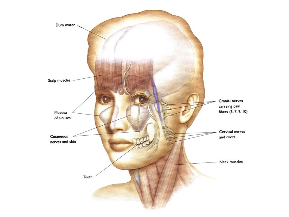
STRUTTURE ALGOGENE DEL CAPO

13
CONTROL OF CRANIO-FACIAL CIRCULATION
Sympathetic innervation Parasympathetic innervation Trigeminovascular system

14
SYMPATHETIC INNERVATION OF CEREBRAL CIRCULATION
STRUCTURES MEDIATORS EFFECTS Hypothalamus Resting conditions: none Counteraction of BP increases (vasoconstriction and reduction of cerebral blood flow) Noradrenaline Neuropeptide Y Serotonin Adenosin-triphosphate Brainstem Spinal cord Superior cervical and stellate ganglia ACA MCA ICA PCA BA BA VA
 Noradrenaline. Neuropeptide Y. Serotonin. Adenosin-triphosphate. Brainstem. Spinal cord. Superior cervical and stellate ganglia. ACA. MCA. ICA. PCA. BA. BA. VA.")
15
PARASYMPATHETIC INNERVATION OF CEREBRAL CIRCULATION
STRUCTURES MEDIATORS EFFECTS . Vasoactive intestinal Pol. . Peptidine histidine isoleucine methionine . Nitric oxide . Acetilcholine Resting conditions: none Neuroprotective role in hypoxia and ischaemia Superior salivatory nucleus Facial nerve Otic and sphenopalatine ganglia Intracarotid microganglia

16
TRIGEMINOVASCULAR INNERVATION OF CEREBRAL CIRCULATION
STRUCTURES MEDIATORS EFFECTS . Substance P . Calcitonin gene-related peptide . Neurokinin . Cholecystokinin . Nitric oxide Resting conditions: none Hyperaemia during hypoxia and post-seizures Protection from vasospasms Brainstem Spinal cord Trigeminal ganglion Trigeminal nerve (I branch) Superficial vessels MCA Middle meningeal artery
 Superficial vessels. MCA. Middle meningeal artery.")
17
Trigeminovascular system
5HT1D Trigeminal neuron 5HT1B/1D/1F 5HT1B Blood vessel Dura mater Brainstem nucleus C1 C2 ganglion

18
TEORIA TRIGEMINO-VASCOLARE
DOLORE Corteccia Neurone trigeminale Talamo TRIGGER Terminazioni trigeminali perivascolari SP SP Brainstem N. trigeminale caudale 5-HT, istamina, BK, PG, CGRP Vasodilatazione Stravaso proteico plasmatico Attivazione macrofagica (adapted from Moskowitz, 80’s)
")
19
Emicrania I meccanismi rigorosi suggeriti nella fisiopatologia dell’emicrania sono teorici

20
Vasi sanguigni del cranio
Emicrania Vasi sanguigni del cranio I meccanismi rigorosi suggeriti nella fisiopatologia dell’emicrania sono teorici

21
Vasi sanguigni del cranio
Emicrania Vasi sanguigni del cranio Ganglio trigeminale I meccanismi rigorosi suggeriti nella fisiopatologia dell’emicrania sono teorici

22
Vasi sanguigni del cranio
Emicrania Vasi sanguigni del cranio Ganglio trigeminale I meccanismi rigorosi suggeriti nella fisiopatologia dell’emicrania sono teorici

23
Vasi sanguigni del cranio
Emicrania Vasi sanguigni del cranio Ganglio trigeminale Nucleo caudale Trigemino I meccanismi rigorosi suggeriti nella fisiopatologia dell’emicrania sono teorici

24
Vasi sanguigni del cranio Nucleo dorsale del rafe
Emicrania Vasi sanguigni del cranio Nucleo dorsale del rafe Locus coeruleus Ganglio trigeminale Nucleo caudale Trigemino I meccanismi rigorosi suggeriti nella fisiopatologia dell’emicrania sono teorici

25
VIP Ach PHI V1 CGRP NKA SP NA NPY SCG VII TCC SSN

26
Fig. 1. Schematic representation of the different types of perivascular nerves. The “extrinsic” nerves to cerebral blood vessels at the surface of the brain come from the peripheral nervous system (PNS) and originate either in the superior cervical (SCG), sphenopalatine (SPG), or otic (OG) or trigeminal (TG) ganglion. Antimigraine drugs like “triptans” act as agonists at prejunctional 5-HT1 receptors on trigeminovascular afferents where they inhibit () the release of CGRP and other peptides. Blood vessels located within the brain parenchyma, or the microcirculation, are innervated by “intrinsic” nerve pathways that find their origin in the central nervous system (CNS). For cortical microvessels, anatomical and/or functional evidence indicate that they receive NA, 5-HT, ACh, or GABAergic afferents from either subcortical neurons from the locus coeruleus, raphe nucleus, basal forebrain, or local cortical interneurons. Inset: schematic representation of the “neurovascular unit” as seen at the electron microscopic level with the vascular [endothelium (medium gray) and smooth muscle or pericyte (dark gray)], astroglial (light gray), and neuronal (axon varicosities are highlighted) compartments (modified from Ref. 6, with permission). ACh, acetylcholine; CGRP, calcitonin gene-related peptide; GABA, -aminobutyric acid; NA, norepinephrine; NKA, neurokinin A; NOS, nitric oxide synthase; NPY, neuropeptide Y; PACAP, pituitary adenylate-cyclase activating polypeptide; SOM, somatostatin; SP, substance P; VIP, vasoactive intestinal polypeptide; 5-HT, serotonin. Hamel et al., 2006
 and originate either in the superior cervical (SCG), sphenopalatine (SPG), or otic (OG) or trigeminal (TG) ganglion. Antimigraine drugs like triptans act as agonists at prejunctional 5-HT1 receptors on trigeminovascular afferents where they inhibit () the release of CGRP. and other peptides. Blood vessels located within the brain parenchyma, or the microcirculation, are innervated by intrinsic nerve pathways that find their origin. in the central nervous system (CNS). For cortical microvessels, anatomical and/or functional evidence indicate that they receive NA, 5-HT, ACh, or GABAergic. afferents from either subcortical neurons from the locus coeruleus, raphe nucleus, basal forebrain, or local cortical interneurons. Inset: schematic representation. of the neurovascular unit as seen at the electron microscopic level with the vascular [endothelium (medium gray) and smooth muscle or pericyte (dark gray)], astroglial (light gray), and neuronal (axon varicosities are highlighted) compartments (modified from Ref. 6, with permission). ACh, acetylcholine; CGRP, calcitonin gene-related peptide; GABA, -aminobutyric acid; NA, norepinephrine; NKA, neurokinin A; NOS, nitric oxide synthase; NPY, neuropeptide Y; PACAP, pituitary adenylate-cyclase activating polypeptide; SOM, somatostatin; SP, substance P; VIP, vasoactive intestinal polypeptide; 5-HT, serotonin. Hamel et al.,")
27
5HT NA vasi cerebrali neuroni midollari Locus Nucleo Coeruleus Dorsale
del Rafe Locus Coeruleus 5HT NA vasi cerebrali neuroni midollari circolazione cerebrale permeabilità BEE nocicezione

28
modulazione globale CBF
arterie piali g. sfenopalatino g. trigeminale g. cervicale superiore Neuroni periferici Neuroni centrali locus coeruleus nucleo dorsale del rafe substantia nigra arteriole penetranti capillari regolazione locale CBF modulazione BEE

29
INTEGRAZIONE AFFERENZE
TEORIE NEUROVASCOLARI Soglia emicranica Stress Stimoli afferenti OROLOGIO BIOLOGICO INTEGRAZIONE AFFERENZE Riflessi assonici LC Ridotto controllo nocicettivo NST DOLORE INFIAMMAZIONE NEUROGENA VASODILATAZIONE (readapted from Lance, 80’s)
")
30
THE CONCEPT OF MIGRAINE THRESHOLD
TRIGGERS Stressors Schedule Shifts Geoclimatic Changes Food Menstrual Cycle MIGRAINE ATTACK THRESHOLD TRAIT Familiarity Dysnociception Neuroendocrine Activity Personality / Mood Disorders Cardiovascular Risk Factors (G. Nappi 1980’s)
")
31
THRESHOLD THEORY OF MIGRAINE INITIATION
ATTACK Threshold breached . Internal and/or external factors THRESHOLD Failure to breach threshold Hormonal trigger Missed lunch Stress overwork Long journey to work Late night MacGregor, 1996

32
Da Toga, UCLA

33
MIGRAINE AS A CHRONIC PAIN CONDITION
Presynaptic Postsynaptic

34
Lashley 1941

35
Blood Flow During Aura and Headache Phase
Key Point: Aura occurs before vasodilation, suggesting that vasodilation is not the sole cause of migraine. One of the most powerful arguments against the vascular theory is that it is in absolute conflict with the blood flow data that should be its greatest support It is clear from Olesen’s studies1, and reinforced by the more recent studies of Cutrer and colleagues2, that the headache phase of migraine with aura starts while blood flow is still reduced. Thus, the headache pain cannot be due simply to vasodilatation Illustration adapted from Olesen et al1 1. Olesen J et al. Timing and topography of cerebral blood flow, aura and headache during migraine attacks. Ann Neurol. 1990;28: Cutrer FM et al. Perfusion-weighted imaging defects during spontaneous migrainous aura. Ann Neurol. 1998;43:25-31. Cortical Spreading Depression CBF=cerebral blood flow Laurizen M. Brain. 1994;118:

36
La spreading depression

37
INTEGRAZIONE AFFERENZE
TEORIE NEUROVASCOLARI Soglia emicranica Stress Stimoli afferenti OROLOGIO BIOLOGICO INTEGRAZIONE AFFERENZE Riflessi assonici LC Ischemia S.D. corticale Ridotto controllo nocicettivo NST DOLORE ALTRI SINTOMI NEUROLOGICI INFIAMMAZIONE NEUROGENA VASODILATAZIONE (readapted from Lance, 80’s)
")
38
FHM: geni e loci Tipo di emicrania Riferimento (i) Gene interessato
Sintomi clinici FHM1, 19p13 Joutel et al. 1993 CACNA1A FHM, MA, SZ rara, PCA, NYS Ophoff et al. 1996 FHM2, 1q23 Marconi et al. 2003 ATP1A2 FHM, MA, MO, MR De Fusco et al. 2003 Terwindt et al. 1997 FHM o BFIC o FHM + BFIC Vanmolkot et al. 2003 FHM Fhm3, 2q24 Dichgans et al., 2005 SCN1A Subnità alfa1 del canale del calcio voltaggio-dip Cav2.1 Subunità alfa2 dell’ATPasi sodio, potassio Canale del sodio neuronale voltaggio-dip Nav1.1 Estevez et al. Hum Genet 2004

39
MIGRAINE AS A CHANNELLOPATHY
FHM-2 ATP1A2 gene 2-subunit Na+/K+ ATPase pump Loss of function FHM-3 SCN1A gene Sodium channel Gain of function? FHM-1 CACNA1A gene Cav2.1Ca2+ Gain of function AURA ↑ GLU/↑ K+ CSD

40
Emicrania 4. Depolarizzazione di membrana cronica 2. Alterata
Riduzione: Buffering del K+ scambio Na+/ Ca++ potenz. di membrana Incremento: Na+ intra Ca++ intra Depolarizzazione corticale 1. Alterato gradiente ionico extracellulare 5. a) Alterato rilascio di neurotrasmettitori, neuropeptidi e neuroormoni con riduzione della soglia della CSD b) Alterazioni dello sviluppo 4. Depolarizzazione di membrana cronica 2. Alterata regolazione genica 3. Alterata sintesi di proteine e neurotramettitori
 Alterato rilascio di neurotrasmettitori, neuropeptidi e neuroormoni con riduzione della soglia della CSD. b) Alterazioni dello sviluppo. 4. Depolarizzazione di membrana cronica. 2. Alterata. regolazione genica. 3. Alterata sintesi di proteine e neurotramettitori.")
41
Fig. 1. Schematic representation of the different types of perivascular nerves. The “extrinsic” nerves to cerebral blood vessels at the surface of the brain come from the peripheral nervous system (PNS) and originate either in the superior cervical (SCG), sphenopalatine (SPG), or otic (OG) or trigeminal (TG) ganglion. Antimigraine drugs like “triptans” act as agonists at prejunctional 5-HT1 receptors on trigeminovascular afferents where they inhibit () the release of CGRP and other peptides. Blood vessels located within the brain parenchyma, or the microcirculation, are innervated by “intrinsic” nerve pathways that find their origin in the central nervous system (CNS). For cortical microvessels, anatomical and/or functional evidence indicate that they receive NA, 5-HT, ACh, or GABAergic afferents from either subcortical neurons from the locus coeruleus, raphe nucleus, basal forebrain, or local cortical interneurons. Inset: schematic representation of the “neurovascular unit” as seen at the electron microscopic level with the vascular [endothelium (medium gray) and smooth muscle or pericyte (dark gray)], astroglial (light gray), and neuronal (axon varicosities are highlighted) compartments (modified from Ref. 6, with permission). ACh, acetylcholine; CGRP, calcitonin gene-related peptide; GABA, -aminobutyric acid; NA, norepinephrine; NKA, neurokinin A; NOS, nitric oxide synthase; NPY, neuropeptide Y; PACAP, pituitary adenylate-cyclase activating polypeptide; SOM, somatostatin; SP, substance P; VIP, vasoactive intestinal polypeptide; 5-HT, serotonin. Hamel et al., 2006
 and originate either in the superior cervical (SCG), sphenopalatine (SPG), or otic (OG) or trigeminal (TG) ganglion. Antimigraine drugs like triptans act as agonists at prejunctional 5-HT1 receptors on trigeminovascular afferents where they inhibit () the release of CGRP. and other peptides. Blood vessels located within the brain parenchyma, or the microcirculation, are innervated by intrinsic nerve pathways that find their origin. in the central nervous system (CNS). For cortical microvessels, anatomical and/or functional evidence indicate that they receive NA, 5-HT, ACh, or GABAergic. afferents from either subcortical neurons from the locus coeruleus, raphe nucleus, basal forebrain, or local cortical interneurons. Inset: schematic representation. of the neurovascular unit as seen at the electron microscopic level with the vascular [endothelium (medium gray) and smooth muscle or pericyte (dark gray)], astroglial (light gray), and neuronal (axon varicosities are highlighted) compartments (modified from Ref. 6, with permission). ACh, acetylcholine; CGRP, calcitonin gene-related peptide; GABA, -aminobutyric acid; NA, norepinephrine; NKA, neurokinin A; NOS, nitric oxide synthase; NPY, neuropeptide Y; PACAP, pituitary adenylate-cyclase activating polypeptide; SOM, somatostatin; SP, substance P; VIP, vasoactive intestinal polypeptide; 5-HT, serotonin. Hamel et al.,")
42
THE PREVALENCE OF MIGRAINE BY GENDER before puberty
12 10 8 Males MIGRAINE PREVALENCE % 6 Females 4 2 3-5 yrs 5-7 yrs 7-9 yrs 9-11 yrs (Mortimer et al, 1992)
")
43
THE PREVALENCE OF MIGRAINE BY GENDER
after puberty 30 25 female 20 Prevalence of migraine (%) 15 10 male 5 20 30 40 50 60 70 80 100 Age (years) Stewart et al.,1994
 male Age (years) Stewart et al.,1994.")
44
Migraine attack onset – Day of menstruation
PREVALENCE OF PURE MENSTRUAL MIGRAINE AND MENSTRUALLY-RELATED MIGRAINE AMONG MIGRAINEURS Attacks occur exclusively on days -2 to +3 of menstruation (ICHD-2004) Prevalence of migraine % Source Migraine attack onset – Day of menstruation Pure Menstrual Menstrually-related Granella et al., 1993 -2 through + 3 9.1 50.8 Mattsson, 2003 21.2 NA McGregor et al., 1990 -2 through +2 7.2 34.5 Granella et al., 2000 3.5 53.5 Dzoljic et al., 2002 12.0 49.0 from Brandes et al., 2006
 Prevalence of migraine % Source. Migraine attack onset – Day of menstruation. Pure Menstrual. Menstrually-related. Granella et al., through Mattsson, NA. McGregor et al., through Granella et al., Dzoljic et al., from Brandes et al.,")
45
SEVERITY OF PURE MENSTRUAL MIGRAINE
Non-menstrual Premenstrual Menstrual * 10 20 30 40 50 60 % of attacks with very severe pain Non-menstrual Premenstrual Menstrual * 0,5 1 1,5 2 2,5 3 number of tablets per attack Granella et al., 2004

46
PAIN THRESHOLD & MENSTRUAL CYCLE
RIII Reflex Threshold Psychophysical Threshold mA mA Tr Tp Tassorelli et al., 2002

47
ESTRADIOL WITHDRAWAL & MIGRAINE
800 Menstrual Cycle 600 Cycle plus estradiol EV 10 mg 400 Estradiol (pg/ml) Migraine Migraine 200 -6 -5 -4 -3 -2 -1 1 2 3 4 5 6 Days of the cycle Somerville, 1972
 Migraine. Migraine Days of the cycle. Somerville,")
48
COURSE OF MIGRAINE IN PREGNANCY
100% 80 No attacks 60 <1 attacks/month Patients 1-3 attacks/month 40 >3 attacks/month 20 Trimesters Pregravid 1st 2nd 3rd Attack frequency during the 3 months preceding pregnancy and during the trimesters of pregnancy in 47 women affected by migraine without aura (MO). First trimester vs pre-gravid period: p=0.0001; second vs first trimester: p=0.0001; third vs second trimester: p=0.02. Sances et al, 2003
. First trimester vs pre-gravid period: p=0.0001; second vs first trimester: p=0.0001; third vs second trimester: p=0.02. Sances et al,")
49
EFFECT OF NATURAL MENOPAUSE ON MIGRAINE
Author/Year Improved (%) Unchanged Worsened Whitty/1968 8 69 23 Neri/1993 67 24 9 Granella/1995 25 45 29 Cupini/1995 30 27 42 Hodson/2000 36 48 16 Mueller/2000 40
 Unchanged. Worsened. Whitty/ Neri/ Granella/ Cupini/ Hodson/ Mueller/")
50
* *** *** ESTRADIOL CHANGES & REPRODUCTIVE LIFE MENSTRUAL CYCLE
ORAL CONTRACEPTION *** *** days days 7 14 21 28 7 7 14 21 28 7 PREGNANCY MENOPAUSE months 1° TRIM 2° TRIM 3° TRIM 3 6 9 12 15 Facchinetti, 2002

51
MIGRAINE & MENSTRUAL CYCLE
ATTACK MIGRAINE ATTACK Estradiol Peak b-EP 5-HT DA NA/A NO PGs Mg++ Others Estradiol Withdrawal & Other Triggers Estradiol Withdrawal & Other Triggers Migraine Window Migraine Window Menstruation Ovulation Menstruation Days (RE Nappi 2001)
")
52
Estrogens and blood vessels
1) regulation of nitric oxide (NO) synthesis and release 2) regulation of ionic fluxes Simoncini et al., 2004
 regulation of nitric oxide (NO) synthesis and release. 2) regulation of ionic fluxes. Simoncini et al.,")
53
Sites of action of estrogens - Relevance for migraine
Endothelium Brain

54
Location of estrogen receptors in the brain
Estrogen receptor-alpha (ER-a) immunocytochemistry Martin & Behbehani, 2006
 immunocytochemistry. Martin & Behbehani,")
55
Activity of different neurotransmitting systems during different phases of the menstrual cycle
Early follicular High sympathetic Low GABA Low serotonergic Mid-cycle High glutamatergic Low sympathetic High serotonergic Mid-luteal High GABAergic High sympathetic Low serotonergic Late luteal Decreasing GABAergic High sympathetic Low serotonergic tone Estrogen Progesterone Martin & Behbehani, 2006

56
Increased vulnerability to migraine attacks
Hormone fluctuations > ? value Mid-cycle, late luteal phases Effect on excitatory neurotransmitter systems Mismatch between nuclear and membrane effects Effect on inhibitory neurotransmitter systems Enhanced glutamatergic tone Decreased opioid, serotonergic and GABAergic tone Sensitization of cortex, brainstem centres and trigeminal system Increased vulnerability to migraine attacks

57
They are often relieved by sleep and are more easily controlled.
Migriane attacks in children tend to be of shorter duration, less severe and easier to treat. They are often relieved by sleep and are more easily controlled. As children move through adolescence, their migraines begin to resemble migraine in adults. (Winner 2005; Guidetti & Canitano, 1992) Da Toga, UCLA
 Da Toga, UCLA.")
58
Fluttuazioni dei livelli di estrogeni Terapia sostitutiva
EMICRANIA E TAPPE DELLA VITA RIPRODUTTIVA Estrogeni erratici Fluttuazioni dei livelli di estrogeni Terapia sostitutiva Perimenopausa Ciclo mestruale Menopausa Gravidanza Menarca Dia Rossella Estrogeni stabili/ settimana sospensione pillola Invecchiamento Estrogeni bassi XX Estrogeni elevati Estrogeni in aumento Fig.

59
Ruolo di fattori psicologici in età infantile-adolescenziale
TRIGGERS Stressors Schedule shifts Geoclimatic changes Menstrual cycle Food TRAIT Familiarity Dysnociception Personality / mood disorders Cardiovascular risk factors Ruolo di fattori psicologici in età infantile-adolescenziale MIGRAINE

60
Emicrania come un disturbo neuropsichiatrico? Balottin et al., 2011
Very good evidence that psychological treatments, principally relaxation and cognitive behavioral therapy, are effective in reducing the severity and frequency of chronic headache in children and adolescents (Eccleston 2003) The efficacy of preventive drug therapy does not seem to be sufficiently proven (Hershey 2010) Emicrania come un disturbo neuropsichiatrico? Balottin et al., 2011 Nappi,
 The efficacy of preventive drug therapy does not seem to be sufficiently proven. (Hershey 2010) Emicrania come un disturbo neuropsichiatrico Balottin et al., Nappi,")
61
La storia naturale dell’emicrania: pietre miliari
INTERVENTO CHIRUGICO MATRIMONIO GRAVIDANZA MENOPAUSA PUBERTA’ LAVORO VIAGGIO Ma sappiamo che ò’emicrani non è solo influenzata da fattori ormonali. In questo schema è rappresentato l’impatto abituale degli eventi della vita sull’andamento dell’emicrania 10 20 30 40 50 60 70 ANNI

62
Estrogens effects

Presentazioni simili
 Valutazione quantitativa dei pazienti sofferenti di (HAE) in Francia, Germania, Italia, Spagna e UK Condotto.>")


>")
 N. Pazienti in Dialisi (2002: 308.910)>")


