Scaricare la presentazione
La presentazione è in caricamento. Aspetta per favore

1
BIOSINTESI DELLE CATECOLAMINE

4
AZIONI PERIFERICHE DELLA DOPAMINA
Recettori D1 (distretti vascolari renale, mesenterico e coronarico) Vasodilatazione; RBF, GFR e secrezione di Na+ Recettori 1 (cardiaci) Effetto inotropo e cronotropo +
 Vasodilatazione; RBF, GFR e secrezione di Na+ Recettori 1. (cardiaci) Effetto inotropo e cronotropo +")
5
Figure 3 | Metabotropic glutamate receptors (mGluRs) and Parkinson's disease. a | Shows the influence of mGluR5 and mGluR2/3 receptors on nigrostriatal degeneration in the 1-methyl-4-phenyl-1,2,3,6-tetrahydropyridine (MPTP) and methamphetamine (METH) models of parkinsonism. MPTP (the active metabolite of which is 1-methyl-4-phenylpyridinium (MPP+)) and METH damage nigrostriatal neurons through different mechanisms that converge on the production of reactive oxygen species (ROS). Endogenous activation of mGluR5 receptors might contribute to ROS formation by facilitating the activity of NMDA (N-methyl-D-aspartate) receptors (NMDARs) through anchoring proteins and stimulating intracellular calcium release Activation of group II mGluRs (mGluR2/3) protects nigrostriatal neurons against MPTP toxicity by inhibiting glutamate release and/or increasing the production of neurotrophic factors in glial cells DA, dopamine; DAG, diacylglycerol; DAT, dopamine transporter; ER, endoplasmic reticulum; GKAP, guanylate-kinase-associated protein; Ins(1,4,5)P3, inositol 1,4,5-trisphosphate; PSD-95, from postsynaptic density 95; PtdIns(4,5)P2, phosphatidylinositol-4,5-bisphosphate; SHANK, from SH3 and multiple ankyrin repeat domains. b | Histological panels show tyrosine hydroxylase (TH) and dopamine transporter (DAT) immunostaining in the substantia nigra of control mice, wild-type mice treated with MPTP, wild-type mice treated with MPTP + MPEP (2-methyl-6-(phenylethynyl)-pyridine), and mGluR5-/- mice treated with MPTP, demonstrating the loss of TH+ cells after treatment with MPTP and their protection by inhibition or lack of mGluR5 receptors
 and methamphetamine (METH) models of parkinsonism. MPTP (the active metabolite of which is 1-methyl-4-phenylpyridinium (MPP+)) and METH damage nigrostriatal neurons through different mechanisms that converge on the production of reactive oxygen species (ROS). Endogenous activation of mGluR5 receptors might contribute to ROS formation by facilitating the activity of NMDA (N-methyl-D-aspartate) receptors (NMDARs) through anchoring proteins and stimulating intracellular calcium release Activation of group II mGluRs (mGluR2/3) protects nigrostriatal neurons against MPTP toxicity by inhibiting glutamate release and/or increasing the production of neurotrophic factors in glial cells DA, dopamine; DAG, diacylglycerol; DAT, dopamine transporter; ER, endoplasmic reticulum; GKAP, guanylate-kinase-associated protein; Ins(1,4,5)P3, inositol 1,4,5-trisphosphate; PSD-95, from postsynaptic density 95; PtdIns(4,5)P2, phosphatidylinositol-4,5-bisphosphate; SHANK, from SH3 and multiple ankyrin repeat domains. b | Histological panels show tyrosine hydroxylase (TH) and dopamine transporter (DAT) immunostaining in the substantia nigra of control mice, wild-type mice treated with MPTP, wild-type mice treated with MPTP + MPEP (2-methyl-6-(phenylethynyl)-pyridine), and mGluR5-/- mice treated with MPTP, demonstrating the loss of TH+ cells after treatment with MPTP and their protection by inhibition or lack of mGluR5 receptors.")
7
MALATTIA DI PARKINSON Segni cardinali Bradicinesia Rigidità muscolare
Tremore a riposo Deficit dell’equilibrio posturale

8
MALATTIA DI PARKINSON

9
Figure 1 | Localization of metabotropic glutamate receptor (mGluR) subtypes in the basal ganglia motor circuit. The figure highlights how dopamine (DA) acting in the neostriatum (caudate nucleus–putamen) influences the activity of the direct and indirect pathways (black and turquoise arrows, respectively). Both pathways converge to regulate the activity of thalamocortical neurons. The activation of D1 dopamine receptors stimulates striatal output neurons of the direct pathway, leading to inhibition of GABA ( -aminobutyric acid) neurons in the internal globus pallidus (GPi) and substantia nigra pars reticulata (SNr). In the indirect pathway, activation of D2 receptors inhibits striatal output neurons that project to the external globus pallidus (GPe). This results in the sequential inhibition of glutamatergic neurons in the subthalamic nucleus (STN) and GABA neurons in the GPi/SNr. The net effect of group I mGluRs is to counterbalance the action of dopamine across the direct pathway, although both mGluR1 and mGluR5 receptors stimulate GPe neurons. Activation of mGluR2/3 receptors mimics the action of dopamine by reducing glutamate release at corticostriatal and STN–GPi and STN–SNr synapses. mGluR4 receptor activation mimics the action of dopamine in the indirect pathway by inhibiting GABA release at the striatum–GPe synapse. For further information see Refs 12, 117, 118, ACh, acetylcholine; CM thalamus, thalamic centromedian nucleus; Glut, glutamate; VA, VL, VM thalamus, ventral anterior, ventrolateral and ventromedial thalamic nuclei.
 acting in the neostriatum (caudate nucleus–putamen) influences the activity of the direct and indirect pathways (black and turquoise arrows, respectively). Both pathways converge to regulate the activity of thalamocortical neurons. The activation of D1 dopamine receptors stimulates striatal output neurons of the direct pathway, leading to inhibition of GABA ( -aminobutyric acid) neurons in the internal globus pallidus (GPi) and substantia nigra pars reticulata (SNr). In the indirect pathway, activation of D2 receptors inhibits striatal output neurons that project to the external globus pallidus (GPe). This results in the sequential inhibition of glutamatergic neurons in the subthalamic nucleus (STN) and GABA neurons in the GPi/SNr. The net effect of group I mGluRs is to counterbalance the action of dopamine across the direct pathway, although both mGluR1 and mGluR5 receptors stimulate GPe neurons. Activation of mGluR2/3 receptors mimics the action of dopamine by reducing glutamate release at corticostriatal and STN–GPi and STN–SNr synapses. mGluR4 receptor activation mimics the action of dopamine in the indirect pathway by inhibiting GABA release at the striatum–GPe synapse. For further information see Refs 12, 117, 118, ACh, acetylcholine; CM thalamus, thalamic centromedian nucleus; Glut, glutamate; VA, VL, VM thalamus, ventral anterior, ventrolateral and ventromedial thalamic nuclei..")
12
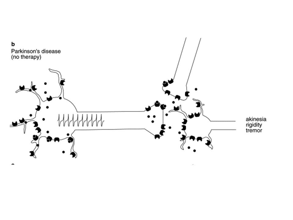
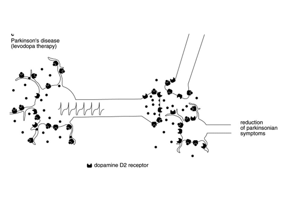
Fig. 1. Schematic representation of the functional activity of the dopaminergic neurones in (a) control conditions, (b) severe dopamine (DA) depletion (Parkinson's disease without therapy) and (c) during the correction of the dopaminergic deficit by levodopa. During the DA-deficient state the firing activity of the spared dopaminergic cells is increased. Consequently, the stores of DA and other neurotransmitters are depleted and the clinical disturbances of Parkinson's disease are evident. The DA substitution therapy could restore the normal functioning of the dopaminergic neurones. Filled circles, dopamine. Electrophysiological pharmacology of the autoreceptor-mediated responses of dopaminergic cells to antiparkinsonian drugs, Trends in Pharmacological Sciences, Volume 18, Issue 7, 1 July 1997, Pages Nicola B. Mercuri, Antonello Bonci and Giorgio Bernardi

15
Tratto GI Sangue e tessuti periferici Cervello LEVODOPA 1% 90% 9% DDC
MAO COMT DDC MAO COMT 90% 9%

16
selegiline X X tolcapone

23
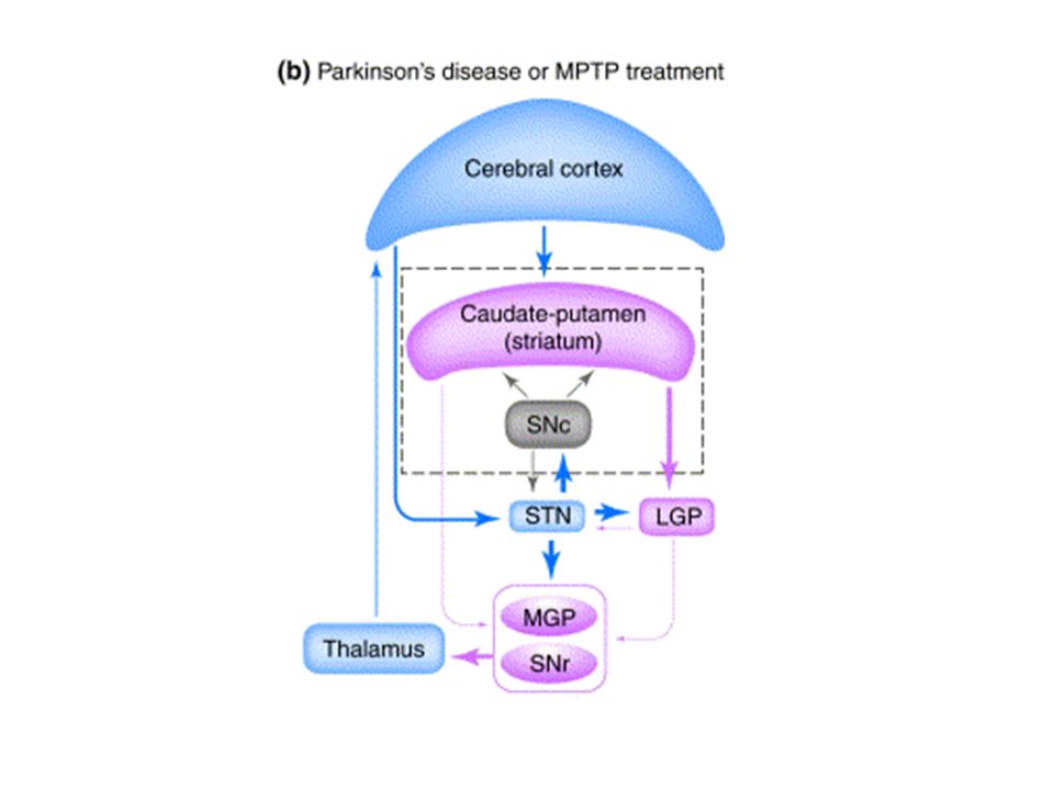
+ - 1 2 3 4 1 2 Fig. 1. Schematic representation of the functional organization of neurons in various brain regions that are responsible for motor control in (a) normal brain function. Glutamate-mediated pathways are indicated in blue, dopamine (DA)-mediated pathways are indicated in orange and GABA-mediated pathways are indicated in pink. (b) Changes in the degree of activation of each signaling component are indicated by arrow thickness in patients with Parkinson's disease (PD) or animals injected with 1-methyl-4-phenyl-1,2,3,6-tetrahydropyridine (MPTP) to induce experimental parkinsonism. A hallmark feature of idiopathic or experimental PD is the injury and loss of DA-containing neurons in the substantia nigra pars compacta (SNc). The loss of signal integration from this nucleus results in disregulation of the circuit leading to disruption of motor control. (c) Kordower and colleagues injected lentiviral vectors to deliver glial cell line-derived neurotrophic factor (GDNF) to the caudate-putamen and substantia nigra. Lenti-GDNF treatment results in increased expression of GDNF, which is taken up by neurons, transported to the nucleus and results in initiation of new gene transcription. Nerve terminals regenerate and DA synthesis is restored. With the restoration of tyrosine hydroxylase (TH) expression in the striatum, there was an associated improvement in functional recovery. These findings suggest that GDNF can not only promote the survival and regeneration of DA-containing neurons but can restore proper signal transduction in the basal ganglia circuit that controls movement. Abbreviations: LGP, lateral globus pallidus; MGP, medial globus pallidus; RF, parvicellular reticular formation; SC, superior colliculus; SNr, substantia nigra pars reticulata; STN, subthalamic nucleus.
 normal brain function. Glutamate-mediated pathways are indicated in blue, dopamine (DA)-mediated pathways are indicated in orange and GABA-mediated pathways are indicated in pink. (b) Changes in the degree of activation of each signaling component are indicated by arrow thickness in patients with Parkinson s disease (PD) or animals injected with 1-methyl-4-phenyl-1,2,3,6-tetrahydropyridine (MPTP) to induce experimental parkinsonism. A hallmark feature of idiopathic or experimental PD is the injury and loss of DA-containing neurons in the substantia nigra pars compacta (SNc). The loss of signal integration from this nucleus results in disregulation of the circuit leading to disruption of motor control. (c) Kordower and colleagues injected lentiviral vectors to deliver glial cell line-derived neurotrophic factor (GDNF) to the caudate-putamen and substantia nigra. Lenti-GDNF treatment results in increased expression of GDNF, which is taken up by neurons, transported to the nucleus and results in initiation of new gene transcription. Nerve terminals regenerate and DA synthesis is restored. With the restoration of tyrosine hydroxylase (TH) expression in the striatum, there was an associated improvement in functional recovery. These findings suggest that GDNF can not only promote the survival and regeneration of DA-containing neurons but can restore proper signal transduction in the basal ganglia circuit that controls movement. Abbreviations: LGP, lateral globus pallidus; MGP, medial globus pallidus; RF, parvicellular reticular formation; SC, superior colliculus; SNr, substantia nigra pars reticulata; STN, subthalamic nucleus.")
Presentazioni simili


>")


 EPATOTOSSICITA’ CRISI IPERTENSIVE EMORRAGIE R.I.P.>")
