Scaricare la presentazione
La presentazione è in caricamento. Aspetta per favore

1
ACETILCOLINA Acido acetico Colina O CH3 +
H3C – C – O – CH2 – CH2 – N – CH3 O CH3 + ACETILCOLINA H3C – C – OH O Acido acetico HO – CH2 – CH2 – N – CH3 CH3 + Colina

2
(Terminazione post-gangliare Terminazioni post-gangliari
SISTEMA NERVOSO CENTRALE IPPOCAMPO, GANGLI DELLA BASE m. di ALzheimer, m. di Parkinson SISTEMA NERVOSO SOMATICO ACh GIUNZIONE NEUROMUSCOLARE Gangli SIMPATICO Midollare surrenale SISTEMA NERVOSO AUTONOMO (Terminazione post-gangliare gh. sudoripare) Gangli PARASIMPATICO Terminazioni post-gangliari organi effettori
 Gangli. PARASIMPATICO. Terminazioni post-gangliari. organi effettori.")
3
Ach NO NO NO NO NO NO NO NO NO NO NO NO NO M3 Rilasciamento della
muscolatura liscia NO NO NO NO NO NO NO NO NO NO NO NO NO Ach

4
EFFETTI DELL’ACETILCOLINA SUI TESSUTI PERIFERICI
Variabile Utero Costrizione; secrezioni Bronchi Erezione Tratto riproduttivo (maschio) Diaforesi Ghiandole sudoripare Contrazione del muscolo detrusore; rilassamento dello sfintere Vescica Tono e secrezioni; rilassamento degli sfinteri Tratto GI Bradicardia; velocità di conduzione; blocco AV ad alte dosi; lieve forza contrattile Cuore Secrezione fluida e acquosa Ghiandole salivari e lacrimali Contrazione e accomodazione del cristallino alla visione ravvicinata Muscolo ciliare Contrazione e miosi Iride (m. sfintere della pupilla) Liberazione di NO e vasodilatazione Vasi (cellule endoteliali) Effetti dell’acetilcolina TESSUTO
 Diaforesi. Ghiandole sudoripare. Contrazione del muscolo detrusore; rilassamento dello sfintere. Vescica. Tono e secrezioni; rilassamento degli sfinteri. Tratto GI. Bradicardia; velocità di conduzione; blocco AV ad alte dosi; lieve forza contrattile. Cuore. Secrezione fluida e acquosa. Ghiandole salivari e lacrimali. Contrazione e accomodazione del cristallino alla visione ravvicinata. Muscolo ciliare. Contrazione e miosi. Iride (m. sfintere della pupilla) Liberazione di NO e vasodilatazione. Vasi (cellule endoteliali) Effetti dell’acetilcolina. TESSUTO.")
6
CARATTERISTICHE CLINICHE DELL’INFEZIONE DA C. botulinum
DEBOLEZZA DEFICIT SENSORIALE PERDITA DEI RIFLESSI TENDINEI EFFETTI SUL SISTEMA NERVOSO AUTONOMO OCCHIO SISTEMA CARDIOVASCOLARE CUTE APPARATO ESCRETORE TRATTO GASTRO-INTESTINALE

7
Responsabile dell’effetto tossico
Responsabile della traslocazione della catena L all’interno delle cellule bersaglio Botulinum toxins Botulism neurotoxin is the single most toxic compound currently known to exist. A dose of to 10-9 grams per kilogram of body weight is lethal. Therefore, for a 150 pound (68.5 kilogram) individual, 6.85 x 10⁻⁹ grams of botulinum toxin is lethal. Thus, botulism is frequently fatal unless therapy is rapidly available. The botulinum neurotoxin acts upon nerve endings which use the neurotransmitter acetylcholine. There are seven types of botulinum toxins: classified as A, B, C1, D, E, F, and G. Each neurotoxin is distinguished by the different monoclonal antibodies which react to the different types of botulinum toxin. The structures of each type of botulinum toxin are relatively comparable, except for type G, which is thought to be encoded by plasmids. The neurotoxins are large molecules (150 kDa) and are formed of polypeptide molecules. The toxin is comprised of two polypeptide chains which are linked by a disulphide bond. The heavy (H) chain is further divided into two subunits, the C-terminal (HC) and the N-terminal (HN) chains. The second polypeptide chain, the light (L) chain, is comprised of Zn++ ions. In terms of toxicity, the C-terminal chain serves to bind the toxin to the peripheral motoneurones and the N-terminal chain provides for penetration of the toxin into the neuron. Once inside the cytoplasm of the neuron, the L chain is released and disrupts the release of the neurotransmitter acetylcholine. Si lega alla superficie delle cellule bersaglio
 individual, 6.85 x 10⁻⁹ grams of botulinum toxin is lethal. Thus, botulism is frequently fatal unless therapy is rapidly available. The botulinum neurotoxin acts upon nerve endings which use the neurotransmitter acetylcholine. There are seven types of botulinum toxins: classified as A, B, C1, D, E, F, and G. Each neurotoxin is distinguished by the different monoclonal antibodies which react to the different types of botulinum toxin. The structures of each type of botulinum toxin are relatively comparable, except for type G, which is thought to be encoded by plasmids. The neurotoxins are large molecules (150 kDa) and are formed of polypeptide molecules. The toxin is comprised of two polypeptide chains which are linked by a disulphide bond. The heavy (H) chain is further divided into two subunits, the C-terminal (HC) and the N-terminal (HN) chains. The second polypeptide chain, the light (L) chain, is comprised of Zn++ ions. In terms of toxicity, the C-terminal chain serves to bind the toxin to the peripheral motoneurones and the N-terminal chain provides for penetration of the toxin into the neuron. Once inside the cytoplasm of the neuron, the L chain is released and disrupts the release of the neurotransmitter acetylcholine. Si lega alla superficie delle cellule bersaglio.")
8
Action of botulinum neurotoxin
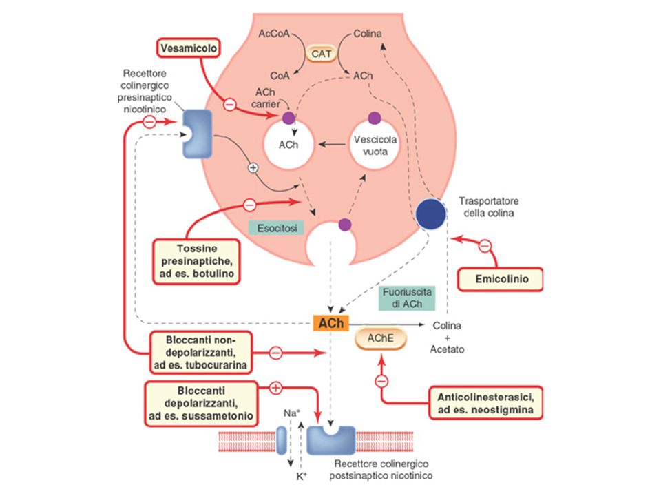
The normal transmission of an impulse to a muscle is initiated when an action potential travels down the axon of a motor neuron. Near the synapse, vesicles (storage organelles) containing the neurotransmitter acetylcholine are stored at the presynaptic nerve terminal. The action potential, the neuronal signal, results in the activation of the vesicles, where they bind to the presynaptic membrane and release the acetylcholine molecules into the synaptic cleft. The acetylcholine then binds to receptors on the surface of the muscle, after traversing the distance between the nerve terminal and muscle membrane. The binding of the neurotransmitters results in a depolarization in the muscle due to the changing in concentration of ions. As the depolarization passes a prescribed threshold, an action potential is created, which subsequently induces the contraction of the muscle. Botulinum toxin disables the nervous system by decreasing the number of acetylcholine containing vesicles that bind to the presynaptic membrane. The toxin does not inhibit the production of acetylcholine. Thus, by reducing the quantity of acetylcholine neurotransmitter, botulism neurotoxin is capable of rendering muscle stimuli ineffective--resulting in paralysis
 containing the neurotransmitter acetylcholine are stored at the presynaptic nerve terminal. The action potential, the neuronal signal, results in the activation of the vesicles, where they bind to the presynaptic membrane and release the acetylcholine molecules into the synaptic cleft. The acetylcholine then binds to receptors on the surface of the muscle, after traversing the distance between the nerve terminal and muscle membrane. The binding of the neurotransmitters results in a depolarization in the muscle due to the changing in concentration of ions. As the depolarization passes a prescribed threshold, an action potential is created, which subsequently induces the contraction of the muscle. Botulinum toxin disables the nervous system by decreasing the number of acetylcholine containing vesicles that bind to the presynaptic membrane. The toxin does not inhibit the production of acetylcholine. Thus, by reducing the quantity of acetylcholine neurotransmitter, botulism neurotoxin is capable of rendering muscle stimuli ineffective--resulting in paralysis.")
9
INDICAZIONI ALL’USO DELLA TOSSINA BOTULINICA DI TIPO A
BLEFAROSPASMO

10
INDICAZIONI ALL’USO DELLA TOSSINA BOTULINICA DI TIPO A
STRABISMO

11
INDICAZIONI ALL’USO DELLA TOSSINA BOTULINICA DI TIPO A
DISTONIA CERVICALE

12
INDICAZIONI ALL’USO DELLA TOSSINA BOTULINICA DI TIPO A
ATTENUAZIONE DELLE LINEE GLABELLARI

13
CLASSIFICAZIONE DEI RECETTORI COLINERGICI MUSCARINICI
Meccanismo di trasduzione G-proteine Antagonisti Agonisti Localizzazione Tipo fosfolipasi C Gq/11 pirenzepina acetilcolina, carbacolo, McN-A-343 SNC; gangli autonomi M1 conduttanza K+; adenilato ciclasi Gi/o AF DX 117 acetilcolina, carbacolo Cuore (t. di conduzione, atri); gangli auonomi M2 come M1 Gq/11 esaidrosil-adifenolo acetilcolina, carbacolo Ghiandole esocrine; muscolatura liscia; endotelio M3 come M2 Gi/o acetilcolina SNC M4 come M1 Gq/11 acetilcolina SNC M5
; gangli auonomi. M2. come M1. Gq/11. esaidrosil-adifenolo. acetilcolina, carbacolo. Ghiandole esocrine; muscolatura liscia; endotelio. M3. come M2. Gi/o. acetilcolina. SNC. M4. come M1. Gq/11. acetilcolina. SNC. M5.")
14
Activation of M1/M3 muscarinic responses
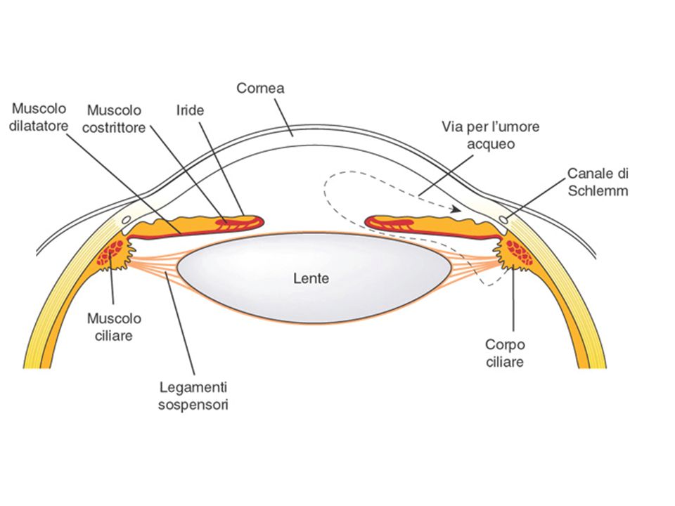
Activation of M1/M3 muscarinic responses. Stimulation of M1-muscarinic receptors by acetylcholine leads to the activation of a Gq coupling protein. The alpha subunit of this G protein activates the effector, phospholipase C, which leads to the release of IP3 (inositol 1,4,5-trisphosphate) and DAG (diacylglycerol) from phosphatidylinositol 4,5-bisphosphate (PtdIns 4,5-P2). IP3 stimulates the release of sequestered stores of calcium, leading to an increased concentration of cytoplasmic Ca2+. Ca2+ may then activate Ca2+-dependent protein kinases, which in turn phosphorylate their substrates. DAG activates protein kinase C. M3-Muscarinic receptors cause contraction of smooth muscle in the following organs: Eye: sphincter of the iris Eye: ciliary muscle Lungs: tracheal and bronchial muscle Stomach and Intestines: increase motility and parasympathetic tone Glands (lacrimal, salivary, bronchial, pancreatic, mucosal, and sweat(sympathetic), adrenal(sympathetic)): increase secretion Bladder: detrusor muscle Sex Organs, Male: M3-receptors cause release of nitric oxide in endothelial cells producing vasodilation (erection)
 and DAG (diacylglycerol) from phosphatidylinositol 4,5-bisphosphate (PtdIns 4,5-P2). IP3 stimulates the release of sequestered stores of calcium, leading to an increased concentration of cytoplasmic Ca2+. Ca2+ may then activate Ca2+-dependent protein kinases, which in turn phosphorylate their substrates. DAG activates protein kinase C. M3-Muscarinic receptors cause contraction of smooth muscle in the following organs: Eye: sphincter of the iris. Eye: ciliary muscle. Lungs: tracheal and bronchial muscle. Stomach and Intestines: increase motility and parasympathetic tone. Glands (lacrimal, salivary, bronchial, pancreatic, mucosal, and sweat(sympathetic), adrenal(sympathetic)): increase secretion. Bladder: detrusor muscle. Sex Organs, Male: M3-receptors cause release of nitric oxide in endothelial cells producing vasodilation (erection)")
15
Transmembrane topology and action of a M2-muscarinic receptor
Transmembrane topology and action of a M2-muscarinic receptor.. The receptor's amino (N) terminal is extracellular (above the plane of the membrane), and its carboxyl (C) terminal intracellular. The terminals are connected by a polypeptide chain that traverses the plane of the membrane seven times. The hydrophobic transmembrane segments (light color) are designated by roman numerals (I-VII). Acetylcholine (Ach) approaches the receptor from the extracellular fluid and binds to a site surrounded by the transmembrane regions of the receptor protein. G proteins (G) interact with cytoplasmic regions of the receptor, especially with portions of the third cytoplasmic loop between transmembrane regions V and VI. The receptor's cytoplasmic terminal tail contains numerous serine and threonine residues whose hydroxyl (-OH) groups can be phosphorylated. This phosphorylation may be associated with diminished receptor-G protein interaction. M2-muscarinic receptor activation is coupled to opening of K+ channels causing potassium to flow outside the cell producing hyperpolarization. The heart becomes more refractory to contraction and the rate slows. M2-muscarinic receptors cause action in the heart in the following manner: Heart SA node: decrease in heart rate. Heart AV node: decrease in conduction velocity Heart Atria: decrease in contractility
 terminal is extracellular (above the plane of the membrane), and its carboxyl (C) terminal intracellular. The terminals are connected by a polypeptide chain that traverses the plane of the membrane seven times. The hydrophobic transmembrane segments (light color) are designated by roman numerals (I-VII). Acetylcholine (Ach) approaches the receptor from the extracellular fluid and binds to a site surrounded by the transmembrane regions of the receptor protein. G proteins (G) interact with cytoplasmic regions of the receptor, especially with portions of the third cytoplasmic loop between transmembrane regions V and VI. The receptor s cytoplasmic terminal tail contains numerous serine and threonine residues whose hydroxyl (-OH) groups can be phosphorylated. This phosphorylation may be associated with diminished receptor-G protein interaction. M2-muscarinic receptor activation is coupled to opening of K+ channels causing potassium to flow outside the cell producing hyperpolarization. The heart becomes more refractory to contraction and the rate slows. M2-muscarinic receptors cause action in the heart in the following manner: Heart SA node: decrease in heart rate. Heart AV node: decrease in conduction velocity. Heart Atria: decrease in contractility.")
16
AGONISTI MUSCARINICI NATURALI
MUSCARINA

17
AGONISTI MUSCARINICI NATURALI
PILOCARPINA

18
AGONISTI MUSCARINICI NATURALI
CH3 COCH3 ARECOLINA

20
AGONISTI MUSCARINICI SINTETICI
H2N – C – O – CH2 – CH2 – N – CH3 O CH3 + CARBACOLO H2N – C – OH O Acido carbammico HO – CH2 – CH2 – N – CH3 CH3 + Colina

21
AGONISTI MUSCARINICI SINTETICI
BETANECOLO (carbamil--metilcolina) H2N – C – O – CH2 – CH2 – N – CH3 O CH3 + METACOLINA (acetil--metilcolina) H3C – C – O – CH2 – CH2 – N – CH3
 H2N – C – O – CH2 – CH2 – N – CH3. O. CH3. + METACOLINA. (acetil--metilcolina) H3C – C – O – CH2 – CH2 – N – CH3.")
22
EFFETTI DI UN’ECCESSIVA STIMOLAZIONE MUSCARINICA

23
AGENTI ANTIMUSCARINICI (PARASIMPATICOLITICI)
")
24
AGENTI ANTIMUSCARINICI (PARASIMPATICOLITICI)
")
25
AGENTI ANTIMUSCARINICI (PARASIMPATICOLITICI)
")
26
AGENTI ANTIMUSCARINICI (PARASIMPATICOLITICI)
")
27
AGENTI ANTIMUSCARINICI (PARASIMPATICOLITICI)
")
28
DEI RECETTORI MUSCARINICI
EFFETTI DEL BLOCCO DEI RECETTORI MUSCARINICI

Presentazioni simili


 Le cellule distanti tra loro comunicano attraverso molecole (MEDIATORI o NEUROTRASMETTITORI)>")

