Scaricare la presentazione
La presentazione è in caricamento. Aspetta per favore

1
Cellule staminali e cellule staminali tumorali Quali cellule sono responsabili per la crescita del tumore?

2
Cellule staminali Sono cellule che hanno la capacitá di perpetuarsi indefinitamente (“self-renewal”) Attraverso il differenziamento, esse danno vita alle cellule “mature” Le cellule differenziate originano dalle cellule staminali del medesimo compartimento Plasticitá delle cellule staminali: apparentemente, le cellule staminali di un tessuto possono dare origine anche a cellule mature di altri tessuti
 Attraverso il differenziamento, esse danno vita alle cellule mature Le cellule differenziate originano dalle cellule staminali del medesimo compartimento Plasticitá delle cellule staminali: apparentemente, le cellule staminali di un tessuto possono dare origine anche a cellule mature di altri tessuti")
3
Cellule staminali e cellule tumorali Il tumore è costituito da cellule con una capacità di self-renewal indefinita La comprensione dei meccanismi di self-renewal delle cellule staminali puó aiutarci a comprendere il tumore

4
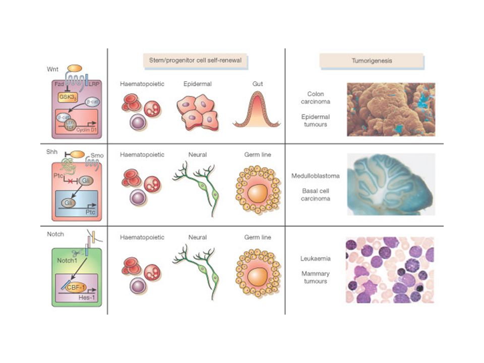
Pathway coinvolti nel self- renewal e nell’oncogenesi Ipotesi: le cellule tumorali -capaci di self- renewal- utilizzano la “machinery” presente nelle cellule staminali Dimostrazione indiretta di tale ipotesi é il fatto che diversi pathway associati all’oncogenesi sono stati coinvolti nel self- renewal delle cellule staminali

5
I pathway di Notch, Shh, Wnt Notch: l’attivazione di questo pathway é associata ad un aumento del pool delle cellule staminali Shh: popolazioni arricchite di cellule staminali umane rispondono in vitro a Shh con un aumentato self-renewal Wnt: la sua attivazione espande il pool di cellule staminali, mentre la sua soppressione inibisce la proliferazione delle cellule staminali

7
1 3 2 Stem cells self renew--?immortal non-stem cells have finite life span Rules of Normal Tissue Growth

8
1 3 2 Rules: 1.) Tumors are clonal – starts in a single cell 2.) All tumor cells have infinite lifespan 3.) All tumor cells divide symmetrically 30 cell divisions = 1 billion cells = 1 cm tumor Traditional View of Tumor Growth
 Tumors are clonal – starts in a single cell 2.) All tumor cells have infinite lifespan 3.) All tumor cells divide symmetrically 30 cell divisions = 1 billion cells = 1 cm tumor Traditional View of Tumor Growth")
9
Non-stem tumor model: every cell in a tumor should initiate a new tumor

10
Experiments showed that very rare cells in a tumor can transplant a new tumor: Tumor Stem Cells

11
Origin of the Theory of Cancer Stem Cells Only a small subset of cancer cells is capable of extensive proliferation Liquid Tumors In vitro colony forming assays: - 1 in 10,000 to 1 in 100 mouse myeloma cells obtained from ascites could form colonies In vivo transplantation assays: - Only 1-4% of transplanted leukaemic cells could form spleen colonies Solid Tumors - A large number of cells are required to grow tumors in xenograft models - 1 in 1,000 to 1 in 5,000 lung cancer, neuroblastoma cells, ovarian cancer cells, or breast cancer cell from cell lines can form colonies in soft agar or in vivo (fewer with 1 0 tumor cells)
")
12
Adult stem cell = undifferentiated Transit amplifying cell Tumor stem cell = tumorigenic Tumor growth is similar to normal tissue growth Normal differentiated cell Non-tumorigenic cell Normal TissuesTumor

13
Cellule staminali tumorali: organogenesi aberrante Il tumore puó essere immaginato come un organo aberrante originato da una cellula trasformata che ha acquisito la capacitá di proliferare indefinitamente attraverso varie mutazioni La popolazione tumorale é eterogenea, e spesso contiene cellule a diversi stadi di differenziamento (seppure anomali): data la clonalitá dei tumori, questo dato implica che la progenie delle cellule tumorali si diversifica (“differenzia”)
: data la clonalitá dei tumori, questo dato implica che la progenie delle cellule tumorali si diversifica ( differenzia )")
14
Evidenze per la presenza di cellule staminali tumorali

15
Hematopoietic Stem Cells Stem Cells Multipotent Progenitors Oligolineage Progenitors Mature Cells Reya et al. 2001 Nature 414:105-111 CD34+ CD38- CD4+ CD35+ CD36+ CD8+ CD20+ CD34- CD38+ CD34- CD38-

16
Cellule staminali ematopoietiche Le cellule caratterizzate con maggiore precisione, grazie ad esperimenti di ripopolamento di topi letalmente irradiati e ricostituiti con popolazioni cellulari altamente purificate a partire dal midollo osseo Le cellule staminali (0.05% delle cellule totali del midollo) danno origine ai progenitori ematopoietici che perdono il loro potenziale di self-renewal
 danno origine ai progenitori ematopoietici che perdono il loro potenziale di self-renewal")
17
Self-renewal Assay in NOD/SCID Mice (Non-obese diabetic/severe combined immunodeficiency) Sublethally irradiated NOD/SCID Mice FACS Cell Sorter Cancer Cells ex: Leukaemia cells CD38 Expression CD34 Expression
 Sublethally irradiated NOD/SCID Mice FACS Cell Sorter Cancer Cells ex: Leukaemia cells CD38 Expression CD34 Expression")
18
Leukaemia stem cells exist in human acute myeloid leukaemia (AML) Leukaemic blasts from AML patients CD34+/ CD38- CD34+/ CD38+ LEUKAEMIA NO LEUKAEMIA John Dick and Dominique Bonnet NOD/ SCIDmiceNOD/ SCIDmice
 Leukaemic blasts from AML patients CD34+/ CD38- CD34+/ CD38+ LEUKAEMIA NO LEUKAEMIA John Dick and Dominique Bonnet NOD/ SCIDmiceNOD/ SCIDmice")
19
Leukaemia is arranged as a hierarchy similar to normal haematopoiesis B-cellT-cellErythrocytePlatelet Monocyte Granulocyte CD34+/ CD38- lymphoid progenitor myeloid progenitor HSC NORMAL LEUKAEMIA Leukaemogenic events Bulk leukaemia cells (CD34+/CD38- and other cells) Block terminal differentiation John Dick and Dominique Bonnet
 Block terminal differentiation John Dick and Dominique Bonnet")
20
Le cellule staminali tumorali come meccanismo di mantenimento del tumore 1.Isolamento di sub-popolazioni cellulari con marcatori di superficie caratteristici delle SC normali (CD34+CD38-), o di cellule piú differenziate, da blasti leucemici di pazienti affetti da varie forme di leucemia mieloide acuta 2.Reinoculo di queste cellule in topi NOD/SCID ed analisi della loro capacitá leuchemogenica 3.Mentre le cellule CD34+38- sono leuchemogeniche, quelle CD34+CD38+ non possono trasferire la leucemia nell’animale immunocompromesso 4.Le cellule tumorali non sono tutte uguali, e le CSC sono responsabili del mantenimento della massa tumorale
, o di cellule piú differenziate, da blasti leucemici di pazienti affetti da varie forme di leucemia mieloide acuta 2.Reinoculo di queste cellule in topi NOD/SCID ed analisi della loro capacitá leuchemogenica 3.Mentre le cellule CD sono leuchemogeniche, quelle CD34+CD38+ non possono trasferire la leucemia nell’animale immunocompromesso 4.Le cellule tumorali non sono tutte uguali, e le CSC sono responsabili del mantenimento della massa tumorale")
21
Aggiornamento dei risultati di Dick e Bonnet Utilizzando topi immunocompromessi con alterazioni genetiche addizionali (> grado di immunocompromissione) il dato originale viene confermato (la frazione CD34+CD38- contiene le cellule staminali della leucemia), ma in una certa percentuale di leucemie anche la frazione CD34+CD38+ contiene cellule staminali in minore proporzione rispetto alle CD34+CD38- –In questi casi I livelli di espressione di alcuni miRNA sono più precisi dei marker di superficie nel definire le cellule staminali leucemiche
 il dato originale viene confermato (la frazione CD34+CD38- contiene le cellule staminali della leucemia), ma in una certa percentuale di leucemie anche la frazione CD34+CD38+ contiene cellule staminali in minore proporzione rispetto alle CD34+CD38- –In questi casi I livelli di espressione di alcuni miRNA sono più precisi dei marker di superficie nel definire le cellule staminali leucemiche")
22
Evidenze da altri tumori Nei tumori solidi si può osservare sperimentalmente una simile struttura gerarchica (i marker sono definiti in maniera meno precisa)
")
23
Self-renewal Assay in NOD/SCID Mice For solid tumors: surgical orthotopic implantation (SOI) FACS Cell Sorter Solid Tumor Single Cell Suspension Mince (small pieces) Surgical Implantation CD24 Expression CD44 Expression
 FACS Cell Sorter Solid Tumor Single Cell Suspension Mince (small pieces) Surgical Implantation CD24 Expression CD44 Expression")
24
CD 44 staining of breast cancer model T. A. Ince 2001

25
Breast Cancer Stem Cells: CD44 + CD24 low Lin - B38.1 + ESA + CD44 and CD24 – adhesion molecules B38.1 – breast/ovarian cancer-specific marker ESA – epithelial specific antigen Al-Hajj (2003) PNAS 100, 3983
 PNAS 100, 3983")
26
Tumor stem cells Non-tumorigenic cells Therapeutic predictions of tumor stem cell model

27
tumor grows back tumor degenerates Therapeutic predictions of tumor stem cell model rapid growing cells killed kill stem cells

28
Therapeutic implications of Cancer Stem Cells Hypothesis: -Most therapies (chemotherapy and radiation) target rapidly proliferating, non-tumorigenic cells and spare the relatively quiescent cancer stem cells -Cell surface pumps -Cancer stem cells have greater invasive and migratory properties and can home to specific tissue niches
 target rapidly proliferating, non-tumorigenic cells and spare the relatively quiescent cancer stem cells -Cell surface pumps -Cancer stem cells have greater invasive and migratory properties and can home to specific tissue niches")
29
Cancer stem cells sono più resistenti alle terapie antitumorali

30
Experimental models in vitro models (ex vivo ) Cultured cell from human gliomas: D456MG D54MG Patient glioblastoma samples in vivo models Human xenograft models in immunocompromised mice
 Cultured cell from human gliomas: D456MG D54MG Patient glioblastoma samples in vivo models Human xenograft models in immunocompromised mice")
31
Brain tumor stem cells: identified by intracranial transplantation of CD133 + cells into adult NOD/SCID mouse forebrain. CD133 + CD133 - Singh et al. 2004 Nature 432: 396-401

32
Resistance to radiation: → given by CD133+

33
Glioma xenograft D456MG: in vivo CD133+ enrichment after radiation →enriched CD133+ population 48h after radiation (3-5x)
")
34
in vitro CD133+ enrichment after radiation Cultures from human glioma xenograft (D54MG): →48h after radiation: 3x enrichment Patient glioblastoma samples:
: →48h after radiation: 3x enrichment Patient glioblastoma samples:")
35
Irradiation effects at molecular level Early DNA damage checkpoint responses (phosphorylation) checked before treatment and after 1h. Higher amount of phosphorylated proteins in CD133+. Early DNA damage checkpoint responses:

36
CD133+ subpopulation has cancer stem cell properties

37
in vivo tumorigenic potential of purified CD133+ tumor cells D456MG CD133- (2 x 10 6 ) formed small tumors in 2 out of 5 xenotransplanted in immunocompromised mice. CD133+ cells (10 4 ) from patient sample or xenograft transplanted into brains of immunocompromised mice. Brain observed at appearence of neurological signs or after 8 weeks. in vitro irradiation
 from patient sample or xenograft transplanted into brains of immunocompromised mice. Brain observed at appearence of neurological signs or after 8 weeks. in vitro irradiation.")
38
Le cellule CD133+ non sono le sole cancer stem cells Molte evidenze suggeriscono che il marcatore CD133 non sia sempre valido nella determinazione delle cancer stem cells (come nelle leucemie)
")
39
Domanda fondamentale È sufficiente attaccare esclusivamente le CSC? Nessuno ha finora dimostrato che l'incapacità di self-renewal delle CSC sia sufficiente ad impedire lo sviluppo di un tumore

40
Promyelocytes Chr 15 Chr 17 t(15;17) Acute Promyelocytic Leukemia (APL) Myeloid differentiation Monoblast
 Acute Promyelocytic Leukemia (APL) Myeloid differentiation Monoblast")
41
Leukemogenesis is a multi-stage process Leukemia-free survival (%) Pre-leukemia At the pre-leukemic stage, hematopoiesis is apparently normal
 Pre-leukemia At the pre-leukemic stage, hematopoiesis is apparently normal")
42
Molecular mechanism of PML-RAR action From DeThe and Chen RA DNMT/HMTs

43
tumor grows back PML-RAR degradation APL Tumor Recurrence LICs Bulk Cells ATRA ATRA acts on bulk APL cells, and on LICs

44
Continuous treatment with HDACi is required for prolonging survival of leukemic mice

45
tumor grows back rapid growing cells killed

46
An assay to measure LICs Leukemic Cells (Ly5.2) Drug treatment Harvest leukemic cells (Ly5.2+) treated/untreated (Ly5.1+) Transplant in Limiting Dilutions Bulk LIC Vehicle Treatment No Effect LIC Expansion LIC Reduction (Ly5.1+))
 Drug treatment Harvest leukemic cells (Ly5.2+) treated/untreated (Ly5.1+) Transplant in Limiting Dilutions Bulk LIC Vehicle Treatment No Effect LIC Expansion LIC Reduction (Ly5.1+))")
47
An assay to measure LICs Bulk LIC Vehicle Treatment No Effect LIC Expansion LIC Reduction Leukemic Cells (Ly5.2) Drug treatment Harvest leukemic cells (Ly5.2+) treated/untreated (Ly5.1+) Transplant in Limiting Dilutions (Ly5.1+) ATRA treatment reduces LIC frequency ≈ 100 fold
 Drug treatment Harvest leukemic cells (Ly5.2+) treated/untreated (Ly5.1+) Transplant in Limiting Dilutions (Ly5.1+) ATRA treatment reduces LIC frequency ≈ 100 fold")
48
VPA spares LICs VehicleVPA LIC Frequency2.5x10^43.9x10^4 Limiting Dilution

49
Short-term inhibition of multiple HDACs with SAHA tackles LICs but does not prolong survival Survival VehicleSAHA LIC Frequency2.5x10^42.3x10^6 LIC assay

50
LeukemiaTumor Recurrence VPA SAHA LICs Bulk Cells ATRA In Summary… ?

51
Eradication of APL by ATRA-SAHA-VPA No leukemic cells detectable

52
In cosa differiscono le cancer stem cells dalle cellule staminali normali?

56
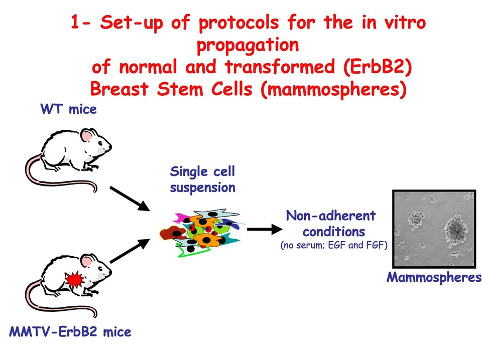
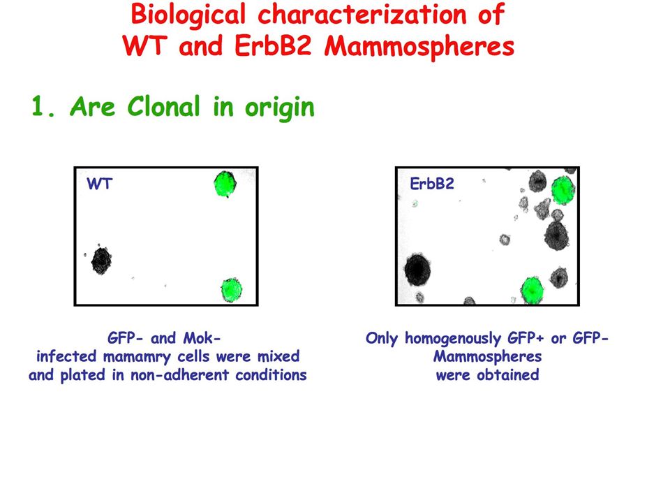
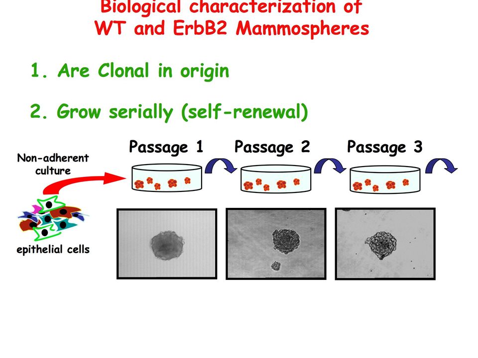
Transplantation into the cleared fat pad of syngenic mice: WT mammospheres form a normal breast tissue ErbB2 mammospheres form tumors 1. Are Clonal in origin 2. Grow serially (self-renewal) 3. Contain SCs Biological characterization of WT and ErbB2 Mammospheres
 3. Contain SCs Biological characterization of WT and ErbB2 Mammospheres.")
57
decrease in number during passages (limited lifespan) Analysis of the replicative potential of Normal and Tumor mammospheres: (serial growth) WT increase in number during passages (near-immortal) ErbB2 R 2: 0,98 502% decrease 10 1 10 5 10 3 10 -1 WT R 2: 0,99 64% decrease ErbB2 : fixed increase at every passage (502%) WT : fixed decrease at every passage (64%) (exponential curves) ErbB2
 Analysis of the replicative potential of Normal and Tumor mammospheres: (serial growth) WT increase in number during passages (near-immortal) ErbB2 R 2: 0,98 502% decrease WT R 2: 0,99 64% decrease ErbB2 : fixed increase at every passage (502%) WT : fixed decrease at every passage (64%) (exponential curves) ErbB2")
58
Stem Cell divisions permit generation of more SCs (‘self-renewal’) and production of cells that differentiate 1.Asymmetric cell division Pr. SC Each SC divides to generate one daughter with SC fate and one that differentiates (progenitror) Mechanisms: 1.Asymmetric localizzation of cell polarity (PINS and aPKC) and cell fate determinants (Numb and Prospero) 2.Asymmetric placement of daughter cells relative to the stem cell niche This strategy leaves stem cells unable to expand in number
 Mechanisms: 1.Asymmetric localizzation of cell polarity (PINS and aPKC) and cell fate determinants (Numb and Prospero) 2.Asymmetric placement of daughter cells relative to the stem cell niche This strategy leaves stem cells unable to expand in number.")
59
2. Symmetric cell division Each SC divides to generate daughter cells that are destined to acquire the same fate SC Pr. SC Limited data available on the modes of division of mammalian SCs: 1.Some mammalian SCs use conserved mechanism to divide asymmetrically; 2.Mammalian SCs can expand in number during development (HSCs, Neural and Epidermal SCs) or after injury (neural SCs after stroke or HSCs after chemotherapy).
 or after injury (neural SCs after stroke or HSCs after chemotherapy)..")
60
ErbB2 Asymmetric 10,3% Symmetric 78,2% Uncertain 11,5% WT Asymmetric 59,5% Symmetric 7,2% Uncertain 33,3% Increased frequency of Symmetric Divisions in tumor cells (ErbB2) vs WT cells
 vs WT cells")
61
Nuovi risultati e incertezze I dati di maggiore rilevanza a supporto della teoria delle CSC derivano da xenotrapianti di cellule tumorali umane in topi immunocompromessi Recentemente è apparso un lavoro molto importante sulla caratterizzazione delle CSC nel melanoma, dove emerge che: –almeno in questo tumore, il numero di cellule con caratteristiche di CSC è altissimo (se si accettano alcune assunzioni, si arriva quasi al 100% delle cellule): se tutte le cellule sono CSC, le CSC non esistono –i protocolli sperimentali per gli xenotrapianti possono influenzare l’attecchimento di determinate sottopopolazioni
: se tutte le cellule sono CSC, le CSC non esistono –i protocolli sperimentali per gli xenotrapianti possono influenzare l’attecchimento di determinate sottopopolazioni")
62
Importanza del topo ricevente e delle condizioni sperimentali

63
I melanomi possono iniziare a partire da una singola cellula

64
Ci sono modelli complementari/alternativi? Plasticità fenotipica: non c’è una vera e propria gerarchia (staminale->non-staminale), ma diversi stati cellulari determinati dalle condizioni “ambientali” (microambiente e segnali) –La stessa cellula può assumere reversibilmente morfologia diversa, espressione di diversi pattern trascrizionali e non di mutazioni irreversibili, manifestando nei suoi diversi fenotipi una maggiore o minore propensione alla “staminalità”
, ma diversi stati cellulari determinati dalle condizioni ambientali (microambiente e segnali) –La stessa cellula può assumere reversibilmente morfologia diversa, espressione di diversi pattern trascrizionali e non di mutazioni irreversibili, manifestando nei suoi diversi fenotipi una maggiore o minore propensione alla staminalità .")
Presentazioni simili


>")


>")

>")


